
We use cookies to ensure our website works properly and to personalise your experience. Cookies policy
Late Adv Dadasaheb Chavan Memorial Institute Of Pharmacy Malwadi Masur.
Persimmon (Diospyros kaki L.) peel, an abundant by product of the fruit processing industry, is increasingly recognized as a valuable source of bioactive compounds with significant health promoting potential. It is particularly rich in phenolic compounds, flavonoids, tannins, carotenoids, dietary fiber, and other phytochemicals that contribute to its strong biological activities. Numerous studies have demonstrated that persimmon peel exhibits potent antioxidant activity, as evidenced by assays such as DPPH, FRAP, hydroxyl radical scavenging, and metal chelating capacity, indicating its ability to reduce oxidative stress and protect biomolecules from oxidative damage. In addition to antioxidant properties, persimmon peel shows promising antidiabetic activity through mechanisms including inhibition of carbohydrate digesting enzymes, improvement of glucose metabolism, and modulation of insulin sensitivity. Various extraction techniques, such as maceration, Soxhlet extraction, and environmentally friendly methods using aqueous alcoholic solvents, have been employed to efficiently recover these bioactive compounds. Overall, persimmon peel represents a sustainable and economical source of functional ingredients with potential applications in nutraceuticals, functional foods, and pharmaceutical formulations
Persimmon Fruits play a very important role in human nutrition and diet. Consumption of these nutrients plays a role in the development of health with the presence of potentially bioactive components, and also the phytochemicals they contain are various bioactive compounds that are widely accepted for their useful roles in human physiology.[1,2]. Persimmon (Diospyros kaki L.) is a member of the Ebenaceae family and is a very popular and important fruit in East Asian countries, being widely produced in China, South Korea, and Japan.[3,4]. The name “persimmon” (Diospyros) originates from the Greek dióspuron, which means “food of Zeus”, while “kaki” comes from the Japanese kaki.[3,5]. Persimmon (Diospyros kaki L.) stands as one of the most fruits globally, particularly dominant in Asia, which encompasses approximately 91 percent of its worldwide production.[6,7]. Herbs, fruits, and vegetables are considered the richest sources of biologically active secondary metabolites, including carotenoids, glucosinolates, polyphenols, and saponins . The consumption of fruits and vegetables globally supports to meet the requirements of vitamins and secondary metabolites for better health . Persimmon (Diospyros kaki L.), a seasonal fruit, has been cultivated for thousands of years in Asia. It is believed to have originated from China and regarded as a traditional fruit . Later it spread out to Korea, Japan, and other parts of the world as an exotic god fruit . The fruit has attracted the attention of several researchers because of its several health‐promoting properties. Persimmon is generally recognized as an outstanding source of biologically active compounds associated with both nutritional and nutraceutical values (Nazir et al., 2013). It has been also noted to contain high levels of water soluble dietary fiber, minerals, trace elements, a significant amount of antioxidants (β‐carotene, β‐cryptoxanthina, lutein, zeaxanthin, and lycopene) and some are active with provitamin A (β‐carotene and β‐cryptoxanthine).[8]. Persimmon is a spherical fruit with a color that ranges from reddish to yellow according to carotene content, the pulp is a viscous orange-red and somewhat rough, depending on the content of tannins. The pulp, regardless of the variety, consists mainly of mucilage and pectin, which is responsible for its characteristic appearance.[3,9]. Generally, over 400 species of persimmon are planted globally. Among these, Diospyros kaki, Diospyros virginiana, Diospyros oleifera, and Diospyros lotus are of significant importance. The popular varieties grown in Japan .[10 ]. The eating quality of persimmon is considered best at the end of the pre-climacteric stage owing to presence of maximum sugars and desired orange color. It is for further info that fruit color is developed just before onset of the ethylene induced respiration . The botanical classification of persimmon fruits is described in Table 1.[10].
Table 1 : Taxonomic Classification :
|
Domain |
Eukaryota |
|
Kingdom |
Plantae |
|
Subkingdom |
Tracheibionta |
|
Division |
Magnoliophyta |
|
Order |
Ebenales |
|
Suborder |
Ebenineae |
|
Family |
Ebenaceae |
|
Genus |
Diosyros |
|
Species |
kaki |
The methodology for this review involved a systematic search and analysis of the literature , focusing on the pharmacological properties of Diospyros kaki, particularly its anti diabetic activities A wide and comprehensive coverage of the available studies was ensured by the retrieval from several reputable databases, including Google Scholar,PubMed, SciSpace, Science Direct, IMMAPT 2.0,PubChem and Web of Science. The search strategy utilized targeted keywords such as D. kaki, anti Diabetic ,pharmacological activity, phytochemicals and TCM . Studies describing the pharmacological characteristics of D. kaki with emphasis on those investigating its anti diabetic effects through in vitro, in vivo or clinical models. Unrelated topics and unavailable full papers were excluded. The extracted data included identified phytochemicals, mechanisms of action, extraction . persimmonfruit ( Diospyros kaki L) contain More Thane 200 identified constituents, including essential nutrients, phenolic compound, carotenoids, vitamins, minerals, and other bioactive molecules, which collectiveiy contribute to their nutritional, antioxidant, and nutraceutical potential.[3].Persimmon fruits (Diospyros kaki L) have been reported to contain approximately 80-100 individual phenolic compounds, comprising phenolic acids, flavonoids, tannins, and anthocyanin’s with tannin representing the predominant fraction.[11].Persimmon fruits (Diospyros kaki) are rich in phenolic compounds, which contribute to their antioxidant properties and astringency.Studies identify dozens of these compound including monomeric phenolic, flavonoids, and polymeric tannin,with variety and ripeness. Key ones are compiled from scientific analyses below. [12].
Fig.1 Persimmon Fruit
Phenolic Acids :-
Table 1: Number of Phenolic Compound of Persimmon Kaki
|
Phenolic acid |
Concentration Range (mg/100g or ppm) |
Relative %of total phenolics |
Uses |
|
Gallic acid |
0.72-2.43 mg/100g;upto 377 mg/kg |
20-50 % |
Antioxidant; Anti inflammatory; Cardiovascular protection |
|
Protocatechuic acid |
Trace to low mg/kg |
<5 % |
Anticancer; Anti diabetic |
|
Ferulic acid |
27.47 ppm |
1-3 % |
Anti inflammatory; Vitamin stabilizer |
|
p-coumaric acid |
Trace to low mg/kg |
2-10 % |
Antimicrobial; Anticancer; |
|
Vanillic acid |
28.84 ppm |
1-3 % |
Antiioxident; Anti inflammintory |
|
Caffeic acid |
0.078-0.1m100gg/ |
2-8%
|
Antiviral; Neuroprotective |
|
Chlorogenic acid |
0.171-0.274 mg/100g |
5-15% |
Blood sugar control; weight loss aid |
Flavonoids :-
Table 2: Number Of Phenolic Compound of Persimmon Kaki
|
Flavonoid |
Concentration range |
Relative % |
Uses |
|
Rutin |
100-500 µg/100g FW |
10-20 % |
Antioxidant; heart protection |
|
Isoquercitrin |
200-800 µg/100g FW |
15-25 % |
Cholesterol lowering; skin health |
|
Hyperoside |
Trace-300 µg/100g |
5-10 % |
Anticancer; enzyme inhibition |
|
Quercetin |
816µg/100g |
15-20 % |
Anti histamine; antiviral |
|
Kaempferol |
Trace-200µg/100g |
~5 % |
Antiobesity; antioxidant |
|
Myricetin |
Trace-150µg/100g |
3-5 % |
Nuroprotection; blood sugar control |
Tannin :
Table 3 : Number Of Phenolic Compound of Persimmon Kaki
|
Tannin |
Concentration range |
Relative % |
Uses |
|
Catechin |
100-500 µg/100g FW |
10-20 % |
Antioxidant; heart protection |
|
Epicatechin |
200-800 µg/100g FW |
15-25 % |
Cholesterol lowering; skin health |
|
Epicatechin gellate |
Trace-300 µg/100g |
5-10 % |
Anticancer; enzyme inhibition |
|
Ellagic acid |
11-20 g/kg |
Variable |
Anticancer; Skin health |
|
Proantocyanidine |
Trimers 1.7-5.1 |
1-7% |
Antidiabetic; anticancer |
Mechanism Of Action :
Free Radical Scavenging: Phenolic compounds like Gallic acid have multiple hydroxyl (-OH) groups on their aromatic rings. These groups can donate a hydrogen atom or an electron to stable free radicals (like DPPH or ABTS radicals), thereby neutralizing them and preventing them from causing cellular damage. The resulting phenolic radical is stabilized by resonance, dampening its reactivity.
Reducing Power: These compounds can reduce oxidized intermediates (e.g., ferric ions to ferrous ions in the FRAP assay) by donating electrons. This reducing ability is a measure of their potential antioxidant activity.
Metal Chelation: Phenolic compounds can chelate transition metal ions, such as iron and copper, which often catalyze the production of harmful free radicals in the body. By binding these metal ions, they inhibit the initiation of oxidative chain reactions.[13].
Persimmon peel is rich in phenolic compounds, including protocatechuic acid, which contribute to its potential antidiabetic effects. These compounds primarily work by inhibiting key enzymes involved in carbohydrate digestion and enhancing glucose metabolism. Protocatechuic acid and related phenolics inhibit pancreatic alpha-amylase, slowing starch breakdown and reducing postprandial glucose spikes. They also suppress alpha-glycosidase activity, further delaying carbohydrate absorption in the gut. Additional actions include boosting insulin sensitivity, promoting glucose uptake in muscles, and lowering oxidative stress linked to diabetes complications. [10].
p-Coumaric acid, a phenolic compound found in persimmon peels, exhibits antidiabetic effects primarily by modulating glucose metabolism and enhancing insulin sensitivity.p-Coumaric acid lowers blood glucose levels by inhibiting gluconeogenic enzymes like glucose-6-phosphatase and fructose-1,6-bisphosphatase while boosting glycolytic enzymes such as hexokinase. It also upregulates insulin levels and activates GLUT2 transporters in the pancreas, improving glucose uptake. Additionally, it reduces lipid accumulation by lowering cholesterol, triglycerides, LDL-C, and VLDL-C while raising HDL-C.[14].
Rutin, a flavonoid phenolic compound found in persimmon peels, offers heart protection through multiple mechanisms. Persimmon peels are rich in such phenolics, contributing to antioxidant and cardiovascular benefits.
Rutin inhibits coronary heart disease by modulating ERK1/2 and Akt signaling pathways, reducing myocardial infarct size and improving cardiac function in animal models. It also suppresses TGF-β1 and SMAD2 expression while enhancing antioxidant defenses to prevent fibrosis and oxidative damage.[15].
Isoquercitrin, a key phenolic compound found in persimmon peels, contributes to cholesterol-lowering effects primarily through antioxidant and metabolic pathways. It activates AMPK signaling to enhance fatty acid oxidation and reduce lipid accumulation in hepatocytes.
Isoquercitrin attenuates free fatty acid-induced oxidative stress, lowering triglycerides, superoxide dismutase, and malondialdehyde levels while reversing proinflammatory cytokines in liver models. It also promotes cholesterol efflux via up regulation of ABCA1 and LDL receptor expression, alongside inhibiting HMG-CoA reductase activity.[16]
Myricetin, a key phenolic flavonoid found in persimmon peel extracts like myricitrin, provides neuroprotection primarily through antioxidant and anti-inflammatory actions. Persimmon peels contain high levels of myricitrin, a glycosylated form of myricetin, which exhibits strong free radical scavenging and metal-chelating properties.
Myricetin reduces oxidative stress by inhibiting reactive oxygen species (ROS) production, lipid peroxidation, and boosting antioxidants like glutathione (GSH) and superoxide dismutase (SOD). It suppresses neuroinflammation by lowering pro-inflammatory cytokines and activates pathways such as Nrf2, Akt, and ERK1/2 for cellular protection. Direct inhibition of caspase-3 prevents apoptosis in models like oxygen-glucose deprivation (OGD).[17].
Persimmon peel contains phenolic compounds like catechin, which act as potent antioxidants. Catechin donates hydrogen atoms to neutralize free radicals and chelates metal ions to prevent oxidative damage
Catechin primarily functions as a radical scavenger by donating electrons or hydrogen to stabilize reactive oxygen species (ROS) like superoxide and hydroxyl radicals. It also chelates pro-oxidant metals such as iron and copper, inhibiting Fenton reactions that generate harmful hydroxyl radicals. These actions reduce lipid peroxidation and cellular oxidative stress in vitro and in vivo.[18].
Persimmon peel contains phenolic compounds like tannins and flavonoids that contribute to cholesterol-lowering effects. Epicatechin, a key flavonoid in persimmon, plays a role through antioxidant and lipid-modulating actions. These compounds reduce cholesterol absorption and synthesis while enhancing excretion.
Phenolics decrease intestinal cholesterol absorption by binding bile acids, increasing fecal excretion. They suppress hepatic fatty acid synthesis via lowered activities of enzymes like FAS, G6PD, and PAP. Combined with fiber, these actions normalize plasma LDL, VLDL, and triglycerides while elevating HDL ratios.[19,20].
Persimmon peel is rich in proanthocyanidins, polyphenolic compounds with notable antidiabetic potential. These oligomers and polymers help regulate blood glucose and mitigate diabetic complications through antioxidant and anti-inflammatory actions.
Proanthocyanidins lower serum glucose and glycosylated proteins by inhibiting oxidative stress and advanced glycation end-products (AGEs) formation in diabetic models. They down regulate proinflammatory genes like NF-κB p65, COX-2, and iNOS, reducing inflammation linked to hyperglycemia.
Oligomeric forms from persimmon peel outperform polymers in decreasing triglycerides, cholesterol, and non-esterified fatty acids while enhancing PPARα expression to promote fatty acid oxidation. This attenuates hyperlipidemia and supports insulin sensitivity in type 2 diabetes.[10,21]
Materials :-
Persimmons sourced from Gwangyang, Korea, were dried and peeled. The peels were cut into small pieces, washed three times with tap water, and then dried in a hot air dryer (Enex-Co-600; Enex, Koyang, Korea) at 50°C for 15 h. The dried peels were pulverized using a blender (KA-2610; Jworld Tech, Ansan, Korea) for 30 s, passed through a 35-mesh sieve, and stored at –20°C until use.[22].
Preparation of Extracts :-
Dried persimmon peel powder (15 g) was extracted twice with 300 mL of distilled water (DW) or ethanol solutions (50%, 70%, and 99%, v/v) by overnight shaking in a VS-8480 orbital shaker (Vision Scientific, Bucheon, Gyeonggi-do, Korea) at room temperature. The resulting extracts were filtered through Whatman No. 1 filter paper, and the solvents were removed under reduced pressure using a CCA-1110 vacuum evaporator (Rikakikai, Tokyo, Japan) at 45 °C until complete dryness. Following ethanol evaporation, the persimmon peel ethanolic extracts were re-dissolved in distilled water to a final concentration of 5% (w/v).[22].
Extraction yield :-
The extraction yield for each solvent was determined by subtracting the dry weight of the plant residue remaining after extraction from the initial dry weight of the original plant material.[22].
Maceration :-
Maceration is an extraction method consisting of a liquid (water, oil, alcohol, etc.) in which the plant is immersed inside an airtight container (Fig. 1). The process is carried out at room temperature with different periods of time depending on the liquid and plant material used (a mixture of ethanol and water (30–90%), a mixture of methanol and water (60–90%), from 20–60°C, from 2– 72 hours). Environmentally Friendly Methods for Flavonoid Extraction from Plant Material: Impact of Their Operating Conditions on Yield and Antioxidant Properties The infusion method in this form has a number of disadvantages: extraction takes a long time; evaporation of the extractant, which causes a decrease in the volume of the tincture; the swollen plant mass cakes at the bottom of the vessel, which requires additional action to eliminate sticking.[23]
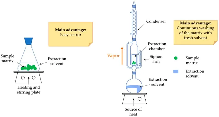
Fig 1.Maceration extraction (ME) with a source of heat (left) and Soxhlet extraction[23]
Percolation :-
Percolation is an extraction method in which the plant is placed in a narrow cone-shaped vessel, open on both sides, called a percolator extractor, and a liquid [16] (solvent: water; ethanol; methanol; a mixture of ethanol and water (50–95%) is passed through it; mixture of methanol and water (80%), from 10–25°C for 24–72 hours) (Fig. 2). This process is carried out at room temperature by passing liquid between solid substances drop by drop, while the extracted substances pass from the raw material to the extractant as a result of their dissolution and diffusion. The percolation rate must be such that the diffusion of the extracted substances into the extract has time to occur. Extracts obtained by infusion or percolation require mandatory purification.[23]
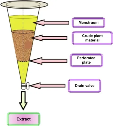
Fig 2.Percolation extraction method[23]
Soxhlet Extraction :-
Compared with other conventional methods used for phenolic compound extraction, Soxhlet extraction requires relatively less solvent and time, while the processing cost remains low. In addition, the extraction apparatus is simple to operate and is suitable for both preliminary and large-scale extractions, offering a high recovery rate . Soxhlet extraction is an improved technique based on reflux extraction that also incorporates the advantages of percolation. Through continuous reflux and siphoning, fresh solvent is repeatedly recycled to extract the target compounds efficiently. The process is largely automated, and compared with conventional extraction methods, Soxhlet extraction generally consumes less solvent and time. However, Soxhlet extraction is a thermal technique, and prolonged extraction at elevated temperatures may lead to thermal degradation of heat-sensitive phenolic compounds. Several studies have successfully employed Soxhlet extraction for phenolic compound recovery. Alara reported that the highest extraction yield from Vitis cinerea (Engelm.) Engelm. ex Millardet leaves was obtained using 60% ethanol. Aspé and Fernández demonstrated that Soxhlet extraction achieved a higher extraction rate than microwave-assisted extraction (MAE), ultrasound-assisted extraction (UAE), and conventional extraction. Nevertheless, compared with UAE and MAE, Soxhlet extraction requires a longer duration to achieve comparable extraction efficiency. Although Soxhlet extraction can yield higher total phenolic and tannin contents than conventional methods, extended extraction periods may result in phenolic degradation due to high temperatures. For example, Quahida reported a total phenolic content of 528.81 mg GAE/g after 6 h of Soxhlet extraction using 70% acetone.[24]
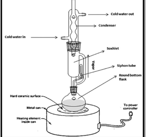
Fig 3. Soxhlet extraction method[23]
Pressurized Liquid Extraction :-
In pressurized liquid extraction (PLE), solid samples are typically packed into a stainless-steel extraction vessel, filled with appropriate solvents, and extracted for 5–15 min under elevated temperature and pressure. Under high pressure, solvents remain in the liquid state at temperatures above their normal boiling points. This condition enhances the solubility and diffusion rate of target compounds in the solvent and facilitates deeper solvent penetration into the sample matrix. Compared with conventional extraction methods, PLE significantly reduces solvent consumption and extraction time while offering improved repeatability. PLE has been widely applied for the extraction of natural products, including anthocyanins and saponins (Ju and Howard, 2003; Zhang et al., 2018). In comparison with conventional techniques, PLE requires an equivalent or even lower volume of solvent and is highly time-efficient, while also minimizing sample handling (Alonso-Salces et al., 2001). Katsinas et al. (2021) reported that, relative to conventional extraction, PLE reduced extraction time by 67% and solvent usage by 38%, while simultaneously increasing phenolic compound yield. However, the enhanced extraction efficiency of PLE is primarily attributed to the use of high temperature and pressure to maintain solvents in the liquid phase, which may lead to thermal degradation of heat-sensitive compounds. Therefore, Ju and Howard (2003) recommended the use of solvents that are less efficient at low temperatures, such as water, in combination with PLE to improve extraction efficiency while minimizing thermal damage.[24].
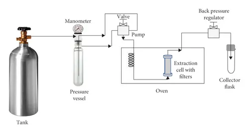
Fig 4.Pressurized Liquid Extraction Equipment [25].
Supercritical Fluid Extraction :-
Supercritical fluid extraction (SFE) is an extraction technique that employs supercritical fluids (SFs) as extraction solvents. Supercritical fluids possess liquid-like solvating power combined with gas-like diffusivity, enabling them to dissolve a wide range of natural compounds efficiently. However, the solvating properties of SFs are highly sensitive to small variations in pressure and temperature near their critical points, which can significantly affect extraction performance. One of the most commonly used supercritical fluids is supercritical carbon dioxide (SC-CO₂), which has been widely applied in extraction processes. The advantages of SC-CO₂ include its low critical temperature (31 °C), high selectivity, chemical inertness, and non-toxic nature, making it suitable for extracting thermolabile compounds. Due to its low polarity, SC-CO₂ is particularly effective for the extraction of non-polar compounds such as lipids. Its solvating power can be further enhanced by the addition of polar modifiers or co-solvents. However, for the efficient extraction of phenolic compounds using SFE, the incorporation of co-solvents is often necessary to increase solvent polarity and improve extraction efficiency. The solubility of phenolic compounds—particularly phenolic acids such as gallic acid, methyl gallate, and caffeic acid—in supercritical fluids is strongly influenced by the choice of co-solvent. Radzali et al. (2020) investigated the effects of various co-solvents, including ethanol, water, methanol, 50% ethanol–water, and 70% methanol–water, on SFE by evaluating the antioxidant capacity of the resulting extracts. Their results demonstrated that a 70% ethanol–water co-solvent significantly enhanced the extraction efficiency and overall extraction quality of SFE. Phenolic compounds are highly sensitive to environmental factors such as temperature, oxygen, and light, which can lead to undesirable degradation. Compared with conventional extraction methods, SFE produces relatively clean extracts and minimizes oxidative degradation of phenolic compounds (Bleve et al., 2008; Vatai et al., 2009). However, the high operational cost of SFE limits its widespread industrial application; consequently, it is primarily employed for the extraction of high-value products (Sunarso and Ismadji, 2009). Liu et al. (2013) demonstrated that SFE is an effective technique for the extraction of phenolic compounds. Key parameters influencing extraction efficiency include temperature, pressure, number of extraction cycles, and extraction time. Among these factors, total phenolic content obtained by SFE is predominantly affected by the number of extraction cycles. Optimal extraction conditions were reported as 2 h per extraction at 50 °C and 350 bar. The extraction efficiency of SFE was further evaluated by Ashfaq et al. (2021), who compared SFE and conventional extraction methods for tea extracts used as preservatives in coriander sauce. The total polyphenol content of SFE extracts (224.40 mg GAE/100 mL) was higher than that of conventional extracts (208.31 mg GAE/100 mL), with similar trends observed across multiple antioxidant assays.[24].
Ultrasounic assisted Extraction :-
Ultrasound is employed in extraction processes to reduce extraction time and enhance quality by inducing solvent-producing cavitation and high shear forces. Ultrasound-assisted extraction (UAE) is a modern method known for its simplicity, energy efficiency, and high reproducibility, offering a substantial yield of active compounds. UAE is more efficient, requiring less solvent and a shorter extraction duration compared to conventional methods .Mainly utilized in solid/liquid systems, UAE disrupts the cellular walls of plant materials, facilitating mass transfer across membranes and increasing solvent access to analytes. Extraction efficiency in UAE is influenced by factors like solvent composition, solvent-to-sample ratio, ultrasound amplitude and cycle, solvent pH, and temperature . While stronger ultrasonic applications can accelerate changes, cost considerations in food industries often lead to optimized applications for the best results with minimal energy usage . To enhance extraction efficiency, factors such as amplifying ultrasound power, minimizing moisture content in food materials, and controlling temperature are considered. Proper selection of ultrasound frequency is essential, impacting the size of bubbles produced during resonance . Solvent selection and temperature were identified as crucial factors impacting UAE efficiency. For highly polar phenolic compounds, extraction with pure organic solvents may exhibit low efficiency, making ethanol/methanol mixtures with water in various proportions commonly used as effective extraction solvents.[26].
The quantification, purification, separation, and identification of individual phenolic compounds, including anthocyanins, often rely on sophisticated and expensive analytical instruments and involve time-consuming sample preparation procedures. Various analytical techniques have been employed for the identification and quantification of anthocyanins, including paper chromatography (PC), thin-layer chromatography (TLC), column chromatography, solid-phase extraction, counter-current chromatography, and high-performance liquid chromatography (HPLC) . In addition, gas chromatography (GC) and HPLC coupled with mass spectrometry (MS) have demonstrated strong capability for the detection and characterization of phenolic compounds in diverse matrices .
These chromatographic techniques offer high accuracy, sensitivity, and efficiency, enabling reliable analysis within relatively short timeframes. Advances in HPLC methodologies have further enabled the simultaneous identification of phenolic compounds across a broad range of polarities . Although HPLC and GC remain the most widely used techniques for phenolic analysis, their high cost and requirement for specialized instrumentation have sustained the use of alternative chromatographic methods for the determination of phenolic compounds.[26]
Most Common Indicators for Phenolic Compound :-
|
Compound Type |
Best Indicator |
|
Phenolic Acids |
FeCl₃, UV |
|
Flavonoids |
AlCl₃, NP/PEG |
|
Anthocyanins |
UV, acidic solvent |
|
Tannins |
FeCl₃ |
Paper Chromatography And Thin Layer Chromatography :-
Paper chromatography (PC) and thin-layer chromatography (TLC) are partition-based techniques commonly used for the separation of phenolic compounds in food matrices. Although PC is a relatively simple and less frequently applied method compared with high-performance liquid chromatography (HPLC) and gas chromatography (GC), it has been successfully employed for the separation and identification of phenolic compounds in tea leaves and green leafy vegetables [26,27]..
Thin-layer chromatography (TLC) is recognized as a simple, rapid, and reliable technique, particularly suitable for the analysis of phenolic compounds in crude plant extracts. Various TLC approaches are cost-effective and enable the simultaneous detection of multiple samples on a single plate within a short analysis time. Moreover, silica gel-based TLC combined with video imaging has been reported as a valuable complementary fingerprinting technique for the identification of phenolic acids and flavonoid fractions from different Salvia (sage) species.[26,28].
Hiigh-Speed counter-current chromatography :-
High-speed counter-current chromatography (HSCCC) is a biphasic liquid–liquid partitioning technique widely used for the isolation and separation of diverse natural compounds [36,41–43]. Because HSCCC operates without a solid stationary phase, it eliminates irreversible adsorption of sample components, enabling efficient isolation and purification of target compounds directly from crude extracts without extensive sample pretreatment[26]
High-Performance liquid chromatography :-
The quantification and identification of phenolic compounds have been extensively investigated using high-performance liquid chromatography (HPLC) coupled with a diode array detector (DAD) . For the analysis of complex matrices, chromatographic techniques—particularly HPLC—are considered highly suitable due to their high sensitivity and resolving power, enabling the effective separation and identification of multiple anthocyanins while providing detailed qualitative and quantitative information. Despite their widespread recommendation, the diversity of available HPLC protocols may complicate the selection of an optimal method for the determination of phenolic compounds or anthocyanins. Nevertheless, HPLC-based flavonoid analysis allows the identification of individual compounds within samples, offering valuable insights into the specific flavonoid profiles of different plant materials, such as grape varieties. However, accurate identification remains challenging because of the limited commercial availability of reference standards .
At present, the most commonly employed methods for phenolic compound analysis involve HPLC coupled with ultraviolet (UV), electrochemical, or mass spectrometric (MS) detection, including particle beam or electron ionization MS. In addition, gas chromatography–mass spectrometry (GC-MS), high-speed counter-current chromatography (HSCCC), chiral capillary electrophoresis (CE), and Fourier transform near-infrared reflectance spectroscopy are frequently applied. Hyphenated techniques such as HPLC-MS and HPLC-MS/MS, which are based on HPLC separation, provide detailed information on molecular mass and structural characteristics of phenolic compounds. These approaches are considered superior in terms of analytical efficiency, suitability for routine analysis, and effectiveness in the separation, identification, and quantification of phenolic constituents . Reversed-phase HPLC is particularly widely used for the analysis of various phenolic groups, while ultra-performance liquid chromatography (UPLC) has been increasingly adopted to enhance the speed, resolution, and sensitivity of phenolic compound analysis in diverse matrices.[26,29]
Gas Chromatography And Mass Chromatography :-
The quantification and identification of phenolic compounds have been extensively investigated using high-performance liquid chromatography (HPLC) coupled with a diode array detector (DAD) . For the analysis of complex matrices, chromatographic techniques—particularly HPLC—are considered highly suitable due to their high sensitivity and resolving power, enabling the effective separation and identification of multiple anthocyanins while providing detailed qualitative and quantitative information. Despite their widespread recommendation, the diversity of available HPLC protocols may complicate the selection of an optimal method for the determination of phenolic compounds or anthocyanins. Nevertheless, HPLC-based flavonoid analysis allows the identification of individual compounds within samples, offering valuable insights into the specific flavonoid profiles of different plant materials, such as grape varieties. However, accurate identification remains challenging because of the limited commercial availability of reference standards .
At present, the most commonly employed methods for phenolic compound analysis involve HPLC coupled with ultraviolet (UV), electrochemical, or mass spectrometric (MS) detection, including particle beam or electron ionization MS. In addition, gas chromatography–mass spectrometry (GC-MS), high-speed counter-current chromatography (HSCCC), chiral capillary electrophoresis (CE), and Fourier transform near-infrared reflectance spectroscopy are frequently applied. Hyphenated techniques such as HPLC-MS and HPLC-MS/MS, which are based on HPLC separation, provide detailed information on molecular mass and structural characteristics of phenolic compounds. These approaches are considered superior in terms of analytical efficiency, suitability for routine analysis, and effectiveness in the separation, identification, and quantification of phenolic constituents . Reversed-phase HPLC is particularly widely used for the analysis of various phenolic groups, while ultra-performance liquid chromatography (UPLC) has been increasingly adopted to enhance the speed, resolution, and sensitivity of phenolic compound analysis in diverse matrices.[26,30].
NMR Spectroscopy :-
Nuclear magnetic resonance (NMR) spectroscopy enables detailed structural elucidation of flavonoid molecules, including their conformational features and electron density distribution. When combined with ultraviolet (UV) and infrared (IR) spectroscopy, NMR provides highly valuable and complementary information for the comprehensive characterization of flavonoid structures. The number of signals observed in an NMR spectrum reflects the number of magnetically distinct proton environments present in the molecule, while the chemical shift positions allow the identification of specific proton types. Due to the low solubility of flavonoid glycosides in low-polar and non-polar solvents commonly used for NMR analysis, these compounds are often examined in the form of acetylated or trimethylsilyl derivatives. NMR spectroscopy not only enables rapid and accurate determination of substituent positions on the A and B rings of flavonoids , but also facilitates the elucidation of the carbohydrate moiety, including the configuration of the glycosidic linkage as well as the nature and conformation of the sugar component.[23]
IR Spectroscopy :-
Infrared (IR) spectroscopy is widely employed in the analysis of flavonoid compounds to establish and confirm molecular structures. In addition to structural elucidation, IR spectroscopy provides valuable information regarding molecular configuration and conformation. IR spectra arise from vibrational motions of atoms within a molecule; these vibrations occur at different energy levels and may involve stretching or bending along valence bonds. The collective vibrations of all atoms in a molecule generate characteristic absorption bands that are specific to each compound.
The fingerprint region (1400–650 cm⁻¹) is particularly useful for identification purposes and is commonly applied for side-by-side comparison of IR spectra of unknown and reference compounds. Functional groups present in flavonoid molecules can be identified based on their characteristic absorption bands within specific regions of the IR spectrum [27,28]. For example, in flavonoids, the stretching vibration of an unsubstituted carbonyl group in flavanones appears in the range of 1660–1690 cm⁻¹, while the C=O stretching vibration of flavonols occurs between 1637 and 1650 cm⁻¹. The presence of a hydroxyl group at the C-7 position lowers the carbonyl stretching frequency by approximately 10–15 cm⁻¹. Furthermore, hydrogen bond formation between the C=O group and a hydroxyl group at the C-5 position accounts for a further decrease in the C=O stretching frequency to around 1640 cm⁻¹.Stretching vibrations of conjugated C=C double bonds are observed as several intense absorption bands in the region of 1600–1470 cm⁻¹. Aromatic C–H stretching vibrations in rings conjugated with a carbonyl group appear in the region of 3130–3110 cm⁻¹. Free aliphatic hydroxyl groups absorb in the range of 3625–3600 cm⁻¹, whereas phenolic hydroxyl groups of flavonoid aglycones are detected in the broader region of 3300–2700 cm⁻¹. Hydroxyl groups of carbohydrate substituents typically exhibit absorption bands in the range of 3600–3300 cm⁻¹. IR spectroscopy also enables the differentiation of α- and β-anomers of monosaccharides and their derivatives. The α-configuration of the C–O bond is characterized by an absorption band at 844 ± 8 cm⁻¹, whereas the β-configuration shows a characteristic band at 891 ± 7 cm⁻¹. When combined with thin-layer chromatography (TLC), IR spectroscopy enhances the selectivity of qualitative analysis. The quantity of compound recovered from a TLC plate is often sufficient to obtain IR spectra of micro-samples, making this technique particularly valuable for the analysis of biologically active substances that occur in low concentrations in plant materials.[23].
The DPPH (2,2-di(4-tert-octylpheny)-1-picrylhydrazyl)test
The DPPH assay is a widely used method for evaluating free radical scavenging activity. The reaction mechanism involves a color change from deep purple (DPPH•) to pale yellow (DPPH-H) as the radical is reduced by antioxidants through hydrogen or electron donation (Benzie and Strain, 1999). DPPH exists as a stable free radical in both solid and liquid states and is readily soluble in organic solvents such as methanol and ethanol, allowing it to react with free radicals that are poorly soluble in water. As reported by Staško et al. (2007), the water content of the reaction medium should be maintained below 60% to ensure adequate solubility and stability of the DPPH radical.
The DPPH radical is neutralized upon accepting electrons or hydrogen atoms from antioxidant compounds, resulting in a measurable decrease in absorbance. Owing to this color change, antioxidant capacity can be quantified spectrophotometrically at 517 nm. Antioxidant activity determined by the DPPH assay is commonly expressed in two ways: EC₅₀, defined as the concentration of antioxidant required to reduce the initial DPPH radical concentration by 50%, and TEC₅₀, which represents the time required to reach the equilibrium state corresponding to the EC₅₀ value.[24]
Hydroxyl radical scavenging activity assay :-
The hydroxyl radical scavenging activity (•OH-RSA) assay is an antioxidant evaluation method based on the Fenton reaction system (Fe³⁺–ascorbate–EDTA–H₂O₂), which generates hydroxyl radicals from hydrogen peroxide. In the presence of iron ions, hydrogen peroxide undergoes a series of reactions known as the Fenton reaction, producing highly reactive hydroxyl radicals (•OH), which can be scavenged by antioxidant compounds.
Quantification in the •OH-RSA assay is based on the measurement of malondialdehyde formed during the degradation of 2-deoxyribose. The degradation products subsequently react with thiobarbituric acid (TBA) to form a colored complex, the absorbance of which is measured spectrophotometrically at 510 nm. A decrease in absorbance indicates effective scavenging of hydroxyl radicals by the tested sample.
Assessment of hydroxyl radical scavenging activity is particularly important in medical and biomedical research, as hydroxyl radicals are among the most reactive oxygen species and can be generated in vivo through iron-mediated reactions, even from relatively non-reactive radicals. Chelation of iron ions can therefore serve as an effective strategy for mitigating oxidative stress-related diseases. However, the toxicity associated with synthetic chelating agents necessitates careful consideration, as therapeutic benefits must be balanced against potential adverse effects.
Franco et al. (2019) demonstrated that antioxidants can protect red blood cells from oxidative damage through mechanisms involving chelation and stabilization of free radicals, which are subsequently eliminated via feces and urine. Owing to the severe side effects reported for many synthetic chelators, the identification of natural chelating agents from plant sources is of considerable scientific and clinical interest. Consequently, the •OH-RSA assay is a valuable tool for evaluating the hydroxyl radical scavenging potential of plant extracts and may facilitate the discovery of novel, less toxic free-radical chelators.[24]
Ferrous ion chelating ability (FICA ) assay :-
The ferrous ion chelating activity (FICA) assay is commonly used to evaluate antioxidant capacity based on the ability of compounds to chelate transition metal ions, such as iron (Fe²⁺) and copper (Cu²⁺). In this assay, ferrous chloride (FeCl₂) is typically used as the source of ferrous ions. The standard procedure involves initially mixing the test sample with FeCl₂, followed by incubation. Subsequently, ferrozine is added to the reaction mixture, where it complexes with unchelated Fe²⁺ to form a colored ferrous–ferrozine complex. The reaction is usually carried out in the dark, and the absorbance is measured spectrophotometrically at 562 nm. The metal-chelating capacity of the sample is quantified by comparing its absorbance to that of a control, with ethylenediaminetetraacetic acid (EDTA) commonly employed as a positive reference standard.
Md. Yusof et al. (2013) applied the FICA assay to evaluate the metal-chelating ability of Pandanus pygmaeus extracts. Their results indicated that only the methanolic extract of P. pygmaeus and the EDTA control exhibited measurable chelating activity. An increase in extract concentration led to a corresponding decrease in the absorbance of the ferrous–ferrozine complex, demonstrating a positive relationship between chelating activity and sample concentration. The maximum chelating activity was observed at a concentration of 10,000 µg/mL, with a chelation percentage of 85.39%. In the FICA assay, chelating ability is typically expressed as an IC₅₀ value, where lower IC₅₀ values indicate stronger metal-chelating activity. In the study by Md. Yusof et al. (2013), EDTA exhibited a lower IC₅₀ value than the P. pygmaeus extract, indicating that while the extract possesses metal-chelating potential, it is less potent than the standard chelating agent.[24].
DISCUSSION
Persimmon (Diospyros kaki) peel exhibits a broad spectrum of biological activities owing to its rich phytochemical composition, including phenolic compounds, flavonoids, tannins, carotenoids, and dietary fiber. Numerous studies have reported its strong antioxidant potential, as demonstrated by DPPH, FRAP, hydroxyl radical scavenging, and metal chelating assays, which collectively contribute to the reduction of oxidative stress and protection of cellular components. Furthermore, persimmon peel shows notable antidiabetic activity through the inhibition of key carbohydrate-digesting enzymes such as α-amylase and α-glucosidase, thereby helping to regulate postprandial blood glucose levels. Its polyphenolic constituents also protect cells against high glucose-induced oxidative damage, a major factor in diabetes-related complications. In vivo studies using diabetic animal models have shown that dietary supplementation with persimmon peel lowers blood glucose, triglyceride, and cholesterol levels, while positively modulating insulin signaling and glucose homeostasis pathways. In addition, anti-inflammatory, hypolipidemic, and antimicrobial activities have been reported, highlighting persimmon peel as a valuable agro-industrial by-product with promising applications in functional foods, nutraceuticals, and pharmaceutical formulations.
CONCLUTION AND RESULT
In this review of the Persimmon peel, it was discovered. that has a wide range of pharmacological properties, including anti-inflammatory, analgesic, antioxidant, anticancer, antibacterial, and so on. However, there is very little evidence on CNS activity.
Persimmon is naturally bestowed with bioactive molecules including proanthocyanidins, flavonoids, tannins, phenolic, carotenoids, dietary fiber, and etc. Persimmon leaves and fruit have imperative significance for coronary health because of hypocholesterolemic, anti-atherosclerosis and antioxidant perspectives. Although hypotensive and anticancer responses have been reported for persimmon and its bioactive especially condensed tannin and flavonoids too but yet demands further probing to unveil their therapeutic mechanisms. In the last, utilization of persimmon and its bioactive components can be effective in reducing the burden of diabetes mellitus. However, coherent and systematic research is still required to bring meticulousness.
REFERENCES


Sangita Shelar, Ananya Dhane, Shraddha Bhosale, Vaishnavi Patil, Anti-Diabetic Efficacy of Persimmon Polyphenols, Int. J. of Pharm. Sci., 2026, Vol 4, Issue 6, 597-614, https://doi.org/10.5281/zenodo.20509174
 10.5281/zenodo.20509174