

We use cookies to ensure our website works properly and to personalise your experience. Cookies policy
1,2,3,4,5,7 School of Pharmacy, RK University, Rajkot, Gujarat, India
6 Faculty of Pharmaceutical Sciences, University of Port Harcourt, Nigeria
Antibody–drug conjugates (ADCs) represent one of the most advanced manifestations of modern prodrug design, combining the target specificity of monoclonal antibodies with the potency of cytotoxic small-molecule payloads. By selectively delivering potent drugs to antigen-expressing cells, ADCs improve efficacy while reducing systemic toxicity relative to conventional chemotherapy. Clinical performance depends on the coordinated function of three components: the antibody, which directs recognition and internalization; the linker, which governs stability and payload release; and the payload, which determines the mechanism and magnitude of cytotoxicity. This review examines ADCs through the lens of prodrug pharmacology, tracing their evolution from Ehrlich's "magic bullet" concept to contemporary approved agents, with emphasis on the causal relationship between target biology, linker chemistry, and payload selection. Current linker technologies, payload classes, conjugation strategies, and FDA-approved ADCs are discussed alongside major challenges (linker instability, off-target toxicity, resistance, and manufacturing complexity) and emerging platforms including bispecific ADCs, ISACs, DACs, and AI-assisted design. Collectively, ADCs achieve classical prodrug goals through a multi-layered targeting strategy, establishing them as a leading paradigm in precision drug delivery.
1.1 The "Magic Bullet" Concept – From Ehrlich to Modern Targeted Therapy
The foundation of modern targeted drug delivery can be traced to the pioneering work of the German scientist Paul Ehrlich, who introduced the concept of the "magic bullet" in the early twentieth century.1 Ehrlich envisioned therapeutic agents that could selectively recognize and destroy disease-causing cells while sparing healthy tissues, thereby maximizing efficacy and minimizing toxicity. Although this concept was initially proposed in the context of infectious diseases, it later became a guiding principle for the development of targeted therapies in oncology.
For decades, the realization of Ehrlich's vision remained elusive due to the lack of technologies capable of achieving highly selective drug delivery. Conventional chemotherapeutic agents exhibited limited specificity, often causing significant damage to normal tissues and resulting in severe systemic toxicities. The advent of monoclonal antibody technology, particularly following the development of the hybridoma technique by Köhler and Milstein in 19752 work for which they shared the 1984 Nobel Prize in Physiology or Medicine. It provided a critical breakthrough by enabling the production of antibodies capable of recognizing specific cellular antigens with high affinity and selectivity.
The integration of monoclonal antibodies with highly potent cytotoxic agents gave rise to antibody–drug conjugates (ADCs), which combine the tumor-targeting capability of antibodies with the cell-killing potency of small-molecule drugs. In this regard, ADCs represent one of the most successful modern implementations of Ehrlich's "magic bullet" concept.
The clinical translation of ADCs, however, was not straightforward. Early generations faced significant challenges, including inadequate target selection, unstable linker chemistries, insufficient payload potency, and dose-limiting toxicities. A major milestone was achieved in 2000 with the approval of the first ADC, gemtuzumab ozogamicin (Mylotarg)3,4, a CD33-targeting antibody conjugated to the potent DNA-damaging agent calicheamicin for the treatment of acute myeloid leukemia. Although initially withdrawn from the market in 20105 due to safety concerns and lack of clinical benefit in confirmatory studies, the drug was subsequently re-approved in 20176 using an optimized fractionated dosing regimen. This experience highlighted the critical importance of antigen selection, linker stability, payload characteristics, and dosing strategy in determining ADC efficacy and safety.
Subsequent advances in antibody engineering, conjugation technologies, linker design7,8 and payload development have transformed ADCs into a rapidly expanding therapeutic class, now spanning multiple regulatory approvals across hematological malignancies and solid tumors.
1.2 Evolution of Prodrug Design – Traditional Prodrugs, The Need for Targeted Delivery, And the Emergence of Biologics
The concept of prodrug design emerged as a rational strategy to overcome the pharmacokinetic and physicochemical limitations of active pharmaceutical ingredients. Traditional prodrugs are pharmacologically inactive or less active derivatives that undergo enzymatic or chemical conversion within the body to release the therapeutically active drug. Historically, prodrug approaches were employed to improve aqueous solubility, membrane permeability, oral bioavailability, metabolic stability, and patient acceptability. Classic examples include enalapril, which enhances the oral delivery of enalaprilat, and valacyclovir, which improves the absorption of acyclovir through transporter-mediated uptake9.
Despite these successes, conventional prodrug strategies offered limited control over the spatial and temporal release of the active drug. Activation typically depended on broadly distributed metabolic enzymes or physiological conditions, resulting in systemic drug exposure following conversion. While such approaches could improve pharmacokinetic properties, they generally failed to address the fundamental challenge of selectively delivering potent therapeutics to diseased tissues while sparing healthy organs10.
This limitation became particularly evident in oncology, where highly effective cytotoxic agents often exhibit narrow therapeutic windows. Although prodrug modification could reduce some adverse effects and improve drug disposition, systemic activation frequently resulted in significant off-target toxicity. Consequently, there was growing recognition that improvements in drug delivery required not only optimized chemistry but also enhanced biological selectivity11.
The emergence of biologics, particularly monoclonal antibodies, represented a transformative advance in this regard. Unlike small-molecule prodrugs, monoclonal antibodies possess the ability to recognize specific cell-surface antigens with high affinity and selectivity, enabling targeted delivery to defined cellular populations. Advances in antibody engineering, recombinant DNA technology, and large-scale biomanufacturing further accelerated their therapeutic application across multiple disease areas, especially cancer12.
The convergence of classical prodrug principles with antibody-mediated targeting ultimately gave rise to antibody–drug conjugates (ADCs). By combining a highly selective antibody, a chemically engineered linker, and a potent cytotoxic payload, ADCs extend the traditional prodrug concept beyond simple pharmacokinetic optimization toward precise, cell-specific drug activation. In this sense, ADCs represent a major evolutionary milestone in prodrug design, transforming a strategy originally focused on modifying drug properties into one capable of achieving targeted therapeutic delivery at the cellular level.
1.3 ADCS as the Pinnacle of Prodrug Engineering – Tripartite Components and Selective Activation
Antibody–drug conjugates (ADCs) represent one of the most sophisticated applications of modern prodrug design, integrating advances in molecular biology, antibody engineering, medicinal chemistry, and drug delivery into a single therapeutic platform. Unlike conventional prodrugs, which rely primarily on physiological or enzymatic activation, ADCs employ a biologically guided activation strategy in which drug release is directed by selective recognition of disease-associated cellular targets.
Structurally, an ADC consists of three essential components: a monoclonal antibody, a chemical linker, and a highly potent cytotoxic payload. The antibody serves as the targeting moiety, recognizing and binding a specific antigen that is preferentially expressed on tumor cells. The linker functions as a molecular bridge connecting the antibody to the payload while maintaining sufficient stability during systemic circulation. The payload, typically several orders of magnitude more potent than conventional chemotherapeutic agents, remains conjugated and largely inactive until released at the target site. Together, these three components determine the efficacy, safety, pharmacokinetics, and therapeutic index of the ADC.
The mechanism of action of ADCs reflects their prodrug-like nature. Following antigen recognition, the antibody–antigen complex undergoes receptor-mediated endocytosis and is transported through the endosomal-lysosomal pathway13,14. Within the intracellular environment, linker cleavage or antibody degradation triggers the release of the active cytotoxic payload. Once liberated, the payload exerts its pharmacological effect through mechanisms such as microtubule disruption, DNA damage15,16, or inhibition of DNA replication, ultimately resulting in tumor cell death. Depending on the physicochemical properties of the released payload, cytotoxic effects may also extend to neighboring antigen-negative tumor cells through the bystander effect17.
From a prodrug design perspective, ADCs offer a unique combination of selective targeting and controlled activation. The antibody provides cellular specificity, the linker governs the timing and location of drug release, and the payload delivers potent anticancer activity. This coordinated tripartite architecture transforms highly toxic small molecules into targeted therapeutics capable of achieving enhanced efficacy with reduced systemic exposure. Consequently, ADCs are widely regarded as the most advanced realization of the prodrug concept, extending traditional strategies for drug optimization into the realm of precision medicine.
1.4 Scope and Objectives of The Review
While numerous reviews have described the clinical development and mechanistic features of ADCs, fewer have examined these agents within the broader conceptual framework of prodrug engineering. This perspective is relevant because ADC activation strategies align closely with classical prodrug objectives, including site-specific delivery, reduced systemic toxicity, and improved therapeutic efficacy.
This review accordingly explores ADCs through the dual lens of prodrug pharmacology and bioconjugation science. After outlining fundamental prodrug principles and positioning ADCs relative to traditional approaches, it develops a design-oriented framework linking target biology, linker engineering, and payload selection as interconnected determinants of ADC performance, emphasizing how antigen characteristics influence intracellular trafficking, how linker chemistry governs payload release, and how payload properties determine efficacy and safety.
Clinically approved ADCs are then examined as practical examples of these principles in action, before evaluating ADCs against classical prodrug objectives to illustrate how advances in antibody engineering, conjugation technologies, and payload development have expanded the scope of targeted drug activation. Current challenges like resistance mechanisms, toxicity, and manufacturing complexity are discussed alongside emerging innovations shaping the next generation of ADC therapeutics.
The overall objective is to provide an integrated understanding of ADCs as advanced targeted prodrugs, with particular focus on the roles of target selection, linker chemistry, and payload strategy in determining clinical success.
2. FUNDAMENTALS OF PRODRUG DESIGN
2.1 Definition and Classification of Prodrugs
A prodrug is a pharmacologically inactive or less-active derivative of a parent drug18,19 that undergoes enzymatic or chemical transformation in vivo to release the active therapeutic moiety. The prodrug concept was developed to overcome limitations associated with drug physicochemical properties, pharmacokinetics, and tissue selectivity while preserving the desired pharmacological activity of the parent compound.
Prodrugs are broadly classified into two categories: carrier-linked prodrugs and bio precursor prodrugs. Carrier-linked prodrugs consist of an active drug covalently attached to a temporary carrier group, commonly referred to as a promoiety, which is removed under specific physiological or enzymatic conditions to release the active drug. Examples include valacyclovir and enalapril, in which the carrier moiety enhances absorption or bioavailability. In contrast, bio precursor prodrugs do not contain a separate carrier group but are instead converted into the active drug through metabolic transformation. Levodopa, which is converted to dopamine following enzymatic decarboxylation, is a classic example of this category20,21.
From a conceptual standpoint, antibody–drug conjugates (ADCs) can be regarded as highly sophisticated carrier-linked prodrugs, in which the antibody and linker together function as an exceptionally large and selective promoiety. Unlike conventional carrier-linked systems, however, ADCs exploit molecular recognition and receptor-mediated uptake to achieve targeted intracellular activation.
2.2 Rationale for Prodrug Design
The development of prodrugs is driven by the need to overcome limitations that restrict the therapeutic usefulness of active pharmaceutical agents. One of the most common objectives is the improvement of aqueous solubility10,22, particularly for highly potent compounds that exhibit poor dissolution characteristics. Enhanced solubility can facilitate formulation development, improve drug exposure, and enable administration of otherwise impractical therapeutic agents.
Another important objective is the enhancement of drug absorption and distribution. Chemical modification can improve membrane permeability, transporter-mediated uptake, or overall bioavailability. Traditional examples include valacyclovir, which exhibits superior oral absorption compared with acyclovir23. In the context of ADCs, however, improved distribution is achieved through a different mechanism: the monoclonal antibody carrier provides prolonged systemic circulation and selective accumulation in antigen-expressing tissues rather than relying on passive diffusion or membrane permeability.24
Prodrug approaches may also be used to prolong drug action by controlling the rate at which the active molecule is released. Conventional systems often achieve this through slow enzymatic or hydrolytic conversion. ADCs similarly exhibit controlled release characteristics, although release is governed by antigen recognition, cellular internalization, intracellular trafficking, and linker cleavage rather than simple hydrolysis kinetics.
Perhaps the most important objective of prodrug design is site-specific drug delivery. Traditional prodrugs attempt to exploit physiological differences such as tissue-specific enzyme expression, pH variation, or metabolic activity18. ADCs represent a major advancement of this principle by incorporating a dedicated targeting mechanism through antibody-mediated antigen recognition, thereby achieving substantially greater selectivity than most conventional prodrug systems.
2.3 Practical Considerations in Prodrug Design
Regardless of the activation strategy employed, successful prodrug development requires a careful balance between stability, activation efficiency, safety, and pharmacokinetic performance. The prodrug must remain sufficiently stable during circulation to prevent premature release of the active drug while retaining the ability to undergo efficient conversion at the intended site of action25.
For ADCs, circulating stability is particularly critical because premature linker cleavage can result in systemic exposure to highly potent cytotoxic payloads, leading to significant toxicity. Equally important is the efficiency of intracellular activation, which depends on antigen binding, cellular uptake, endosomal trafficking, and payload release mechanisms. The toxicity profile of both the intact conjugate and any released metabolites must also be considered, as off-target effects can compromise the therapeutic index.
Finally, favorable pharmacokinetic behavior is essential for maximizing therapeutic benefit. An effective ADC must combine the prolonged circulation characteristics of monoclonal antibodies with efficient delivery and release of the payload within target cells. Consequently, stability, activation, toxicity, and pharmacokinetics serve as the key design parameters against which linker chemistries, conjugation strategies, and payload classes are evaluated throughout this review.
3. ADCS AS ADVANCED TARGETED PRODRUGS
3.1 Traditional Prodrug Vs. Antibody–Drug Conjugate
Although antibody–drug conjugates (ADCs) share the fundamental objective of traditional prodrugs (namely, the controlled delivery of an active therapeutic agent), they differ substantially in their mechanisms of targeting, activation, and pharmacokinetic behavior. Classical prodrugs rely primarily on chemical modification of a parent drug and subsequent activation by endogenous physiological or enzymatic processes. In contrast, ADCs integrate molecular recognition, intracellular trafficking, and controlled payload release into a single therapeutic platform26. Consequently, ADCs can be viewed as an advanced form of carrier-linked prodrug in which selective activation is achieved through antigen-directed targeting rather than solely through differences in tissue metabolism.
TABLE 1:COMPARISON BETWEEN TRADITIONAL SMALL-MOLECULE PRODRUGS AND ANTIBODY–DRUG CONJUGATES
|
Feature |
Traditional Small-Molecule Prodrug |
Antibody-Drug Conjugate |
References |
|
Targeting |
Tissue-specific enzyme or metabolic differences |
Antigen-specific antibody binding |
18, 26 |
|
Activation trigger |
Esterases, phosphatases, oxidoreductases |
Receptor-mediated endocytosis, lysosomal proteases, acidic pH, or reductive intracellular environment |
18, 26 |
|
Molecular size |
Small molecule (<1 kDa) |
Large biologic conjugate (~150 kDa + payload) |
15, 27 |
|
Selectivity basis |
Differential enzyme expression |
Differential antigen expression (tumor vs normal) |
15, 18 |
|
Pharmacokinetics |
Governed by small-molecule promoiety |
Governed by antibody (FcRn recycling, prolonged t½) |
24, 28 |
|
Bystander effect |
Generally absent |
Present with membrane-permeable payloads |
17 |
|
Manufacturing |
Relatively low complexity |
High; biologic production, conjugation, extensive characterization |
18, 29 |
The comparison highlights a fundamental shift in prodrug philosophy. Whereas traditional prodrugs primarily address physicochemical and pharmacokinetic limitations through chemical modification, ADCs combine these objectives with precise biological targeting. By coupling highly potent cytotoxic agents to antigen-specific antibodies through engineered linkers, ADCs achieve a level of spatial control over drug activation that is generally unattainable with conventional prodrug systems. This evolution from metabolism-dependent activation to target-guided activation underpins the growing recognition of ADCs as one of the most advanced forms of modern prodrug engineering.
3.2 Structure of Antibody–Drug Conjugate
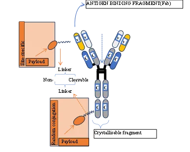
Figure 1: General structure of an ADC illustrating the monoclonal antibody scaffold, conjugation sites, chemical linker, cytotoxic payload, and drug-to-antibody ratio (DAR).
The therapeutic activity of an antibody–drug conjugate (ADC) is determined by the coordinated function of three fundamental components: a monoclonal antibody, a chemical linker, and a cytotoxic payload. These elements are integrated into a single molecular construct through covalent conjugation, enabling selective delivery of highly potent drugs to antigen-expressing target cells while minimizing systemic exposure.
The monoclonal antibody serves as the targeting component of the ADC. Most clinically approved ADCs employ humanized or fully human immunoglobulin G (IgG) antibodies15,27 that recognize tumor-associated antigens with high specificity and affinity. In addition to directing the ADC toward target cells, the antibody contributes significantly to pharmacokinetic behavior through interactions with the neonatal Fc receptor (FcRn), which promotes antibody recycling and prolongs systemic circulation28,30.
The linker functions as a molecular bridge between the antibody and the payload. Its primary role is to maintain stability during circulation while permitting efficient release of the cytotoxic agent after internalization into the target cell. Depending on the design strategy, linkers may be classified as cleavable or non-cleavable, each offering distinct advantages with respect to stability, payload release, and bystander activity.31 Because linker performance directly influences both efficacy and safety, linker chemistry is widely regarded as one of the most critical determinants of ADC success.
The payload constitutes the pharmacologically active component of the conjugate. Since only a limited quantity of drug molecules can be attached to each antibody, ADC payloads are typically selected from classes of highly potent cytotoxic agents capable of inducing cell death at sub nanomolar concentrations. Common payload categories include microtubule inhibitors, DNA-damaging agents, and topoisomerase inhibitors.
Another important structural parameter is the drug-to-antibody ratio (DAR), defined as the average number of payload molecules attached to each antibody molecule. DAR influences potency, pharmacokinetics, stability, and toxicity. While higher DAR values can increase drug delivery to target cells, excessive payload loading may promote aggregation, rapid clearance, and reduced therapeutic performance32. Consequently, optimization of DAR has become a key aspect of ADC engineering.
Collectively, the antibody, linker, payload, and DAR form an integrated design framework in which each component contributes to the overall therapeutic index of the ADC. Understanding the structural roles of these elements provides the foundation for examining the mechanisms of action, linker strategies, and payload selection principles discussed in subsequent sections.
3.3 Mechanism of Action of Antibody–Drug Conjugates
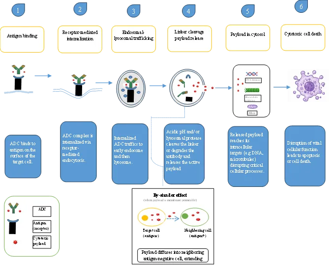
Figure 2: Mechanism of action of antibody–drug conjugates: antigen binding → receptor-mediated internalization → endosomal-lysosomal trafficking → linker cleavage → payload release → cytotoxic cell death.
The therapeutic activity of antibody–drug conjugates (ADCs) depend on a coordinated sequence of biological and chemical events that culminate in selective delivery of a highly potent cytotoxic agent to tumor cells. Although the precise mechanism varies with target antigen, linker chemistry, and payload class, the overall process follows a common multistep pathway33.
The first step involves selective binding of the monoclonal antibody to a tumor-associated cell-surface antigen. Effective target antigens are typically characterized by high expression on malignant cells, limited expression in normal tissues, and the ability to undergo receptor-mediated internalization following antibody engagement34.
Following antigen binding, the ADC–antigen complex is internalized through receptor-mediated endocytosis and trafficked through the endosomal–lysosomal pathway. The acidic environment and enzymatic activity of endosomes and lysosomes facilitate either linker cleavage or degradation of the antibody–linker complex, depending on ADC design35. This step is critical because it determines when and where the active payload is released.
The liberated payload then accesses its intracellular target. Depending on payload class, cytotoxic effects may arise through disruption of microtubule dynamics, induction of DNA damage, inhibition of DNA replication, or interference with other essential cellular processes, triggering cell-cycle arrest, apoptosis, or other forms of programmed cell death.
In ADCs containing membrane-permeable payloads, released drug molecules may diffuse across cellular membranes into neighboring cells (the bystander effect) enabling killing of adjacent tumor cells that express low levels of, or lack, the target antigen. While this can enhance efficacy in heterogeneous tumors, it may also contribute to off-target toxicity depending on the physicochemical properties of the released payload.
Collectively, the mechanism of action can be summarized as: antigen binding → internalization → intracellular trafficking → linker cleavage or degradation → payload release → cytotoxic activity → tumor cell death. This pathway forms the central framework linking target biology (Section 3.4), linker engineering (Section 4), and payload pharmacology (Section 5).
3.4 Target Selection Principles
Target selection is the foundational decision in ADC design, since the biological characteristics of the chosen antigen directly constrain antibody selection, linker chemistry, payload properties, and overall therapeutic strategy. Successful ADC development therefore requires understanding not only that an antigen is tumor-associated, but also its expression profile, internalization behavior, shedding characteristics, and distribution within the tumor microenvironment.
Antigen density and internalization kinetics. Efficient ADC activity depends on the antigen binding the antibody with sufficient density and subsequently undergoing receptor-mediated internalization, transporting the complex through the endosomal–lysosomal pathway where payload release occurs. Antigens that internalize rapidly and efficiently are generally preferred for ADCs relying on intracellular linker cleavage or lysosomal degradation. Clinically relevant targets such as HER2, TROP2, CEACAM5, and mesothelin exhibit distinct expression levels and internalization kinetics that influence ADC performance: higher antigen density can enhance payload delivery per cell, whereas slow or inefficient internalization limits intracellular drug release and reduces efficacy14,36.
Tumor selectivity versus normal-tissue expression. The therapeutic index of an ADC depends heavily on differential antigen expression between tumor and healthy tissue. Because ADC payloads are typically orders of magnitude more potent than conventional chemotherapeutics, even low-level antigen expression in healthy tissue can cause significant on-target/off-tumor toxicity. While linker stability and payload design can mitigate systemic toxicity, the antibody's ability to discriminate malignant from healthy cells remains the primary safeguard, making target selection often the principal determinant of an ADC's safety profile.
Antigen shedding and the antigen sink effect. Some tumor-associated antigens undergo extracellular shedding, releasing soluble fragments into circulation that can bind the ADC before it reaches the tumor — the antigen sink effect. This reduces the ADC available for tumor targeting, may alter pharmacokinetics, and can increase non-target tissue exposure to the conjugate. Antigens with limited shedding are therefore generally favored37.
Expression heterogeneity and bystander-effect implications. Tumors rarely show uniform antigen expression; substantial intratumoral heterogeneity often leaves some cells with little or no target antigen, allowing them to escape direct ADC-mediated killing. The bystander effect (diffusion of membrane-permeable payloads from antigen-positive cells into neighboring antigen-negative cells) can address this limitation, with its magnitude governed by linker cleavability, payload membrane permeability, release kinetics, and tumor architecture38. ADCs capable of a robust bystander effect have shown enhanced activity in heterogeneous tumors, though this must be balanced against increased risk of off-target toxicity from payload diffusion beyond intended cells.
How target biology constrains linker and payload selection. Antigens with rapid internalization, limited shedding, and homogeneous expression favor stable, non-cleavable linkers paired with membrane-impermeable payloads that maximize target-cell specificity. Tumors with heterogeneous antigen expression instead favor cleavable linkers paired with membrane-permeable payloads capable of generating a therapeutically useful bystander effect
4. LINKER CHEMISTRY AND CONJUGATION ENGINEERING
4.1 Role of Linkers
Among the three core components of an ADC, the linker occupies a uniquely important position as the functional bridge between target recognition and payload delivery. While the antibody determines where the ADC is delivered and the payload determines the cytotoxic mechanism, the linker controls when, where, and how the active drug is released, making linker design a major determinant of ADC efficacy, safety, pharmacokinetics, and therapeutic index.
An ideal linker must satisfy two seemingly contradictory requirements: sufficient stability during systemic circulation to prevent premature payload release (which would expose healthy tissue to potent cytotoxic agents and reduce drug reaching the tumor), and efficient payload liberation once the ADC reaches its intracellular destination. Excessive instability risks systemic toxicity; excessive stability limits release and reduces antitumor activity.39
The linker's chemical structure also determines the release trigger such as intracellular proteases, acidic pH, reducing conditions, or degradation of the antibody itself and consequently the physicochemical form of the liberated payload, including its membrane permeability, intracellular retention, and potential for bystander killing40. As discussed in Section 3.4, this creates a direct dependency between linker selection, target biology, and payload properties such as potency and hydrophobicity.
Advances in linker chemistry have been a major driver of the clinical success of modern ADCs. Many limitations of early-generation conjugates like premature payload release, insufficient efficacy, unacceptable toxicity were directly attributable to suboptimal linker design, establishing linker engineering as a specialized field balancing stability, release efficiency, and payload pharmacology.
Based on their mechanism of payload release, ADC linkers are generally classified as cleavable or non-cleavable, each with distinct advantages and limitations, examined in the following sections.
4.2 Cleavable Linkers
Cleavable linkers are engineered to remain sufficiently stable during systemic circulation while releasing the cytotoxic payload in response to specific intracellular or tumor-associated stimuli. By exploiting biological differences between the tumor environment and normal tissues, these linkers enable controlled payload liberation after the ADC reaches its target cell. The choice of cleavage mechanism influences not only payload release efficiency but also intracellular retention, bystander activity, and overall therapeutic index41.
Acid-Cleavable Linkers
Acid-cleavable linkers are designed to exploit the pH gradient that exists between the bloodstream (pH ~7.4) and intracellular endosomal or lysosomal compartments (pH ~4.5–5.5)42. Among the earliest examples are hydrazone-based linkers, which undergo hydrolysis under acidic conditions following ADC internalization43. This strategy was employed in the first-generation ADC gemtuzumab ozogamicin (Mylotarg), in which acid-mediated cleavage releases the calicheamicin payload44.
Although hydrazone linkers demonstrated proof-of-concept for pH-triggered drug release, their relatively limited plasma stability sometimes resulted in premature payload liberation and systemic toxicity. Consequently, more stable linker technologies have largely replaced hydrazone systems in modern ADC development.
Protease-Cleavable Linkers
Protease-cleavable linkers are among the most widely used linker systems in contemporary ADCs. These linkers incorporate short peptide sequences that are selectively recognized and cleaved by lysosomal proteases, particularly cathepsin B and related enzymes. Common examples include valine–citrulline (Val–Cit) and valine–alanine (Val–Ala) linkers45,46
A well-known example is the Val–Cit–PABC linker employed in brentuximab vedotin (Adcetris). Following lysosomal cleavage of the dipeptide sequence, the para-aminobenzyloxycarbonyl (PABC) self-immolative spacer undergoes spontaneous fragmentation, releasing free monomethyl auristatin E (MMAE)47,48. Protease-cleavable linkers generally provide an effective balance between plasma stability and efficient intracellular drug release, contributing significantly to the success of many clinically approved ADCs49.
Disulfide Linkers
Disulfide linkers exploit differences in redox potential between extracellular and intracellular environments. While plasma contains relatively low concentrations of reducing agents, the intracellular compartment is enriched in glutathione and other thiol-containing molecules capable of reducing disulfide bonds48. Cleavage of the disulfide bond releases the attached payload within the target cell.
Disulfide-based systems have been widely explored with maytansinoid payloads such as DM450. However, linker stability can be influenced by the local redox environment, making careful structural optimization necessary to prevent premature cleavage and maintain an acceptable therapeutic index.
Collectively, cleavable linkers provide a versatile platform for controlled drug release and often facilitate bystander activity when paired with membrane-permeable payloads. Their design reflects a central principle of ADC engineering: exploiting tumor-associated biological conditions to achieve selective activation of highly potent cytotoxic agents.
4.3 Non-Cleavable Linkers
In contrast to cleavable systems, non-cleavable linkers do not rely on a specific chemical trigger for payload release. Instead, drug liberation occurs only after complete intracellular degradation of the antibody component within the lysosome. As a result, payload release depends on proteolytic processing of the entire ADC rather than cleavage of the linker itself.
The most widely recognized example is the thioether-based linker SMCC51 (succinimidyl-4-(N-maleimidomethyl)cyclohexane-1-carboxylate), which is used in ado-trastuzumab emtansine (Kadcyla)52,53. Following lysosomal degradation of the antibody, an active metabolite containing the payload, linker fragment, and amino acid residue (typically lysine-MCC-DM1) is generated. Because this metabolite retains a charged linker-derived moiety, it exhibits limited membrane permeability and is largely confined to the target cell52.
The principal advantage of non-cleavable linkers is their exceptional plasma stability, which minimizes premature payload release and reduces systemic toxicity. However, because the released metabolite generally remains intracellular, non-cleavable linkers typically produce little or no bystander effect. Consequently, they are often favored in tumors with relatively homogeneous antigen expression, where selective killing of antigen-positive cells is sufficient to achieve therapeutic efficacy50.
The choice between cleavable and non-cleavable linkers ultimately represents a balance between stability and diffusibility. Cleavable linkers may enhance efficacy in heterogeneous tumors through bystander killing, whereas non-cleavable linkers maximize target-cell specificity and plasma stability. This trade-off has become a central consideration in modern ADC design54.
4.4 Cleavable Versus Non-Cleavable Linkers
The choice between cleavable and non-cleavable linker systems represents one of the most important design decisions in ADC development. Cleavable linkers prioritize efficient payload release and bystander activity, whereas non-cleavable linkers emphasize plasma stability and target-cell specificity. Neither approach is universally superior; the optimal linker depends on target biology, tumor heterogeneity, payload properties, and the desired therapeutic profile.
TABLE 2:COMPARISON OF CLEAVABLE AND NON-CLEAVABLE ADC LINKERS
|
Property |
Cleavable Linkers |
Non-Cleavable Linkers |
References |
|
Release mechanism |
Chemical (pH) or enzymatic (protease/reductive) cleavage |
Complete lysosomal degradation of antibody backbone |
42,46,51 |
|
Plasma stability |
Generally lower; risk of premature cleavage |
Generally higher |
42,54 |
|
Released species |
Free payload, often membrane-permeable |
Payload-linker-amino acid adduct; charged, membrane-impermeable |
46,52 |
|
Bystander effect |
Frequently present (especially with hydrophobic payloads) |
Typically minimal |
17,50 |
|
Representative ADC |
Brentuximab vedotin (Val-Cit-MMAE) |
Ado-trastuzumab emtansine (SMCC-DM1) |
47,53 |
|
Preferred application |
Heterogeneous tumors benefiting from local diffusion |
Homogeneous tumors where maximal target specificity is desired |
49,50 |
This distinction reflects a recurring theme in ADC engineering: the trade-off between payload diffusibility and systemic stability, which carries direct consequences for both efficacy in heterogeneous tumors and control over off-target toxicity.
4.5 Emerging Linker Technologies
Although contemporary linker platforms have significantly improved ADC performance, limitations such as premature payload release, incomplete intracellular activation, and off-target toxicity continue to drive innovation.
Self-immolative linker systems incorporate a spacer that undergoes spontaneous fragmentation following cleavage of an upstream trigger. The para-aminobenzyloxycarbonyl (PABC) spacer used in Val-Cit linker systems is a widely used example: after protease-mediated cleavage of the dipeptide, the spacer rapidly decomposes, releasing the active payload in its native form and minimizing accumulation of partially processed intermediates55.
Dual-responsive linkers require two independent biological stimuli before release, for example, both acidic pH and lysosomal enzyme activity, or enzymatic activity combined with intracellular reducing conditions. By requiring multiple activation signals, these linkers aim to improve release specificity and further reduce premature liberation in non-target tissues.
Additional innovations include enzyme-selective linkers, extracellularly activatable linkers, and tunable architectures capable of modulating release kinetics. Collectively, these approaches seek to combine the stability of non-cleavable linkers with the efficient release of cleavable systems, expanding the therapeutic window of future ADCs56.
4.6 Conjugation Strategy and Drug-To-Antibody Ratio (Dar) Optimization
Beyond linker chemistry, the method used to attach the linker-payload construct to the antibody significantly influences product homogeneity, drug-loading distribution, pharmacokinetics, stability, and therapeutic index.
Stochastic conjugation. Early-generation ADCs relied on conjugation to naturally occurring lysine or cysteine residues. Lysine conjugation exploits the large number of accessible lysine residues on IgG molecules but generates heterogeneous populations with variable conjugation patterns and DAR. Cysteine-based conjugation, via reduction of interchain disulfide bonds, offers somewhat better control over drug loading but still produces heterogeneous mixtures. Several clinically approved ADCs continue to use these approaches because of their simplicity and established manufacturing processes57.
Site-specific conjugation. To overcome this heterogeneity, site-specific technologies generate more homogeneous products. THIOMAB technology58, in which engineered cysteine residues are introduced at predefined positions, was among the earliest and most influential approaches, enabling precise payload attachment while preserving antibody structure and antigen-binding activity. THIOMAB conjugates have demonstrated improved pharmacokinetics, enhanced therapeutic index, and reduced off-target toxicity relative to randomly conjugated ADCs — frequently achieving high efficacy despite carrying fewer payload molecules, illustrating that optimized placement can matter as much as overall payload quantity.
A further refinement uses unnatural amino acids (UAAs) bearing biorthogonal reactive groups, permitting precise conjugation at predetermined sites with tight stoichiometric control. Preclinical studies show UAA-based ADCs can achieve favorable pharmacokinetics, potent antitumor activity, and superior homogeneity relative to conventional conjugation59.
DAR: finding the sweet spot. The drug-to-antibody ratio (DAR) (the average number of payload molecules per antibody) is one of the most important quantitative parameters in ADC design, directly influencing potency, stability, clearance, and toxicity60,61. Increasing DAR generally enhances payload delivery and cytotoxic potential, but excessive loading increases hydrophobicity, promotes aggregation, accelerates clearance, and may increase toxicity; DAR values that are too low may limit efficacy through insufficient intracellular drug delivery. Most clinically successful ADCs operate within a DAR range of approximately 2–8, with many modern site-specific platforms converging on highly homogeneous values near 2–442. The objective is not to maximize drug loading but to achieve the optimal balance between potency, pharmacokinetics, stability, and safety. Therefore, making DAR optimization a central principle of modern ADC engineering.
5. PAYLOAD STRATEGIES
5.1 Ideal Payload Characteristics
An ideal ADC payload must satisfy a unique set of pharmacological and chemical requirements that distinguish it from conventional anticancer agents. Because only a small fraction of the administered ADC dose ultimately reaches and is internalized by tumor cells, the payload must possess exceptionally high cytotoxic potency, typically in the sub-nanomolar to picomolar range, ensuring that even limited intracellular drug release produces meaningful antitumor effects15,62.
The payload must also contain a chemically accessible site for linker attachment without significantly compromising biological activity, while the resulting conjugate remains stable during manufacturing and circulation and allows efficient release after intracellular processing. Physicochemical balance matters too: excessive hydrophobicity may promote ADC aggregation and rapid clearance, whereas excessive hydrophilicity may impair cellular activity after release32.
Mechanism of action is a further consideration. Since many tumors contain slowly proliferating or quiescent cell populations, payloads that exert profound cytotoxic effects at very low concentrations are generally preferred. Consequently, most clinically successful ADC payloads belong to a limited number of highly potent classes: microtubule inhibitors, DNA-damaging agents, and topoisomerase inhibitors which is discussed in the sections that follow.
5.2 Microtubule Inhibitors
Microtubule-disrupting agents were among the first payload classes successfully incorporated into ADCs and remain among the most widely used in approved products. These agents interfere with microtubule dynamics, leading to mitotic arrest and apoptotic cell death, with extraordinary potency well suited to ADC applications.
Auristatins. Auristatins are synthetic derivatives of the marine natural product dolastatin 10 that bind tubulin, preventing microtubule polymerization and disrupting mitotic spindle formation. The two most widely used auristatin payloads are monomethyl auristatin E (MMAE) and monomethyl auristatin F (MMAF)63,64. MMAE is membrane-permeable and can diffuse across cellular membranes following intracellular release; ADCs containing MMAE, such as brentuximab vedotin and enfortumab vedotin, often exhibit a significant bystander effect, which can be advantageous in tumors with heterogeneous antigen expression47,65. MMAF, by contrast, carries a charged C-terminal phenylalanine residue that markedly reduces membrane permeability, so released MMAF tends to remain confined within the targeted cell, limiting bystander killing but potentially reducing off-target toxicity46. This distinction illustrates how small structural modifications can profoundly influence ADC pharmacology and therapeutic strategy.
Maytansinoids. Maytansinoids are semisynthetic derivatives of maytansine, a highly potent natural-product microtubule inhibitor66 that, like auristatins, disrupts microtubule function and induces cell-cycle arrest, though binding a distinct tubulin site with different physicochemical properties. The most clinically important maytansinoids are DM1 and DM462. DM1 is the payload in ado-trastuzumab emtansine, attached via the non-cleavable SMCC linker; following lysosomal degradation, the active metabolite remains largely intracellular, contributing to the conjugate's limited bystander activity. DM4 is frequently combined with cleavable disulfide-based linkers and is used in agents such as mirvetuximab soravtansine67; depending on linker design and released metabolite, DM4-containing ADCs can exhibit greater bystander activity than DM1-based conjugates.
5.3 DNA-Damaging Agents
DNA-damaging payloads are among the most potent cytotoxic agents used in ADC development. Unlike microtubule inhibitors, which primarily affect actively dividing cells, these agents can induce irreversible genomic damage at extremely low intracellular concentrations, making them attractive payloads, though their narrow therapeutic window places stringent demands on linker stability and target selectivity.
Calicheamicins. Calicheamicins are enediyne antibiotics among the first payloads incorporated into ADCs68. Following intracellular release, calicheamicin binds the DNA minor groove and undergoes activation to generate reactive radical intermediates that induce double-strand breaks, triggering apoptosis. Calicheamicin was used in gemtuzumab ozogamicin, the first FDA-approved ADC, and later in inotuzumab ozogamicin69, a clinical history that highlighted both the extraordinary efficacy and significant toxicity risks of highly potent DNA-damaging payloads.
Pyrrolobenzodiazepines (PBDs). PBDs are highly potent DNA cross-linking agents that bind the minor groove and form covalent interstrand cross-links, inducing minimal helix distortion and making the resulting lesions difficult for repair systems to detect. This contributes to extraordinary potency, allowing effective activity even at low DAR, though the same potency has been associated with delayed, sometimes severe toxicities during clinical development70,71.
Duocarmycins. Duocarmycins are natural-product-derived DNA minor-groove alkylating agents of exceptionally high potency, acting through covalent DNA alkylation72,73. A particularly attractive feature is their compatibility with prodrug-style masking: structural modifications can render the molecule relatively inactive until intracellular activation, improving selectivity and reducing systemic toxicity, a property closely aligned with ADC-mediated targeted delivery and of considerable interest for next-generation development.
Collectively, DNA-damaging payloads show how extreme potency can compensate for limited intracellular drug delivery, though their application requires careful optimization of target selection, linker stability, and dosing to avoid unacceptable toxicity.
5.4 Topoisomerase Inhibitors
Topoisomerase I inhibitors represent a newer generation of ADC payloads that have significantly expanded the field's therapeutic potential. Unlike microtubule inhibitors and many DNA-damaging agents, these payloads often combine high potency with favorable membrane permeability, enabling substantial bystander effects.
SN-38, the active metabolite of irinotecan, inhibits topoisomerase I, an enzyme that relieves torsional stress during DNA replication74 and transcription, by stabilizing the transient DNA-topoisomerase cleavage complex, preventing re-ligation of single-strand breaks and ultimately causing replication-associated double-strand breaks and cell death. The most notable ADC employing this payload is sacituzumab govitecan, in which SN-38 is linked via the CL2A linker to an anti-TROP-2 antibody]75,76, demonstrating that moderately potent payloads can achieve substantial clinical efficacy when combined with optimized targeting and release, challenging the earlier assumption that only ultra-potent cytotoxins suit ADC development.
Deruxtecan (DXd), a derivative of exatecan, is currently one of the most clinically successful ADC payloads77. Like SN-38 it inhibits topoisomerase I, but with exceptionally high potency and favorable membrane permeability following release. DXd is used in trastuzumab deruxtecan78 and datopotamab deruxtecan; after linker cleavage, released DXd can diffuse into neighboring tumor cells, generating a robust bystander effect believed to contribute to remarkable efficacy in tumors with heterogeneous antigen expression, including HER2-low cancers. However, the same diffusibility may contribute to off-target toxicities, most notably interstitial lung disease (ILD) and pneumonitis79, which occur in a meaningful proportion of treated patients and have emerged as important safety considerations. DXd-based ADCs thus illustrate the central ADC design trade-off between maximizing tumor penetration and minimizing unintended tissue exposure.
5.5 Bystander Effect
The bystander effect (diffusion of a released payload from an antigen-positive cell into neighboring cells regardless of their antigen status) is a major determinant of ADC efficacy in tumors with heterogeneous antigen expression (Section 3.4), where direct killing of antigen-positive cells alone may be insufficient for durable control.
Its magnitude depends on payload physicochemistry and linker design. Membrane-permeable payloads such as DXd and SN-38, combined with cleavable linkers38,79, can extend an ADC's therapeutic reach beyond the initially targeted cell. In contrast, payloads remaining attached to charged linker fragments after release — such as lysine-MCC-DM1 from the non-cleavable SMCC linker in T-DM80, show minimal membrane permeability and remain largely confined to antigen-positive cells.
While bystander activity can enhance efficacy in heterogeneous tumors, diffusion beyond the intended target population may increase normal-tissue exposure and off-target toxicity. The choice between maximizing bystander killing and maximizing target-cell specificity is thus a fundamental ADC design trade-off, tightly linked to target biology, linker selection, and payload chemistry — reinforcing the target → linker → payload framework developed throughout this review.
5.6 Emerging Payload Classes
Recent advances have expanded ADC payloads beyond microtubule- and DNA-targeting cytotoxins toward agents that degrade proteins, modulate immunity, deliver radiation, or regulate gene expression.
Degrader-antibody conjugates (DACs) incorporate proteolysis-targeting chimeras (PROTACs)81, exploiting the ubiquitin-proteasome pathway to induce selective degradation of intracellular proteins rather than merely inhibiting them , offering potential access to historically "undruggable" oncogenic drivers.
Immune-stimulating antibody conjugates (ISACs) deliver immune-activating molecules such as TLR or STING agonists to the tumor microenvironment rather than cytotoxic payloads, aiming to convert immunologically "cold" tumors "hot" by enhancing antigen presentation and effector-cell recruitment82. However, excessive immune activation risks systemic inflammation and cytokine-release syndrome, a distinct therapeutic-index challenge from conventional ADCs.
Radioimmunoconjugates deliver a radioactive isotope to tumor tissue83, with cytotoxicity mediated by emitted radiation rather than a released drug, potentially useful against heterogeneous antigen expression, since radiation affects neighboring cells within its path length.
Antibody-RNA conjugates, delivering siRNA, antisense oligonucleotides, or related gene-silencing agents, combine antibody targeting with RNA-mediated gene suppression, though intracellular delivery, endosomal escape, and stability remain substantial challenges.
6. CLINICAL TRANSLATION: FDA-APPROVED ADCS
As of 2025, the FDA had approved 15 ADCs spanning hematologic malignancies84 and solid tumors, with recent approvals such as datopotamab deruxtecan and telisotuzumab vedotin illustrating continued expansion into new targets and indications. Table 3 summarizes how different combinations of target antigen, linker platform, and payload class have been tailored to specific disease settings.
TABLE 3:SELECTED FDA-APPROVED ANTIBODY–DRUG CONJUGATES
|
ADC (Brand Name) |
Target |
Linker |
Payload |
Major Indication |
Approval Year |
References |
|
Gemtuzumab ozogamicin (Mylotarg) |
CD33 |
Hydrazone (acid-cleavable) |
Calicheamicin |
Acute myeloid leukemia |
2000* |
3,4,44 |
|
Brentuximab vedotin (Adcetris) |
CD30 |
Val-Cit |
MMAE |
Hodgkin lymphoma |
2011 |
47,48 |
|
Ado-trastuzumab emtansine (Kadcyla) |
HER2 |
SMCC (non-cleavable) |
DM1 |
HER2+ breast cancer |
2013 |
52,53 |
|
Inotuzumab ozogamicin (Besponsa) |
CD22 |
Hydrazone (acid-cleavable) |
Calicheamicin |
B-cell ALL |
2017 |
69,85 |
|
Polatuzumab vedotin (Polivy) |
CD79b |
Val-Cit |
MMAE |
DLBCL |
2019 |
86 |
|
Enfortumab vedotin (Padcev) |
Nectin-4 |
Val-Cit |
MMAE |
Urothelial carcinoma |
2019 |
87 |
|
Trastuzumab deruxtecan (Enhertu) |
HER2 |
GGFG (cleavable) |
DXd |
HER2+ breast cancer |
2019 |
78,79 |
|
Sacituzumab govitecan (Trodelvy) |
TROP-2 |
CL2A |
SN-38 |
Triple-negative breast cancer |
2020 |
75,76 |
|
Belantamab mafodotin (Blenrep) |
BCMA |
Maleimidocaproyl |
MMAF |
Multiple myeloma |
2020 |
88 |
|
Loncastuximab tesirine (Zynlonta) |
CD19 |
Val-Ala |
PBD dimer |
DLBCL |
2021 |
89 |
|
Tisotumab vedotin (Tivdak) |
Tissue factor |
Val-Cit |
MMAE |
Cervical cancer |
2021 |
90 |
|
Mirvetuximab soravtansine (Elahere) |
FRα |
Disulfide |
DM4 |
Ovarian cancer |
2022 |
67 |
|
Datopotamab deruxtecan (Datroway) |
TROP-2 |
GGFG (cleavable) |
DXd |
Breast cancer |
2025 |
75 |
Several trends emerge from this landscape: a shift from early hydrazone linkers toward protease-cleavable peptide systems (Val-Cit, Val-Ala) offering improved plasma stability; reliance on a small number of payload families like auristatins, maytansinoids, calicheamicins, PBD dimers, and topoisomerase I inhibitors and reflecting the stringent potency demands discussed in Section 5; expansion from hematologic malignancies, where circulating malignant cells are readily accessible, toward solid tumors including breast, ovarian, cervical, lung, and urothelial cancers; and the growing prominence of topoisomerase-I payloads (SN-38, DXd), whose bystander activity helps address the antigen-heterogeneity limitations of earlier generations.
7. ADCS THROUGH THE LENS OF PRODRUG DESIGN
Returning to the classical objectives outlined in Section 2, the target–trafficking–linker–payload framework developed in Sections 3–6 shows that ADCs do not merely replicate traditional prodrug goals, in several respects they substantially extend them.
Solubility. Traditional prodrugs mask hydrophobic or ionizable groups with small, cleavable promoieties to improve aqueous solubility. ADCs achieve the same end differently: payloads such as the auristatins and maytansinoids are highly hydrophobic and would be clinically impractical as free drugs, but conjugation to a ~150 kDa hydrophilic antibody scaffold masks these properties at a scale unattainable by conventional chemistry. The antibody itself functions as an extraordinarily large carrier-linked promoiety91,92.
Distribution and pharmacokinetics. Classical prodrugs alter tissue distribution through lipophilicity or metabolic susceptibility, with limited control over where activation occurs. ADCs instead achieve distribution through molecular recognition: FcRn-mediated recycling confers prolonged circulation, while antigen binding drives selective accumulation and internalization at the target site93. The classical challenge of "distribution" is thereby transformed into a problem of target selection, antigen density, and internalization kinetics (Section 3.4).
Sustained release. Where traditional sustained-release prodrugs depend on predictable hydrolysis or metabolic kinetics, ADCs layer multiple sequential delays (internalization, endosomal trafficking, lysosomal processing, and linker cleavage) each acting as a kinetic barrier before payload activation. Linker chemistry adds a further programmable layer: hydrazone linkers respond to acidic pH, Val-Cit/Val-Ala linkers require proteolytic cleavage, and non-cleavable linkers require complete antibody degradation. This combination of biological trafficking and tunable chemistry exceeds the temporal control achievable by most conventional prodrugs94,95.
Site-specific delivery. This is where ADCs most clearly surpass traditional approaches. Conventional prodrugs rely on tissue differences in enzyme expression, pH, or metabolism. These mechanisms offering only modest spatial precision. ADCs instead encode selectivity directly into the construct via antigen-specific recognition, as reflected in the clinical success of agents targeting HER2, CD30, TROP2, CD79b, and Nectin-4. The bystander effect extends this further: payloads such as DXd and SN-38 diffuse from antigen-positive to antigen-negative neighboring cells, trading strict cellular specificity for the ability to overcome intratumoral heterogeneity, allowing ADCs to balance precision and diffusion according to tumor biology96.
Beyond the classical framework. Conventional prodrugs typically depend on a single activation event. ADCs distribute selectivity across multiple sequential, independent steps — antibody binding, internalization, trafficking, linker processing, and payload release — any of which can gate successful activation. The agents discussed throughout this review illustrate how these layers can be tuned toward different goals: gemtuzumab ozogamicin pairs antibody targeting with acid-sensitive release; brentuximab vedotin uses protease-cleavable release of MMAE; ado-trastuzumab emtansine maximizes specificity via a non-cleavable, membrane-impermeable metabolite; and trastuzumab deruxtecan deliberately exploits a diffusible payload for bystander activity in heterogeneous tumors97. ADCs are thus integrated biological-chemical delivery systems in which target selection, trafficking, linker engineering, and payload pharmacology function as interconnected determinants of activity, the most advanced manifestation of prodrug design developed to date.
8. CHALLENGES AND LIMITATIONS
The major challenges facing ADCs arise at multiple points along the target–trafficking–linker–payload cascade, and failure at any stage can reduce efficacy, narrow the therapeutic window, or drive resistance.
Linker instability. Premature cleavage of cleavable linkers exposes healthy tissue to potent payloads, reducing tumor-specific delivery. Early hydrazone linkers sometimes cleaved under mildly acidic physiological conditions, and disulfide linkers can be susceptible to circulating reducing agents; the clinical history of gemtuzumab ozogamicin (including its withdrawal and re-approval with optimized dosing) underscored the importance of linker stability to safety and efficacy.
Off-target toxicity. Toxicity arises through several mechanisms: low-level target antigen expression on healthy tissue causing on-target/off-tumor effects; nonspecific Fc-receptor-mediated uptake by reticuloendothelial cells; and premature deconjugation exposing tissues to free payload. The bystander effect compounds this risk, the interstitial lung disease and pneumonitis seen with DXd-containing ADCs illustrate how payload diffusibility can translate into clinically significant toxicity79.
Antigen heterogeneity and resistance. Intratumoral variation in antigen expression allows antigen-negative or antigen-low cells to evade direct killing. Acquired resistance can also arise through antigen downregulation98, impaired internalization, altered trafficking, enhanced lysosomal degradation, increased efflux, or compensatory survival signaling. While bystander-capable payloads partially address heterogeneity, resistance remains a major active challenge likely requiring combination strategies.
Manufacturing complexity and cost. ADC production requires coordinated antibody expression, linker synthesis, payload preparation, conjugation chemistry, and extensive analytical characterization of DAR distribution, aggregation, free-payload content, and stability29. Site-specific conjugation improves homogeneity but can add complexity through engineered antibodies or non-natural amino acids, contributing to high production costs that may limit accessibility as ADC use expands.
9. FUTURE DIRECTIONS
Future ADC generations are likely to extend well beyond the traditional antibody–linker–cytotoxin paradigm.
Bispecific ADCs, recognizing two antigens simultaneously, may improve selectivity, enhance internalization, and reduce antigen escape; HER2-directed bispecifics have shown accelerated internalization and lysosomal trafficking preclinically, potentially helping address intratumoral heterogeneity99.
Immune-stimulating antibody conjugates (ISACs) deliver TLR or STING agonists to convert immunologically "cold" tumors "hot," though cytokine-mediated toxicity remains a therapeutic-index challenge under active optimization.
Degrader-antibody conjugates (DACs), delivering PROTACs for targeted intracellular protein degradation, may extend therapy to previously undruggable targets — alongside dual-payload ADCs, probody-drug conjugates, and antibody fragments with improved tissue penetration.
AI-assisted design, leveraging spatial transcriptomics, single-cell sequencing, and machine learning, is being applied to predict antigen density, internalization kinetics, linker performance, and bystander behavior, though regulatory acceptance and interpretability remain open questions100.
Beyond oncology, antibody-antibiotic conjugates are being explored for difficult bacterial infections, antibody-delivered immunomodulators for autoimmune disease, and antibody-RNA conjugates for gene-silencing therapeutics, suggesting ADC-targeting principles may extend across therapeutic areas101.
Personalized therapy is expected to grow as immunohistochemistry, RNA profiling, and molecular diagnostics enable patient selection based on antigen density, spatial distribution, and internalization behavior rather than histology alone, integrating ADC therapy with companion diagnostics.
Collectively, these developments suggest the next ADC generation will involve not merely improved versions of existing drugs, but novel targeting strategies, payload classes, computational tools, and personalized paradigms.
CONCLUSION
ADCs represent the convergence of nearly a century of prodrug theory with advances in antibody engineering, medicinal chemistry, and targeted delivery. From Ehrlich's "magic bullet" to the fifteen FDA-approved ADCs available by 2025, the field demonstrates that effective targeted therapy requires coordinated optimization of multiple interconnected components that includes target selection, intracellular trafficking, linker chemistry, and payload pharmacology, rather than a single pharmacological principle. Unlike conventional prodrugs, which depend on one activation event, ADCs layer antigen recognition, internalization, intracellular processing, and controlled release into a sophisticated activation cascade. Challenges such as linker instability, off-target toxicity, antigen heterogeneity, resistance, manufacturing complexity remain, but emerging directions (bispecific ADCs, ISACs, DACs, AI-assisted design, non-oncology applications) suggest ADCs, as biologically programmed prodrugs, will remain among the most sophisticated manifestations of modern prodrug design.
CONFLICT OF INTEREST
The authors declare that they have no known competing financial interests or personal relationships that could have appeared to influence the work reported in this manuscript.
ACKNOWLEDGEMENT
The authors first and foremost give thanks to God Almighty for His grace, wisdom, strength, and guidance throughout the preparation of this manuscript. The authors sincerely appreciate the encouragement, collaboration, and unwavering commitment of all co-authors throughout the development of this review. We also acknowledge the support and resources provided by our respective institutions and express our gratitude to the researchers and scientists whose published work formed the foundation of this review.
REFERENCES

G. E. Ebimo-Moko, F. O. Oladele, J. D. Joel, J. E. Sampson, W. C. Madu, V. Ebimo-Moko, T. Ganatra, Antibody-Drug Conjugates in Modern Prodrug Design: Linker Chemistry and Payload Strategies, Int. J. of Pharm. Sci., 2026, Vol 4, Issue 8, 2631-2659. https://doi.org/10.5281/zenodo.21972505
 10.5281/zenodo.21972505