

We use cookies to ensure our website works properly and to personalise your experience. Cookies policy
Department of Pharmacology, Faculty of Pharmacy, M. S. Ramaiah University of Applied Sciences, Bengaluru, Karnataka - 560054, Indi.
Bee venom has anticancer activity that is largely attributed to melittin, but its non-selective membrane toxicity limits clinical use. This study combined computational target prediction with the formulation and cell-based testing of bee venom-loaded niosomes. Network pharmacology of melittin was done with SuperPred and GeneCards, followed by protein–protein interaction analysis, selection of ten hub targets and HDOCK docking, and ADMET properties were predicted with pkCSM. Niosomes were prepared by thin-film hydration with Span 60, Tween 60 and cholesterol, and were evaluated by FTIR, UV spectrophotometry, dynamic light scattering, TEM and dialysis-based release. Cytotoxicity against MCF-7 cells was measured by the MTT assay. Docking gave the most favourable scores for MAPK1 (?233.01) and PIK3R1 (?232.85). ADMET prediction indicated poor intestinal absorption and possible hepatotoxicity. FTIR showed no evident incompatibility between venom and excipients. The optimised vesicles measured 208.7 nm, with a zeta potential of ?21.51 mV and an entrapment efficiency of 84.73%, and TEM showed rounded to oval vesicles. In the first 6 h, 28.17% of the venom was released from the niosomes against 71.32% for pure venom. The formulation reduced MCF-7 viability in a concentration-dependent manner, with an IC50 of 150.35 µg/mL at 48 h. These findings support bee venom-loaded niosomes as a candidate nanocarrier for breast cancer therapy and justify in vivo evaluation.
Honeybee venom is a mixture of peptides, enzymes and small amines, of which melittin, phospholipase A2 and apamin are the best studied.1-3 Melittin makes up the largest share of the dry venom and is a cationic, amphipathic peptide that inserts readily into lipid bilayers.4 Bee venom and melittin have slowed the growth of several cancer cell types and triggered apoptosis in them, and reports in breast cancer models include MCF-7 cells and HER2-enriched and triple-negative disease.5-9
The membrane activity that makes melittin cytotoxic to tumour cells is not selective. Free melittin and whole venom also damage normal cells, so the margin between an effective and a harmful dose is narrow, and a peptide is further limited by poor stability and uncontrolled release.4,9 Encapsulating the venom in a carrier is one way to address these problems.
Niosomes are vesicles assembled from non-ionic surfactants and cholesterol. Their size, entrapment and release behaviour can be adjusted through the surfactant type, the surfactant-to-cholesterol ratio and the preparation conditions.10,11 Melittin-loaded niosomes have been tested against breast cancer,12 whole bee venom has been loaded into niosomes and into nanoliposomes,13,14 and the venom nanoformulations were cytotoxic to cancer cell lines in those studies. What is still lacking is a stepwise assessment that links computational prediction of the likely targets to the formulation and its cell-level activity.
We therefore combined three steps in this work. First, melittin, the principal peptide of the venom, was examined by network pharmacology, molecular docking and ADMET prediction to identify breast cancer-related targets. Second, whole bee venom was formulated into Span 60/Tween 60/cholesterol niosomes and characterised for compatibility, size, surface charge, entrapment, morphology and release. Third, the formulation was tested for cytotoxicity against MCF-7 breast cancer cells.
MATERIALS AND METHODS
MATERIALS
Whole bee venom was purchased from One Bee Organic LLC. Span 60, Tween 60, cholesterol, chloroform and phosphate-buffered saline (PBS, pH 7.4) were used for vesicle preparation, and the remaining reagents were of analytical grade. MCF-7 human breast cancer cells were used for the cytotoxicity study.
In silico analysis
Melittin was chosen for the computational work because it is the main active peptide of the venom and its structure is available. Its structure was taken from PubChem15 and its potential targets were predicted with SuperPred. Genes associated with breast cancer were collected from GeneCards using the search term “breast cancer”.16 The two lists were intersected in Venny 2.1.0 to obtain the shared targets. These were submitted to STRING17 and the resulting protein–protein interaction network was opened in Cytoscape,18 where the CytoHubba plugin ranked the nodes and the ten top-ranked hub targets were retained.
Crystal structures of the ten hub proteins were downloaded from the Protein Data Bank. Melittin was docked to each protein separately on the HDOCK web server.19 The docking scores were compared across targets and the best-scoring complexes were inspected visually. Absorption, distribution, metabolism, excretion and toxicity properties of melittin were predicted with the pkCSM server.20
Preparation of niosomes
Niosomes were made by thin-film hydration.10,11 Span 60, Tween 60 and 1:1:2 (w/w/w) were dissolved in chloroform in a round-bottom flask, and the solvent was evaporated under reduced pressure in a rotary evaporator to leave a thin film. Bee venom, 5 mg, was dissolved in PBS (pH 7.4) and used to hydrate the film with magnetic stirring for 60 minutes. The dispersion was then sonicated with a probe sonicator for about 15 mintues to reduce vesicle size. Blank niosomes were prepared in the same way without venom.
Preformulation studies
The solubility of bee venom in distilled water and PBS (pH 7.4) was judged by visual inspection after mixing and equilibration. The absorption maximum was found by scanning a venom solution in a UV-visible spectrophotometer, and a calibration curve was drawn from solutions of 20–100 µg/mL read at that wavelength. For the compatibility study, FTIR spectra of bee venom, Span 60, Tween 60 and cholesterol were recorded individually and for their physical mixture, and the position of the characteristic bands was compared.
Characterisation of the niosomes
Mean vesicle size and polydispersity index (PDI) were measured by dynamic light scattering, and zeta potential by electrophoretic light scattering, on suitably diluted dispersion. To determine entrapment efficiency, unentrapped venom was separated from the vesicles by centrifugation at 2 - 4°c, 14000 rpm and measured at the absorption maximum by using UV-Visible spectrophometry. Entrapment efficiency (%) was calculated as (total venom − unentrapped venom)/total venom × 100. Vesicle morphology was examined by transmission electron microscopy.
Release was studied by the dialysis-bag method. Pure venom and the niosomal dispersion were placed in separate dialysis membranes immersed in the release medium (PBS, Ph 7.4) and 1ml of samples were withdrawn and replaced with fresh medium at 0.5, 1, 2, 4, 6, 8, 12 and 24 h. The venom released was read spectrophotometrically, and the cumulative release was fitted to zero-order, first-order, Higuchi and Korsmeyer–Peppas models.
Cytotoxicity assay
MCF-7 cells were grown in a humidified CO2 incubator, seeded into 96-well plates and allowed to attach. They were exposed for 48 h to the niosomal formulation at 10, 50, 100, 200 and 400 µg/mL, with untreated cells as the control and camptothecin (7.8 µg/mL) as the reference drug. Viability was determined by the MTT assay;21 formazan crystals were dissolved and absorbance was read at 570 nm. Viability (%) was calculated as the absorbance of treated cells divided by that of control cells × 100, and cytotoxicity (%) as 100 minus viability. The IC50 was obtained from the logarithmic regression of viability on concentration.
RESULTS AND DISCUSSION
Network pharmacology and target selection
Melittin targets predicted by SuperPred overlapped with the breast cancer gene set, giving a group of shared proteins (Fig. 1). The protein–protein interaction network built from these showed a densely connected core, and ranking by CytoHubba selected ten hub targets for docking (Fig. 2 and 3). The overlap suggests that melittin may act on several nodes of the disease network at once, a pattern in line with its reported effects on proliferation, apoptosis, invasion and angiogenesis.9 These are predicted associations only and have not been shown experimentally.
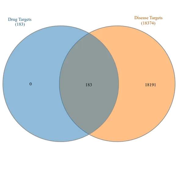
` Figure 1: Venn Diagram of Targets Shared Between Melittin and Breast Cancer.
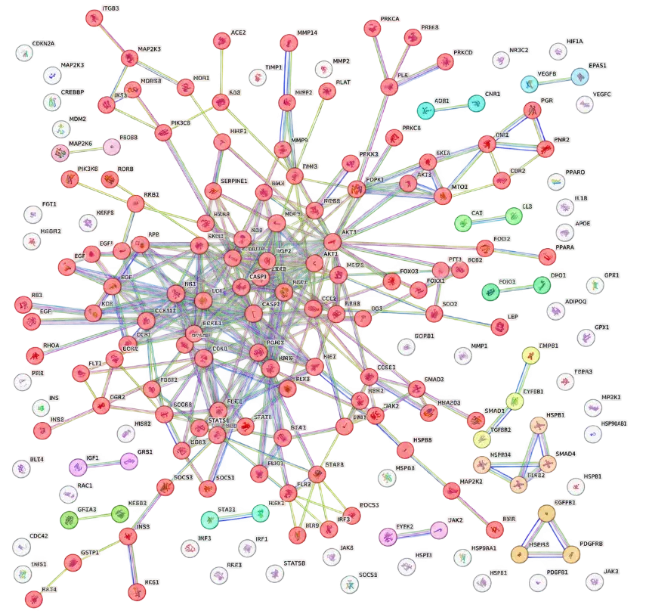
Figure 2: Protein–Protein Interaction Network of the Shared Targets.
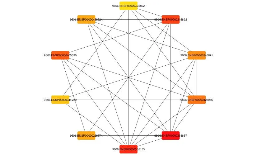
Figure 3: Top Ten Hub Targets Identified by CytoHubba.
Molecular docking
Melittin gave negative HDOCK scores with all ten hub proteins (Table 1). The two most favourable were MAPK1 (−233.01) and PIK3R1 (−232.85), followed by NFKB1 (−216.70) and STAT3 (−216.14); ESR1 gave the weakest score (−197.04). The poses of melittin with MAPK1 and PIK3R1 are shown in Fig. 4 and 5. MAPK1 belongs to the MAPK cascade and PIK3R1 encodes a regulatory subunit of PI3K, and both pathways control cell growth and survival. Melittin has been reported to suppress PI3K/Akt/mTOR signalling in breast cancer cells22 and PI3K/AKT/mTOR and MAPK signalling in melanoma cells,23 which is consistent with these docking results. A favourable docking score indicates a predicted fit, not inhibition of the protein, so these targets need experimental confirmation.
Table 1: Docking Scores of Melittin with the Ten HUB Targets
| Sr. No. | Target protein | PDB ID | HDOCK score |
|---|---|---|---|
| 1 | MAPK1 | 2OJJ | −233.01 |
| 2 | PIK3R1 | 4OVU | −232.85 |
| 3 | NFKB1 | 1SVC | −216.70 |
| 4 | STAT3 | 6NJS | −216.14 |
| 5 | HSP90AA1 | 3T0H | −210.42 |
| 6 | PDPK1 | 1UU9 | −208.16 |
| 55 | AKT2 | 1O6K | −205.29 |
| 8 | FYN | 2DQ7 | −204.95 |
| 9 | LYN | 3A4O | −202.77 |
| 10 | ESR1 | 1A52 | −197.04 |
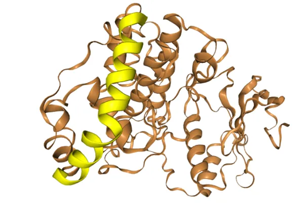
Figure 4: Predicted Interaction of Melittin with MAPK1 (PDB ID 2OJJ).
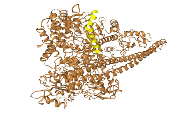
Figure 5: Predicted Interaction of Melittin with PIK3R1 (PDB ID 4OVU).
ADMET prediction
The pkCSM output for melittin is summarised in Table 2. Intestinal absorption was predicted as 0%, which is expected for a peptide and supports a non-oral delivery strategy and a carrier. Melittin was predicted to be a P-glycoprotein substrate and a CYP3A4 substrate, with no inhibition of the five CYP isoforms tested, no AMES toxicity and no hERG I inhibition. Hepatotoxicity and hERG II inhibition were, however, predicted. Because pkCSM was developed on small molecules, its values for a 26-residue peptide should be read as indicative, and the predicted toxicity flags are a reason for experimental safety testing rather than evidence of harm. The computation also used melittin alone, so it speaks to the likely contribution of this peptide and not to the whole venom.
Table 2: Selected Predicted Admet Properties of Melittin (pkCSM)
| Property | Predicted value | Unit / type |
|---|---|---|
| Water solubility | −2.892 | log mol/L |
| Caco-2 permeability | −1.143 | log Papp |
| Intestinal absorption (human) | 0 | % absorbed |
| P-glycoprotein substrate | Yes | Yes/No |
| VDss (human) | 0.011 | log L/kg |
| Fraction unbound (human) | 0.381 | Fu |
| CYP3A4 substrate | Yes | Yes/No |
| CYP inhibition (1A2, 2C19, 2C9, 2D6, 3A4) | No | Yes/No |
| Total clearance | −1.294 | log mL/min/kg |
| AMES toxicity | No | Yes/No |
| hERG I inhibitor | No | Yes/No |
| hERG II inhibitor | Yes | Yes/No |
| Hepatotoxicity | Yes | Yes/No |
| Oral rat acute toxicity (LD50) | 2.482 | mol/kg |
Preformulation: solubility, UV method and FTIR
Bee venom dissolved in both distilled water and PBS (pH 7.4), and PBS was chosen as the hydration medium. The venom had an absorption maximum at 278 nm (Fig. 6). Absorbance was linear over 20–100 µg/mL, with the regression y = 0.00517x − 0.039 and R² = 0.9983 (Fig. 7), which was adequate for estimating venom in the entrapment and release experiments. In the FTIR study, the characteristic amide, ester and alkyl bands of the individual components were present in the physical mixture without notable shifts or loss of the main venom peaks (Fig. 8), so no evident interaction between venom and excipients was seen.
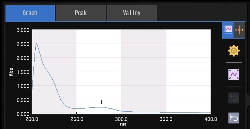
Figure 6: UV-visible Absorption Spectrum of Bee Venom (Λmax 278 nm).
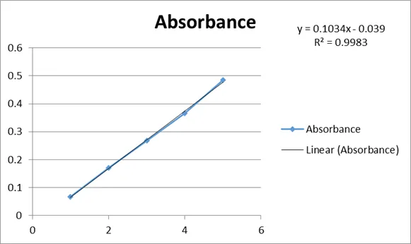
Figure 7: Calibration Curve of Bee Venom, 20–100 Μg/ml.
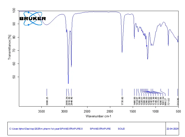
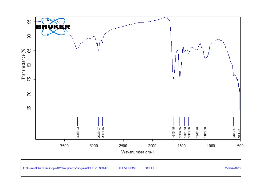
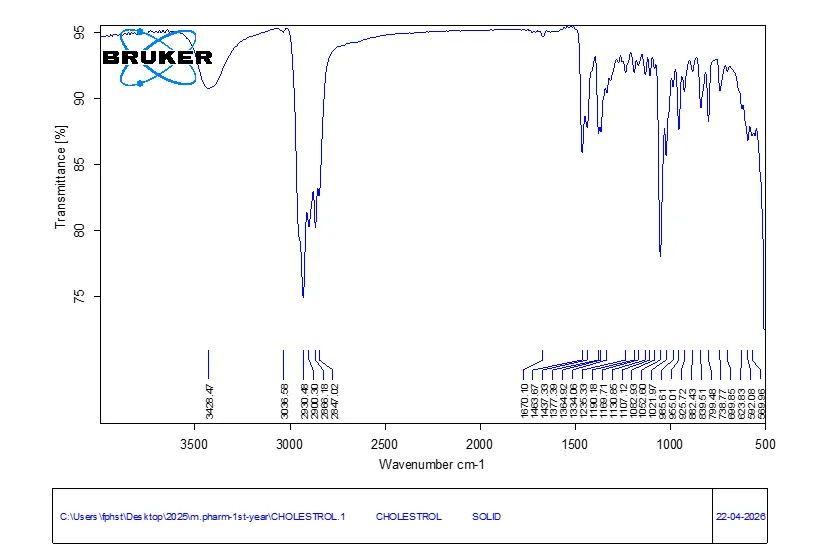
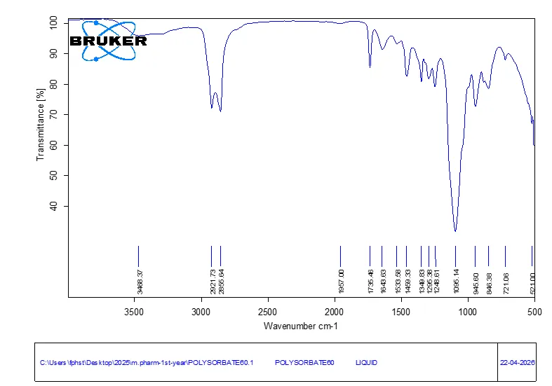
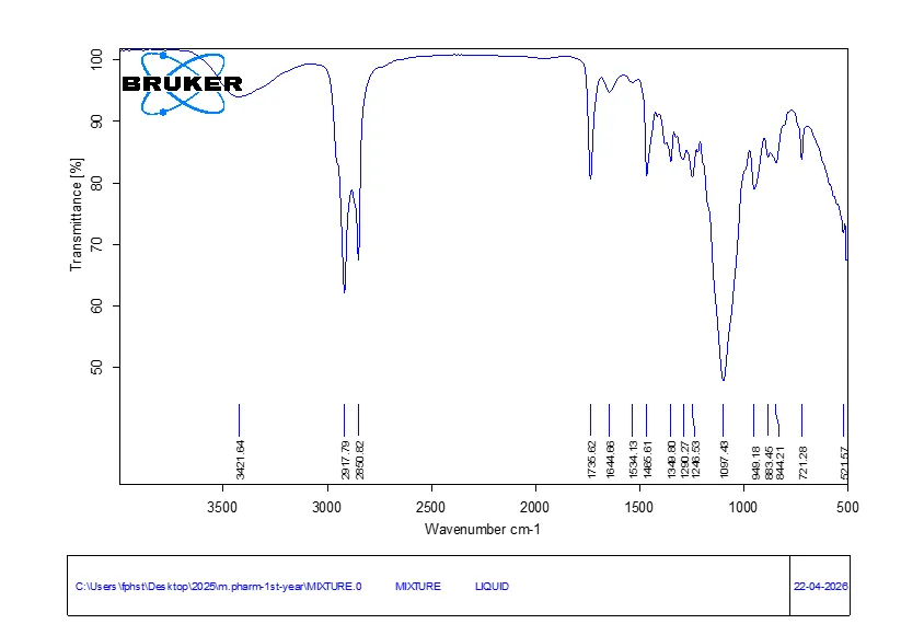
Figure 8: FTIR Spectra of Bee Venom, Span 60, Tween 60, Cholesterol and Their Physical Mixture.
Physicochemical characteristics of the niosomes
The dispersion was milky in appearance, as expected for vesicle formation. The optimised formulation had a mean size of 208.7 nm and a PDI of 0.3219, which indicates a moderately broad distribution within the nanometre range (Fig. 9). The zeta potential was −21.51 mV (Fig. 10); a negative surface charge of this size can provide some electrostatic repulsion between vesicles. The unentrapped venom gave an absorbance of 0.209, corresponding to 47.72 µg/mL, or 0.764 mg in 16 mL, out of 5 mg added, so the entrapment efficiency was 84.73% (Table 3). TEM showed rounded to oval vesicles with distinct boundaries, some variation in size and shape, and clustering in places (Fig. 11). The sizes seen by TEM were smaller than the hydrodynamic diameter from DLS, which is commonly seen because DLS measures the hydrated vesicle in suspension and TEM the dried sample.
Table 3: Physicochemical Characteristics of Bee Venom-Loaded Niosomes
| Parameter | Observed value |
|---|---|
| Mean particle size | 208.7 nm |
| Polydispersity index | [0.30 / 0.3219 – confirm] |
| Zeta potential | −21.51 mV |
| Absorbance of unentrapped venom | 0.209 |
| Unentrapped venom (16 mL) | 0.764 mg |
| Entrapment efficiency | 84.73% |
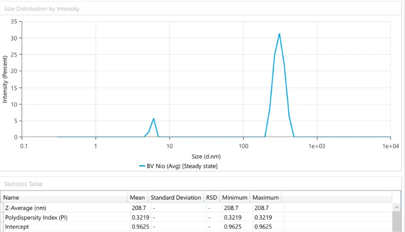
Figure 9: Particle Size Distribution and Polydispersity Index (PDI) of Bee Venom-Loaded Niosomes.
`
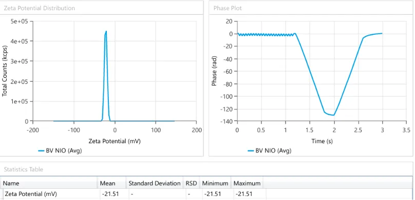
Figure 10: Zeta Potential Distribution of Bee Venom-Loaded Niosomes.
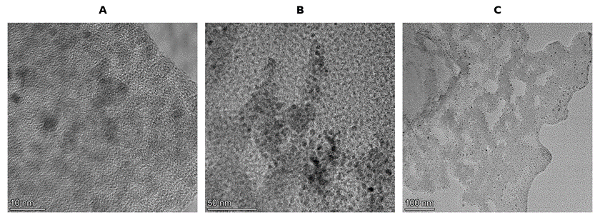
Figure 11: TEM Images of Bee Venom-Loaded Niosomes.
In vitro release
Pure venom released 16.70% at 0.5 h, 71.32% at 6 h and 93.92% at 24 h, whereas the niosomes released 8.43%, 28.17% and 89.18% at the same times (Table 4). Release from the niosomes was therefore much slower over the first 6 h, which is the clearest effect of encapsulation, but it rose sharply between 6 and 8 h (28.17% to 63.36%) and by 12 h was close to that of the pure venom (82.33% against 83.99%). The vesicle bilayer thus delayed early release but did not sustain it over the full 24 h. A different surfactant-to-cholesterol ratio could be explored if a longer plateau is required.
Table 4: Cumulative in Vitro Release of Bee Venom from Pure Venom and Niosomes
| Time (h) | Pure bee venom (%) | Bee venom-loaded niosomes (%) |
|---|---|---|
| 0.5 | 16.70 | 8.43 |
| 1 | 29.56 | 11.95 |
| 2 | 43.80 | 16.25 |
| 4 | 60.10 | 21.52 |
| 6 | 71.32 | 28.17 |
| 8 | 80.90 | 63.36 |
| 12 | 83.99 | 82.33 |
| 24 | 93.92 | 89.18 |
Of the four models, Korsmeyer–Peppas gave the highest coefficient of determination for both pure venom (R² = 0.936) and niosomes (R² = 0.984), and the Higuchi model came next (0.876 and 0.885) (Table 5, Fig. 13). Diffusion therefore appears to contribute to release, suggesting that the release mechanism may involve both diffusion and structural relaxation, as indicated by the Korsmeyer-Peppas model.
Table 5: Coefficients of Determination (R²) for Release Kinetic Models
| Kinetic model | Pure bee venom | Bee venom-loaded niosomes |
|---|---|---|
| Zero-order | 0.683 | 0.819 |
| First-order | 0.735 | 0.825 |
| Higuchi | 0.876 | 0.885 |
| Korsmeyer–Peppas | 0.936 | 0.984 |
[Figure - see the formatted PDF]
[Figure - see the formatted PDF]
[Figure - see the formatted PDF]
[Figure - see the formatted PDF]
Figure 13: Release Kinetic Plots: (A) Zero-Order, (B) First-Order, (C) Higuchi and (D) Korsmeyer–Peppas.
Cytotoxicity against MCF-7 cells
After 48 h the formulation lowered MCF-7 viability in a concentration-dependent way (Table 6, Fig. 14). Viability was 91.00% at 10 µg/mL and 84.20% at 50 µg/mL, fell to 58.32% at 100 µg/mL and 49.74% at 200 µg/mL, and reached 21.31% at 400 µg/mL. The IC50 from the logarithmic fit (y = −18.47 ln x + 142.59, R² = 0.8689) was 150.35 µg/mL (Fig. 15). The reference drug camptothecin at 7.8 µg/mL left 59.02% of cells viable. The MTT assay reports on metabolic activity and cannot by itself show how the cells died, and the study did not include free bee venom or blank niosomes as comparators, so the contribution of the carrier and any gain over the free venom remain to be established. The IC50 here is also not directly comparable with values from other niosomal venom studies that used different assays and exposure times.13,24
Table 6: Effect of Bee Venom-Loaded Niosomes on MCF-7 Cell Viability at 48 H
| Treatment | Concentration (µg/mL) | Viability (%) | Cytotoxicity (%) |
|---|---|---|---|
| Untreated control | – | 100.00 | 0.00 |
| Camptothecin | 7.8 | 59.02 | 40.98 |
| Formulation | 10 | 91.00 | 9.00 |
| Formulation | 50 | 84.20 | 15.80 |
| Formulation | 100 | 58.32 | 41.68 |
| Formulation | 200 | 49.74 | 50.26 |
| Formulation | 400 | 21.31 | 78.69 |
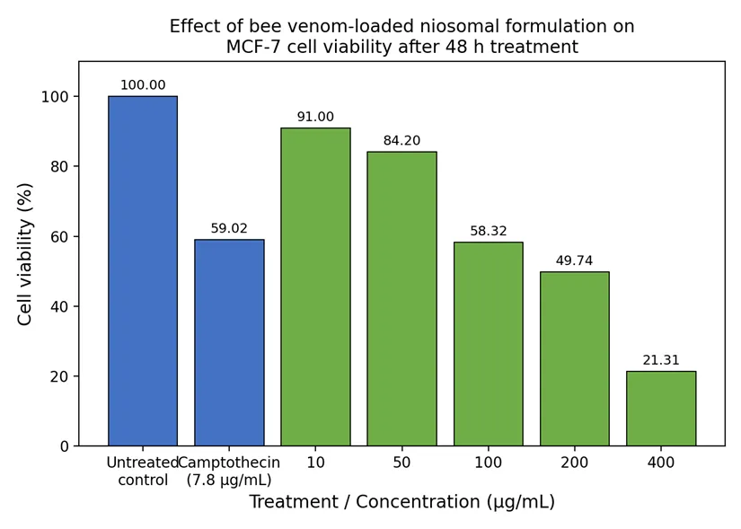
Figure 14: Viability of MCF-7 Cells After 48 H Exposure to Bee Venom-Loaded Niosomes.
[Figure - see the formatted PDF]
Figure 15: Determination of the IC50 of Bee Venom-Loaded Niosomes Against MCF-7 Cells.
CONCLUSION
Computational analysis pointed to MAPK1 and PIK3R1, among ten hub targets, as the best-scoring partners of melittin and flagged poor oral absorption and possible hepatotoxicity, which supports a delivery system and further safety work. Bee venom was successfully formulated into Span 60/Tween 60/cholesterol niosomes of about 209 nm with an entrapment efficiency of 84.73%, and no evident incompatibility with the excipients. The vesicles slowed the early release of venom and reduced MCF-7 viability in a concentration-dependent manner, with an IC50 of 150.35 µg/mL. These results justify testing the formulation in vivo; comparison with free venom and blank vesicles and mechanistic studies are needed to define the benefit of the carrier.
ACKNOWLEDGEMENT
The authors thank the Faculty of Pharmacy, M. S. Ramaiah University of Applied Sciences, Bengaluru, for providing the facilities needed for this work.
FUNDING
This research received no external funding.
CONFLICT OF INTEREST
The authors declare that the research was conducted in the absence of any commercial or financial relationships that could be construed as a potential conflict of interest.
REFERENCES

Daneshwari Y., Alin Bose J., Bee Venom-Loaded Niosomes for Breast Cancer: In Silico Target Prediction, Formulation, Characterization and in Vitro Cytotoxicity Against MCF-7 Cells, Int. J. of Pharm. Sci., 2026, Vol 4, Issue 11, 205-218. https://doi.org/10.5281/zenodo.23877275
 10.5281/zenodo.23877275