

We use cookies to ensure our website works properly and to personalise your experience. Cookies policy
1 Vinayaka College of Pharmacy.
2,3,4,5,6,7 Dreamz College of Pharmacy
Doxorubicin (DOX) is a popular chemotherapeutic agent, but is limited in clinical use because of its systemic toxicity and non-specific distribution. The aim of the current research was to design and test PLGA-based nanoparticles to deliver DOX to the target site and thereby enhance therapeutic effectiveness and minimize side effects. The nanoprecipitation method was used to prepare nanoparticles and optimized through the use of different concentrations of polymer and stabilizer. The optimized formulation had a particle size of 145.6 ± 5.2 nm, PDI of 0.212 ± 0.03 and zeta potential of -23.4 + 2.1 mV which was indicative of good stability and uniformity. High drug entrapment efficiency (78.5 ± 3.4%) and drug loading (12.3 ± 1.2%) were observed. In vitro drug release experiments revealed a biphasic and pH-sensitive release profile with augmented drug release amid acidic circumstances. Kinetics of release were based on the Higuchi model, which denotes the release is diffusion controlled. The anticancer activity of the DOX-loaded nanoparticles was found to be significantly higher than that of free DOX and the IC?? value was lower. The findings indicate that nanoparticles made of PLGA have a significant improvement in cytotoxicity (IC??: 3.2 µg/mL vs 6.8 µg/mL) and they possess pH-responsive drug release properties, which depict them as an effective tumor-targeted drug delivery system.
Cancer is still a major worldwide health concern and a substantial contributor to death and morbidity, despite decades of scientific advancement. Although conventional chemotherapy remains the mainstay of care for many cancers, it suffers from low solubility, restricted absorption and lack of selectivity between healthy and malignant tissues. These therapeutic limitations sometimes manifest in devastating systemic side effects, including myelosuppression and organ damage, that significantly limit the maximum acceptable dosage and therapeutic index of strong anticancer drugs. Hence, there is an urgent clinical need to create advanced next-generation delivery technologies that provide spatiotemporal control over medication administration(Yao et al., 2020).
The advent of the nanotechnology has transformed the scene in oncological pharmacology, by providing a strong platform on which the inherent limitations of traditional pharmacotherapy may be overcome. Drug delivery systems based on nanoparticles that include liposomes and polymeric micelles as well as inorganic metallic nanoparticles are highly versatile drug delivery systems that greatly impact the pharmacokinetic and pharmacodynamic profiles of the encapsulated drugs(Makadia & Siegel, 2011). By controlling the physicochemical properties of these carriers, such as particle size, surface charge, and morphology, researchers can protect the cargo against premature degradation, enhance cellular uptake, and increase blood circulation time. This structural engineering enables the accurate encapsulation of various chemical modalities, enabling the delivery of hydrophobic drugs that otherwise would not be soluble in physiological fluids(Hong et al., 2023).
In addition to simple encapsulation, the layout of contemporary nanocarriers focuses on the attainment of site-selective delivery via passive and active modalities of targeting. Passive targeting is based on the enhanced permeability and retention (EPR) effect, which is a phenomenon that is characterized by the disorganized, leaky vasculature and impairment of lymphatic drainage of solid tumors which encourages the preferential accumulation of nanocarriers within the tumor microenvironment. To further localize this, active targeting strategies are used where the surface of the nanoparticle is conjugated with molecular ligands such as monoclonal antibodies, aptamers and small molecules. These ligands recognize and bind to overexpressed receptors, or unique biomarkers on the surface of malignant cells and thereby internalizes the therapeutic payload and optimizes intracellular accumulation whilst avoiding exposure of healthy tissues to unintended exposure(Yu et al., 2016).
The emerging new frontier in targeted oncology is the development of so-called smart or stimuli-responsive nanocarriers. These systems react to external or internal stimuli to release drug at the target site, so that the therapeutic payload is only released when there is a reaction to stimuli. Structural changes or degradation of the nanocarrier can be induced by internal stimuli, such as the localized acidic pH of the tumor interstitium, redox gradients (e.g., glutathione concentrations) or elevated levels of specific enzymes (Dang & Guan, 2020). This on-demand release system has a strong negative impact on off-target cytotoxicity, which is a key factor that increases patient compliance and overall therapeutic efficacy(Yu et al., 2016).
The historical context of the therapeutic environment of oncology has been historically limited by the constraints of traditional chemotherapy. Although cytotoxic drugs are still key in the management of cancer, they lack specificity and thus tend to cause a lot of off-target toxicity and reduced efficacy because the agents are distributed throughout the body instead of being concentrated in tumor tissues. The presence of these clinical challenges requires the design of sophisticated pharmacological approaches that can enhance therapeutic index by maximizing tumor accumulation and minimizing toxicity to normal physiological systems (Dutta et al., 2025).
Nanotechnology is a versatile system that can be used to overcome these traditional constraints by serving as a carrier of a controlled and targeted delivery of anticancer therapeutics. These systems, often between 1 to 100 nm, utilize special physicochemical properties, such as a large surface/volume ratio and adjustable surface chemistry, to improve drug stability, solubility, and half-life of circulation. Nanocarriers can effectively shield normal tissues against untimely exposure, thereby reducing systemic side effects, which are prevalent with standard chemotherapeutic regimens (Bahrami et al., 2017).
Targeting and Microenvironment
Successful tumor-targeted delivery is based on two fundamental mechanisms: passive and active targeting. Passive targeting takes advantage of increased permeability and retention (EPR) effect whereby, the unorganized, leaky vascular architecture of tumors allows the preferential extravasation and accumulation of nanoparticles within the tumor interstitium. To enhance this process, active targeting strategies include conjugation of a specific ligand (i.e., antibodies, peptide, or aptamers) to the nanoparticle surface, which facilitate high-affinity binding to biomarkers overexpressed on the surface of malignant cells(Gavas et al., 2021).
Stimuli-Responsive Innovation
Recent developments have shifted towards so-called smart nanocarriers that can respond to endogenous stimuli that exist within the tumor microenvironment (TME). These types of systems are designed to detect biological signals, such as the typical acidic pH of the tumor interstitium, redox gradients, or the presence of tumor-associated enzyme activity, which act as triggers for the local release of the therapeutic cargo. By combining these stimuli-responsive properties with highly accurate active targeting, researchers hope to develop highly selective delivery platforms that not only overcome traditional drug resistance mechanisms but also greatly improve clinical outcomes in cancer patients.
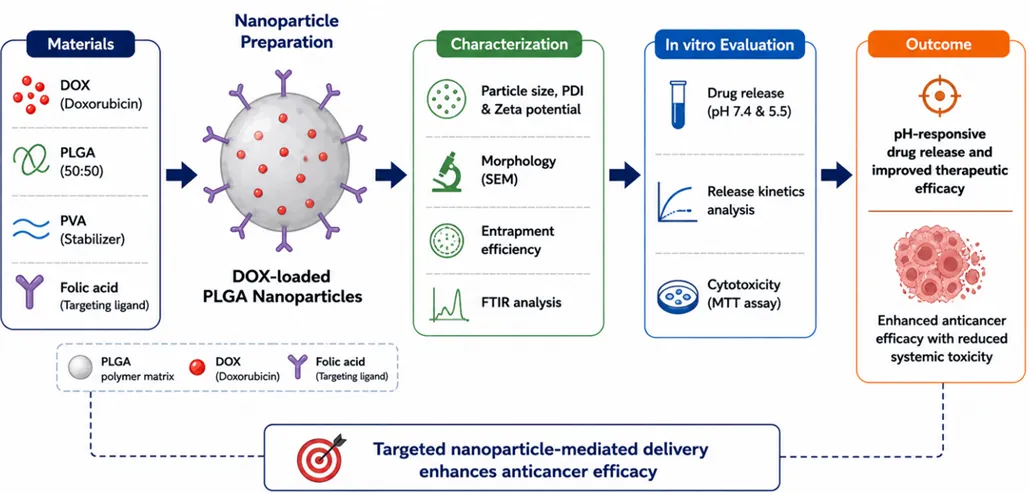
Figure 1: Schematic representation of preparation and evaluation of DOX-loaded PLGA nanoparticles.
Aim & Objectives
To develop and evaluate PLGA-based nanoparticles for targeted delivery of doxorubicin to enhance anticancer efficacy and achieve controlled drug release.
Objectives
MATERIALS AND METHODS
Materials
Doxorubicin hydrochloride (DOX) was chosen as the model anticancer drug because it has a broad-spectrum activity(Barenholz, 2012). Poly(lactic-co-glycolic acid) (PLGA; ; 50:50, Sigma-Aldrich, USA) was taken as a biodegradable polymer because it is an excellent biocompatible polymer that is also acceptable by regulations (Danhier et al., 2012). Polyvinyl alcohol (PVA; Himedia, India) was used as a stabilizing agent. There was the use of folic acid (Merck, Germany) as a targeting ligand. They were used without any additional purification in organic solvents like acetone and ethanol (analytical grade). All the reagents were of pharmaceutical grade.
Preparation of Nanoparticles
Nanoparticles were prepared using the nanoprecipitation method as previously reported. Briefly, PLGA and DOX were dissolved in acetone to form the organic phase. This organic phase was added dropwise into an aqueous phase containing PVA under constant magnetic stirring at 1000 rpm. Nanoparticles were formed due to rapid solvent diffusion, leading to polymer precipitation.
The nanoprecipitation technique was used to prepare nanoparticles as reported elsewhere (Fessi et al., 1989). In a nutshell, acetone was used to dissolve PLGA and DOX to form organic phase. The drop-wise addition of this organic phase into an aqueous phase containing PVA under constant magnetic stirring at 1000 rpm was performed. The nanoparticles were formed because of the quick diffusion of solvents into a polymer, causing it to precipitate.
The resultant solution was agitated for 4 h to achieve full evaporation of the organic solvent. The nanoparticles were centrifuged (Remi R-24, India) at 15,000 rpm for 20 minutes and rinsed with distilled water to remove excess stabilizer and were lyophilized for future usage.
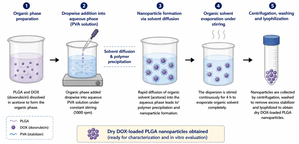
Figure 2: schematic illustration of preparation of DOX-loaded PLGA nanoparticles by nanoprecipitation method
Formulation Optimization
Various batches were made by changing the polymer and stabilizer concentrations to obtain an optimum formulation.
Table 1: Composition of Formulations
|
Formulation |
PLGA (mg) |
DOX (mg) |
PVA (%) |
Particle Size (nm) |
|
F1 |
50 |
10 |
0.5 |
210 ± 6.3 |
|
F2 |
75 |
10 |
1.0 |
165 ± 5.8 |
|
F3 |
100 |
10 |
1.0 |
145 ± 5.2 |
|
F4 |
100 |
10 |
1.5 |
160 ± 4.9 |
F3 was chosen as the optimized formulation out of the prepared formulations due to its small particle size and narrow size distribution.
Surface Functionalization
Surface modification of nanoparticles was performed using folic acid via carbodiimide (EDC/NHS) chemistry as reported in previous studies (Hermanson, 2013). The activated nanoparticles were reacted with folic acid under controlled conditions, followed by purification through centrifugation.
Characterization of Nanoparticles
Particle Size, PDI and Zeta Potential
Dynamic Light Scattering was used to determine particle size, polydispersity index (PDI) and zeta potential (Malvern Zetasizer Nano ZS, UK). Samples were appropriately diluted prior to analysis.
Morphological Analysis
Scanning Electron Microscopy was used to study the surface morphology of nanoparticles (JEOL JSM-7610F, Japan).
Drug Entrapment Efficiency
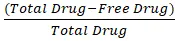
The quantity of free drug in the supernatant was measured using a UV-Visible spectrophotometer (Shimadzu UV-1800) at λmax = 480nm.
EE% = (Total Drug-Free Drug)Total Drug
FTIR Analysis
The spectra were measured (Bruker FTIR) in the range of 4000-400 cm⁻¹ to assess potential interactions between the drug and polymer.
In Vitro Drug Release Study
The drug release was examined by the dialysis bag diffusion method, a commonly used methodology for nanoparticle-based drug delivery systems (Soppimath et al., 2001), in phosphate buffer solutions (pH 7.4 and pH 5.5) at 37°C with constant stirring. Samples were collected at predefined intervals and replaced with new buffer to keep sink conditions. The concentration of drug was measured spectrophotometrically at 480nm.
In Vitro Cytotoxicity Study
The MTT test was used to measure the level of cytotoxicity on to the MCF-7 cell lines of breast cancer as stated earlier(Mosmann, 1983). Different concentrations (1-100 µg/mL) of free DOX and DOX-loaded nanoparticles were used to treat cells. The incubation was followed by measuring the absorbance of the microplate at 570 nm using a microplate reader (Bio-Rad).
Statistical Analysis
Each experiment was conducted three times, and data were presented in terms of mean + standard deviation. GraphPad Prism 8.0 was used to statistically analyse the data and trio of differences were considered statistically significant at p < 0.05.
RESULTS
Particle Size, PDI and Zeta Potential
Table 2: Physicochemical Properties
|
Parameter |
Value |
|
Particle Size (nm) |
145.6 ± 5.2 |
|
PDI |
0.212 ± 0.03 |
|
Zeta Potential (mV) |
−23.4 ± 2.1 |
The size of the particles less than 200 nm is the one that allows an efficient tumor accumulation using the EPR effect, and the zeta potential (−23.4 mV) guarantees the tumor accumulation through the EPR effect in the nanoparticles (Maeda et al., 2000). The low PDI value is a sign of uniform size distribution whereas the negative zeta potential is a sign of good colloidal stability.
Morphological Analysis
SEM analysis revealed the nanoparticles to be of a spherical shape with smooth surface morphology and had no significant aggregation, which is an indication that there was successful formulation.
Drug Entrapment Efficiency
Table 3: Entrapment Efficiency and Drug Loading
|
Parameter |
Value |
|
Entrapment Efficiency (%) |
78.5 ± 3.4 |
|
Drug Loading (%) |
12.3 ± 1.2 |
The high entrapment efficiency can be explained by effective encapsulation of the drug into the polymeric matrix which can be ascribed to the presence of hydrophobic interactions between DOX and PLGA.
In Vitro Drug Release
Table 4: Drug Release Profile
|
Time (hrs) |
% Release (pH 7.4) |
% Release (pH 5.5) |
|
1 |
12.5 ± 1.1 |
18.3 ± 1.3 |
|
4 |
25.2 ± 1.5 |
38.6 ± 2.1 |
|
8 |
38.4 ± 2.0 |
55.2 ± 2.5 |
|
24 |
52.6 ± 2.4 |
72.8 ± 3.0 |
|
48 |
65.3 ± 2.8 |
85.4 ± 3.2 |
The formulation had a biphasic release pattern, which is typical of polymeric nanoparticle systems (Kumari et al., 2010). The higher rate of drug release during the acidic pH may be explained by the greater extent of polymer swelling and the faster diffusion of the drug during the acidic tumor-mimicking conditions. This pH-sensitive release behavior is beneficial to cancer treatment, because the tumor microenvironment is relatively acidic as compared to normal physiological environment (Maeda et al., 2000),(Danhier et al., 2012).
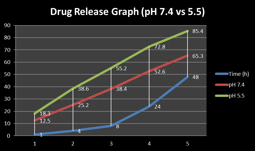
Figure 3: In vitro drug release profile of DOX-loaded PLGA nanoparticles at pH 7.4 (physiological) and pH 5.5 (tumor microenvironment), showing pH-responsive and biphasic release behavior (mean ± SD, n = 3).
Release Kinetics
Table 5: Kinetic Model Analysis
|
Model |
R² Value |
|
Zero Order |
0.912 |
|
First Order |
0.934 |
|
Higuchi |
0.982 |
|
Korsmeyer-Peppas |
0.967 |
The release kinetics data was best characterized by the Higuchi model, demonstrating that drug release is mostly regulated by a diffusion-controlled mechanism as indicated by (Higuchi, 1963).
Cytotoxicity Study
Table 6: Cell Viability Results
|
Treatment |
Cell Viability (%) |
|
Control |
100 ± 2.1 |
|
Free DOX |
52.3 ± 2.5 |
|
DOX Nanoparticles |
28.4 ± 2.0 |
The difference was statistically significant (p < 0.05), which indicates increased therapeutic efficiency of the nanoparticle system, which is in line with the previously reported nanoparticle-based anticancer systems (Peer et al., 2007).
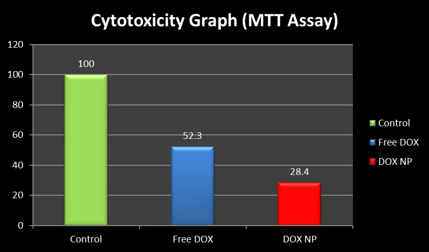
Figure 4: In vitro cytotoxicity of free DOX and DOX-loaded PLGA nanoparticles in MCF-7 cells. Data are expressed as mean ± SD (n = 3). p < 0.05 vs control; #p < 0.05 vs free DOX.
IC₅₀ values:
Reduced IC₅₀ value of nanoparticle formulation is an excellent indicator of enhanced anticancer efficacy, which can be explained by the enhanced cellular uptake and sustained drug release. This increase in cytotoxic effect indicates a better intracellular delivery and retention of DOX via uptake through nanoparticles.
DISCUSSION
The current research was able to prepare nanoparticles of the chosen drug, doxorubicin, using the nanoprecipitation method with the help of the nanoparticle, PLGA. The optimized formulation (F3) had a particle size less than 200 nm which is regarded as the optimum particle size in passive tumor targeting using the enhanced permeability and retention (EPR) effect. The small value of PDI was used to confirm even distribution of the particles and the negative value of zeta potential was taken to confirm good colloidal stability which reduces the chances of aggregation.
The reason behind the high entrapment efficiency observed in the study is because it is possible to incorporate DOX effectively within the PLGA matrix. This is explained by the hydrophobic forces of attraction between the drug and the polymer that increases retention of the drug in the nanoparticles.
In vitro drug release experiments showed that the drug release had a biphasic release pattern, which was characterized by an initial burst release and a sustained drug release. The initial burst can be attributed to the release of the surface-adsorbed drug, but the sustained one is controlled by the diffusion of the drug through the polymer matrix. The change in drug release at low pH conditions (pH 5.5) relative to the physiological pH (pH 7.4) confirms the pH-responsive characteristic of the formulation. This feature is especially advantageous in cancer treatment, where tumor microenvironment is more acidic, thus allowing site-specific drug delivery.
The analysis of the release kinetics revealed that the drug release process was in agreement with the Higuchi model, indicating that the process could be attributed to a diffusion-controlled drug release process. This is in support of the sustained release profile that was observed in the formulation.
Cytotoxicity experiments showed that DOX loaded nanoparticles had a much higher anticancer activity than unloaded DOX. The reduced IC₅₀ of nanoparticle formulation implies an increased uptake by the cell and better therapeutic efficacy. This increased effect could be attributed to the enhanced intracellular delivery and longer retention of the drug in cancer cells. Altogether, these results indicate the potential of the PLGA nanoparticles as an effective drug delivery system to treat cancer, which offers controlled release, enhanced targeting and better therapeutic effects. The obtained entrapment efficiency (78.5%) and particle size (~145 nm) are in agreement with previously reported systems of nanoparticle systems based on PLGA, which typically shows a particle size range of 100-200 nm and an entrapment efficiency of 70-85% (Kumari et al., 2010; Danhier et al., 2012). Likewise, the experimental bipolar drug release profile is consistent with previous reports, and confirms the theoretical diffusion-controlled release behavior in polymeric nanoparticle systems. This system can permit decrease in dose and lower cardiotoxicity in the systemic system when using doxorubicin.
CONCLUSION
In conclusion, the current paper has been able to develop and optimize DOX-loaded PLGA nanoparticles with desirable physicochemical characteristics and increased anticancer effects. The nanoparticles displayed targeted and pH-sensitive drug delivery behavior, which is beneficial to targeted cancer therapy. The positive cytotoxic activity and the lower IC50 value of the nanoparticle system relative to free drug confirms the increased therapeutic activity of the nanoparticle system. Thus, nanoparticles made of PLGA can be considered a promising and effective delivery method to the targeted delivery of anticancer drugs. In vivo experiments should be furthered to confirm their clinical utility. The results indicate that this could be translated to preclinical in vivo and future clinical implementations. The developed nanosystem shows great potential in translation into preclinical cancer models and may help in reducing systemic toxicity that comes with conventional chemotherapy.
REFERENCES






Dr. Puneet Kumar, Vikrant Kumar, Sita Ram, Vijay Sharma, Nisha Devi, Neha, Shivani, Design, Development and In-vitro Characterization of a Sensitive Nanoparticles Drug Delivery System for Targeted Delivery of an Anticancer Drug, Int. J. of Pharm. Sci., 2026, Vol 4, Issue 5, 8167-8176. https://doi.org/10.5281/zenodo.20471708
 10.5281/zenodo.20471708