

We use cookies to ensure our website works properly and to personalise your experience. Cookies policy
1,2,4 Hindu College of Pharmacy, Gunter, 522002.
3 Nirmala College of Pharmacy, Atmakur, 522503.
Tuberculosis (TB), caused by Mycobacterium tuberculosis, is a formidable global health threat due to the raising prevalence of multidrug-resistant strains (MDR) and extensively drug-resistant (XDR) strains posing a major challenge to effective treatment. Conventional anti-tubercular therapy is associated with prolonged treatment duration, adverse drug reactions, and increasing drug resistance, highlighting the need for safer and more effective alternatives. Discovering alternative biomolecules with low toxic profiles and effective mechanism of action has become the priority of this research. The present study aimed to develop and evaluate binary Polyherbal formulations prepared from Aegle marmelos, Moringa oleifera, Vitex negundo, and Alstonia scholaris for their in vitro anti-tubercular activity. Ethanolic extracts of the selected medicinal plants were subjected to preliminary phytochemical screening and formulated in equal proportions. Anti-tubercular activity was assessed using the Microplate Alamar Blue Assay (MABA) against Mycobacterium tuberculosis. The phytochemical analysis confirmed the presence of alkaloids, flavonoids, phenolics, terpenoids, tannins, and other bioactive constituents. The formulations exhibited varying degrees of inhibitory activity, with pH-1.4 showing the most promising antimycobacterial effect. The findings suggest that binary Polyherbal formulations possess significant anti-tubercular potential due to synergistic action of their phytoconstituents and may serve as promising complementary agents for tuberculosis management. Further phytochemical characterization, toxicity evaluation, and in vivo studies are recommended to validate their therapeutic potential.
Tuberculosis is a contagious airborne bacterial infection that commonly affects lungs, but can also affect other areas of the body like spine, brain or kidneys. It is caused by Mycobacterium tuberculosis. Every infected person will not get sick. Some may have the infection but no symptoms, which is called inactive tuberculosis or latent tuberculosis. Some people can have latent TB infection for lifetime without ever developing symptoms. TB can become active if immune system becomes weakened. [1][2]
Causes
A mycobacterium tuberculosis bacterium causes TB. It spreads through the air when an infected person coughs, sneezes, or talks and can infect your lungs when you breathe them in. it is a main causative agent which can also cause the disease through M. bovis and M. africanum. It is a strict aerobe and an intracellular parasite. It has a unique, waxy cell that is rich in mycolic acid, makes it resistant to weak disinfectants and allows it to survive in the air or dry surfaces for extended periods. People infected with HIV/AIDS are at much greater risk of developing active TB because of weakened immunity. [1][3]
Symptoms
Active TB symptoms include:
Some people won’t have symptoms but might have a positive TB test. Such TB is known as inactive TB. [1][2]
Transmission
TB can spread through a person with active TB coughs, sneezes or talks. People with active lung infection are contagious. Being in close contact with the infected for long time transmits TB. Most people can fight and stop the bacteria from growing. This causes a latent TB infection. The bacteria can remain dormant for years but may reactivate and cause active disease if the host’s immune system becomes compromised (due to HIV, malnutrition, or aging). [1][2][4]
Types
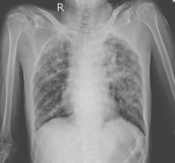
Tuberculosis can affect multiple organs. Based on the site of infection TB is classified into pulmonary TB and extra pulmonary TB. [1][2]
Pulmonary Tuberculosis (PTB)
This is the most common form of tuberculosis which involves lungs. [3][4][5]

Key features
Transmission
Diagnosis
Extra pulmonary Tuberculosis (EPTB)
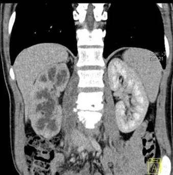
This refers to TB affecting organs other than the lungs. [1]
Common sites
Symptoms
Transmission
Global Burden of TB
Tuberculosis is one of the world’s leading infectious diseases. Despite the availability of effective treatment, TB continues to be a major public health problem, especially in low and middle-income countries.
According to World Health Organization Global TB Report, TB affects millions of people every year and is among the top causes of death from a single infectious agent worldwide.
High-Burden Countries
The countries contributing largest number of TB cases include:
Among these India accounts for the highest number of TB cases globally. [6]
Factors Contributing to High Prevalence
Drug-Resistant TB
A major global concern is multidrug-resistant tuberculosis (MDR-TB) and extensively drug-resistant tuberculosis (XDR-TB). These forms are difficult to treat and require longer therapy with more toxic and expensive drugs. [1]
Global Control Measures
Causative Organism Characteristics
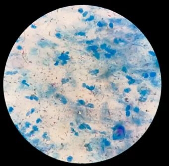
Acid-Fast Bacillus
Slow-Growing Organism
Cell Wall Structure
Virulence Factor
Pathogenesis of Tuberculosis (TB)
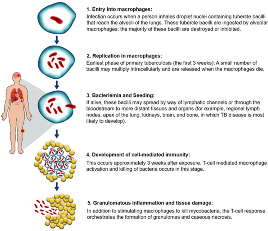
Pathogenesis of Tuberculosis is multi-stage process driven by the interplay between the causative bacterium, Mycobacterium tuberculosis, and the host’s cell-mediated immune response. The infection progresses through distinct clinical and pathological phases, shifting from an initial cellular invasion to either containment or destructive tissue disease. [2][4][5]
|
Clinical Form |
Pathogenic Mechanism |
Clinical Status |
|
Latent TB Infection (LTBI) |
The solid granuloma completely sequesters the visible bacilli, forcing them into a dormant metabolic state. |
Asymptomatic; Non-infectious; Positive skin/blood tests. |
|
Active/Post-Primary TB |
If the immune system weakens, the Caseous center liquefies, granting the bacilli a rich environment to rapidly multiply. |
Symptomatic (cough, hemoptysis); Highly contagious; Cavitary lung lesions. |
If the initial immune response fails to contain the primary focus altogether, unchecked local replication can cause bronchopneumonia, or widespread vascular rupture leading to systemic miliary TB. [7][10]
Current Anti-TB Therapy
First-line anti-tuberculosis therapy relies on a combination of four primary medications. The standard regimen for drug-susceptible tuberculosis (TB) consists of an initial 2-month intensive phase using all four drugs to rapidly decrease the bacterial load, followed by a 4-month continuation phase usually using only two. [1][2][4]
First-Line Anti-TB Drugs [11][12]
Key Clinical and Operational Discussions
The high rate of adverse drug reactions is a leading cause of poor patient adherence. Beyond liver issues, specific first- line drugs cause distinct toxicities:
The escalation of multidrug-resistant and extensively drug-resistant (MDR and XDR) strains of Mycobacterium tuberculosis remains a critical hurdle for global healthcare networks. Conventional short-course anti-TB therapeutic regimens involve combining multiple synthetic drugs, such as Isoniazid, Rifampicin, Pyrazinamide, and Ethambutol. However, the side effects of these therapies are reportedly high. These often trigger poor patient compliance due to their systemic side effects, such as drug-induced liver injury (DILI) and nephrotoxicity, accelerating the evolution of resistant bacteria populations. [2][11]
Polyherbal formulations (PHFs) utilizes the therapeutic synergy of multiple medicinal plants to achieve a distinct multi-target pharmacological action while overcoming the metabolic toxicity associated with the chemical compounds present in the synthetic medicines. Aegle marmelos, Moringa oleifera, Alstonia scholaris, and Vitex nirgundi are recognized historically in ethno pharmacological literature for treating chronic respiratory disorders, pulmonary infections, and deep-seated tissue inflammation. After performing the phytochemical screening of these botanical sources indicates wide source of active secondary metabolites, including coumarins, alkaloids, flavonoids, and terpenes which shows varying degrees of antimicrobial activity. [13][14]
Limitations of current anti-TB therapy
1. Prolonged treatment duration and High pill burden
One of the major drawbacks of present Anti-TB treatment is prolonged duration of treatment. Standard treatment requires at least 6months of multi drug administration. Although shorter four month routine is recently introduced for some patients and they are not implemented globally. The longer period of treatment is accompanied by every day pill burden, and sometimes patients may have to administer multiple medications simultaneously. This may lead to fatigue and gastrointestinal discomfort. [2][11]
2. Emergence of drug resistance
The increasing prevalence of drug resistant mycobacterium tuberculosis represents one of the greatest obstacles to control TB. Resistance develops through spontaneous genetic mutations that are due to ineffective therapy. The emergences of multidrug resistant and extensively drug resistant tuberculosis, these resistant forms require longer, more expensive and more toxic treatment and have lower success rates. [2][12]
3. Drug toxicity and adverse drug reactions
Hepatotoxicity is the most serious complication and is commonly linked with Isoniazid, Rifampicin, and Pyrazinamide. Drug induced liver injury frequently necessitates interruption or modification of treatment, there by compromising therapeutic success. Patients also commonly experience gastrointestinal disturbances, including nausea, vomiting, abdominal pain, anorexia, and diarrhea, which affects the quality of life. [2][11]
Second line anti TB agents used for drug resistant tuberculosis is having even more toxicity. Older inject able drugs caused irreversible hearing loss frequently. Linezolid which is a newer agent which produce peripheral neuropathy, optic neuropathy, and bone marrow suppression during prolonged therapy. [2][11][12]
The limitations associated with current TB chemotherapy have increased interest in exploring of medicinal plants as therapeutic agents. Several studies have demonstrated that these bioactive constituents such as alkaloids, flavonoids, terpenoids, phenolic compounds, tannins, and saponins, possess inhibitory effects against Mycobacterium tuberculosis, including drug-resistant strains, while also exhibiting comparatively lower toxicity in experimental models. [11][12]
Prolonged treatment duration and high pill burden: Plant-derived bioactive compounds may possess potent antimycobacterial activity and could serve as supporting treatments. They may enhance treatment efficacy, enables drug dose reduction and shortened treatment courses. [2]
Drug resistance: Many medicinal plants contain multiple bioactive compounds (alkaloids, flavonoids, terpenoids, phenolics, tannins, etc.) which have different mechanisms. These compounds may inhibit drug-resistant M. tuberculosis interfere with biofilm formation, or increasing the activity of existing antibiotics, potentially reducing resistance development. [11][12]
Drug toxicity and adverse effects: some medicinal plants contain antioxidant, anti-inflammatory, and hepatoprotective properties. These activities may decrease oxidative stress and protect organs such as the liver from drug causing injury. Some plant extracts have also been reported to have gastrointestinal inflammation and improve treatment tolerability. [11][12]
Although number of medicinal plants have showed potential antimycobacterial activity only few research have explored standardized Polyherbal formulations combining Aegle marmelos, Moringa oleifera, Alstonia scholaris, and Vitex negundo. The synergistic potential, phytochemical profile and therapeutic efficacy of such a formulation remain underexplored. Therefore, the present study was undertaken to develop and evaluate a Polyherbal formulation against tuberculosis. [13][14]
Selected Medicinal Plants
1. Aegle marmelos
Aegle marmelos, commonly known as Bael, is a medicinal tree belonging to the family Rutaceae and is widely distributed across India, Sri Lanka, Nepal, Bangladesh, and Southeast Asia. The plant has been extensively used in traditional systems of medicine, including Ayurveda, Siddha, and Unani, for the treatment of gastrointestinal disorders, diabetes, respiratory diseases, inflammation, and various microbial infections. Different parts of the plant, particularly the leaves, fruits, bark, and roots, are valued for their diverse therapeutic properties. [13][14]
Phytochemical studies have revealed that A. marmelos is rich in biologically active constituents such as alkaloids, flavonoids, coumarins, tannins, phenolic compounds, terpenoids, and essential oils. Important bioactive compounds including aegeline, marmelosin, skimmianine, rutin, quercetin, and lupeol contribute to its broad pharmacological activities. These phytochemicals exhibit potent antioxidant, antimicrobial, anti-inflammatory, immunomodulatory, antidiabetic, hepatoprotective, and anticancer properties. [13][14]
Several studies have demonstrated that extracts of A. marmelos possess significant antimicrobial activity against both Gram-positive and Gram-negative bacteria as well as certain fungal pathogens. The antimicrobial efficacy is primarily attributed to the synergistic action of flavonoids, alkaloids, coumarins, and phenolic compounds, which interfere with microbial cell membrane integrity, enzyme activity, and cellular metabolism. Although direct studies against Mycobacterium tuberculosis are comparatively limited, the presence of these bioactive constituents suggests promising antimycobacterial potential. Furthermore, its antioxidant and immunomodulatory effects may enhance host defense mechanisms by reducing oxidative stress and regulating inflammatory responses during infection. Owing to its rich phytochemical profile and broad-spectrum biological activities, A. marmelos represents a valuable medicinal plant for incorporation into binary Polyherbal formulations aimed at improving anti-tubercular efficacy through synergistic interactions. [15][16]
Moringa oleifera
Moringa oleifera, commonly known as the drumstick tree or horseradish tree, belongs to the family Moringaceae and is widely cultivated in tropical and subtropical regions. It is recognized as one of the most nutritionally and medicinally valuable plants due to its rich content of vitamins, minerals, proteins, and bioactive phytochemicals. Traditionally, various parts of the plant, including the leaves, seeds, bark, flowers, and roots have been used to treat infections, inflammation, malnutrition, diabetes, hypertension, and gastrointestinal disorders. [13][14]
Phytochemical investigations have identified a wide range of bioactive constituents such as flavonoids, phenolic acids, alkaloids, glucosinolates, isothiocyanates, tannins, saponins, and terpenoids. Major compounds including quercetin, kaempferol, chlorogenic acid, niazimicin, and benzyl isothiocyanates contribute significantly to its pharmacological activities. These constituents possess strong antioxidant properties that protect cells against oxidative stress and support immune function. [13][14]
Extensive pharmacological studies have demonstrated that M. oleifera exhibits antimicrobial, anti-inflammatory, antioxidant, antidiabetic, hepatoprotective, immunomodulatory, and anticancer activities. Several reports have shown that leaf and seed extracts inhibit growth of various pathogenic bacteria and fungi through disruption of microbial cell membranes, inhibition of enzyme activity, and interfere with essential metabolic pathways. Moreover, preliminary investigations have indicated promising antimycobacterial activity against Mycobacterium tuberculosis and related mycobacterial species. The combined antimicrobial, antioxidant, and immunomodulatory properties of M. oleifera make it an attractive candidate for incorporation into binary Polyherbal formulations, where synergistic interactions among phytochemicals may enhance anti-tubercular efficacy while minimizing the development of drug resistance. [17]
Vitex negundo
Vitex negundo, commonly known as the five-leaved chaste tree or Nirgundi, belongs to the family Lamiaceae and is widely distributed throughout India, China, Sri Lanka, and other tropical regions of Asia. The plant has been extensively employed in Ayurveda, Siddha, And traditional folk medicine for the management of inflammation, pain, fever, respiratory disorders, arthritis, skin diseases, and microbial infections. Leaves are the most commonly utilized part because of their rich phytochemical composition and therapeutic significance. [13][14]
Phytochemical investigations have revealed the presence of flavonoids, iridoid glycosides, alkaloids, phenolic compounds, terpenoids, volatile oils, tannins, and lignans. Major constituents such as casticin, vitexin, negundoside, agnuside, and ursolic acid contribute to its diverse pharmacological activities. These compounds possess potent antioxidant, antimicrobial, anti-inflammatory, analgesic, and immunomodulatory properties. [13][14]
Numerous studies have reported that V. negundo exhibits broad-spectrum antimicrobial activity against several Gram-positive and Gram-negative bacteria as well as fungal pathogens. The antimicrobial action is attributed to its ability to alter microbial membrane permeability, inhibit enzyme function, and suppress microbial growth. In addition, preliminary investigations have demonstrated promising antimicrobial activity of leaf extracts against Mycobacterial tuberculosis. The antioxidant and anti-inflammatory effects of plants may further support host defense by reducing oxidative damage and modulating immune responses during infection. Considering its rich phytochemical profile and documented biological activities, V. negundo is considered a promising medicinal plant for inclusion in binary Polyherbal formulations designed to enhance anti-tubercular activity through synergistic phytochemical interactions. [18]
Alstonia scholaris
Alstonia scholaris, commonly known as the Devil’s tree or Saptaparni, belongs to the family Apocynaceae and is widely distributed throughout India, Southeast Asia, and Australia. The plant has long been used in traditional medicine for the treatment of respiratory disorder, fever, malaria, chronic cough, asthma, diarrhea, skin diseases, and infectious conditions. The bark and leaves are particularly valued for their therapeutic properties and have been extensively investigated for their pharmacological activities. [13][14]
The plant contains a diverse array of phytochemicals, including indole alkaloids, flavonoids, iridoids, triterpenoids, phenolic compounds, tannins, and steroids. Important alkaloids such as echitamine, scholaricine, picrinine, and alstonine are considered responsible for many of its biological activities. These compounds exhibit significant antioxidant, antimicrobial, anti-inflammatory, antimalarial, and immunomodulatory effects. [13][14]
Several studies have demonstrated the antimicrobial efficacy of A. scholaris extracts against a wide range of bacterial and fungal pathogens. The antimicrobial activity is mainly attributed to indole alkaloids and phenolic compounds, which disrupts microbial cell integrity and inhibits essential metabolic processes. Notably extracts of A. scholaris have shown encouraging antimicrobial activity against Mycobacterium tuberculosis in invitro studies, highlighting its potential as a natural source of anti-tubercular agents. Furthermore, its antioxidant and immunomodulatory properties may enhance host immune response during tuberculosis infection. Owing to its potent phytochemical profile and documented antimycobacterial activity, A. scholaris represents an important component of binary Polyherbal formulations developed to improve therapeutic efficacy through synergistic interactions among medicinal plants. [19]
MATERIALS AND METHODS
Plant Materials
Plant materials of Aegle marmelos, Moringa oleifera, Vitex negundo and Alstonia scholaris are collected and dried for 7 days. All four plant materials are separately titurated into fine powder and stored in airtight containers at room temperature until extraction.
Preparation of Methanolic Extracts
Methanolic extracts of each medicinal plant were prepared individually using the cold maceration technique. 20g of each powdered plant material was transferred into separate clean conical flasks containing 200ml of ethanol. These mixtures are covered with aluminum foil and kept at room temperature for 72 hours with intermittent shaking to facilitate efficient extraction of phytoconstituents. After maceration, the extracts were filtered through Whattman No. 1 filter paper to remove insoluble plant residues. The filtrates were concentrated by allowing the solvent to evaporate at room temperature until semisolid crude extracts were obtained. The extracts were stored in airtight containers under refrigerated conditions (4˚C) until further use.
Preliminary Phytochemical Screening
The ethanolic extracts of Aegle marmelos, Moringa oleifera, Vitex negundo and Alstonia scholaris were subjected to preliminary qualitative phytochemical screening using standard phytochemical procedures. The extracts were tested for the presence of major classes of phytoconstituents, including carbohydrates, glycosides, alkaloids, flavonoids, phenols, tannins, saponins, steroids, and terpenoids. The tests were performed using standard qualitative method based on characteristic colour change or precipitate formation. The intensity of reactions was recorded as highly present (+++), moderately present (++), weakly present (+), or absent (-) according to the observed response.
|
Phytochemicals |
Aegle marmelos |
Moringa oleifera |
Vitex negundo |
Alstonia scholaris |
|
Carbohydrates |
+ |
++ |
++ |
++ |
|
Glycosides |
+ |
++ |
++ |
++ |
|
Alkaloids |
+ |
+++ |
+++ |
+++ |
|
Flavonoids |
+ |
+++ |
+++ |
+++ |
|
Phenols |
+ |
+++ |
+++ |
+++ |
|
Tannins |
+ |
++ |
++ |
+ |
|
Saponins |
- |
++ |
++ |
++ |
|
Steroids |
+ |
++ |
+++ |
+++ |
|
Terpenoids |
+ |
- |
+++ |
+++ |
The qualitative phytochemical screening demonstrated that all four medicinal plants possessed diverse classes of bioactive secondary metabolites, although their abundance varied. Alkaloids, flavonoids, terpenoids and phenolic compounds were consistently detected indicating their potential contribution to the antimicrobial and antioxidant properties of the formulations. The combination of these extracts in binary Polyherbal formulations may therefore provide synergistic therapeutic effects through multiple mechanisms, including microbial growth, antioxidant activity, modulation of inflammatory responses, and enhancement of host immune defense. These phytochemical findings provide a scientific basis for the anti-tubercular activity observed in the MABA assay.
Preparation of Polyherbal Formulations
Four binary Polyherbal formulations were prepared by mixing the individual ethanolic extracts in an equal ratio (1:1, w/w). The formulations prepared were:
|
Formulation Code |
Composition |
|
PH – 1.2 |
Aegle marmelos + Moringa oleifera |
|
PH – 2.3 |
Moringa oleifera + Vitex negundo |
|
PH – 3.4 |
Vitex negundo + Alstonia scholaris |
|
PH – 1.4 |
Alstonia scholaris + Aegle marmelos |
The extracts were thoroughly mixed until a homogenous Polyherbal formulation was obtained. The prepared formulations were stored in sterile containers under refrigerated conditions until evaluation.
Anti tubercular activity by Microplate Alamar Blue Assay (MABA)
The in vitro anti-tubercular activity of the prepared binary Polyherbal formulations was evaluated against Mycobacterium tuberculosis using the MABA. The assay was performed in sterile 96-well Microplate containing Middlebrook 7H9 broth supplemented with 10% OADC (oleic acid-albumin-dextrose-catalase) enrichment and 0.2% glycerol. A standardized bacterial inoculum was prepared from cultures grown on Lowenstein-Jensen (LJ) medium and diluted appropriately before inoculation.
Serial two-fold dilutions of each binary Polyherbal formulation were prepared to obtain final concentrations of 25, 12.5, 6.25, 3.125, 1.56, and 0.78µg/mL. Rifampicin was used as the reference anti-tubercular drug. After inoculation, the microplates were sealed and incubated at 37˚C for 5 days. Following incubation, 25µL of a freshly prepared 1:1 mixture of Alamar Blue reagent and 10% Tween 80 was added to each well, and the plates were incubated for an additional 24 hours.
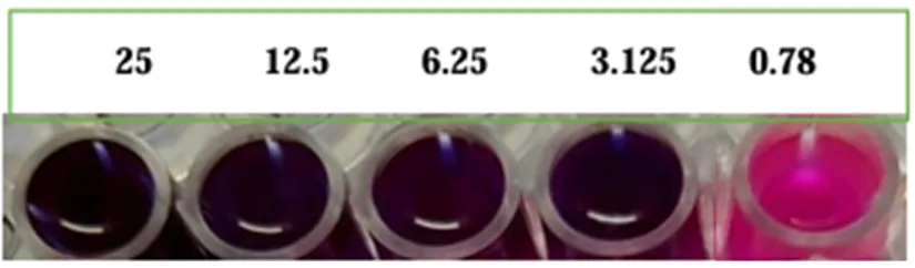
The results were interpreted based on colour change of the Alamar Blue indicator. Wells that remained blue indicated inhibition of bacterial growth (sensitive), whereas wells that changed to pink indicated bacterial growth (resistant). The minimum inhibitory concentration (MIC) was defined as the lowest concentration of the formulation that inhibited visible bacterial growth, as indicated by the absence of a colour change.
RESULTS
Anti TB activity by Alamar Blue Assay (MABA)[5][7][8]
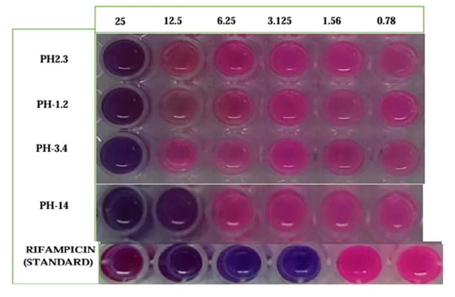
Anti TB activity results of the compounds
|
SR. NO |
Samples and Standard |
25 |
12.5 |
6.25 |
3.125 |
1.56 |
0.78 |
|
1 |
PH-1.2 |
S |
R |
R |
R |
R |
R |
|
2 |
PH-2.3 |
S |
R |
R |
R |
R |
R |
|
3 |
PH-3.4 |
S |
R |
R |
R |
R |
R |
|
4 |
PH-1.4 |
S |
S |
R |
R |
R |
R |
|
5 |
Rifampicin (standard) |
S |
S |
S |
S |
R |
R |
NOTE:
S – Sensitive
R – Resistant
OBSERVATION
The present study evaluated the in vitro anti-tubercular activity of binary Polyherbal formulations prepared from four medicinal plants, namely Aegle marmelos, Moringa oleifera, Vitex negundo, and Alstonia scholaris. The results demonstrated that the formulations exhibited varying degrees of inhibitory activity against Mycobacterium tuberculosis, indicating that combining medicinal plant extracts can enhance their therapeutic potential. Among all the formulations tested, those containing higher concentrations of phytochemical rich extracts produced greater inhibition, showing a concentration-dependent antimicrobial effect.
The activity observed may be attributed to the presence of bioactive phytoconstituents such as alkaloids, flavonoids, tannins, saponins, terpenoids, phenolic compounds, and glycosides. The combined action of these phytochemicals likely contributed to the overall inhibitory effect observed against M. tuberculosis. When combined, these phytochemicals may act synergistically by targeting multiple biochemical pathways simultaneously, thereby improving antimicrobial efficacy compared to individual extracts.
Aegle marmelos provides coumarins, marmelosin, tannins, and phenolic compounds that have demonstrated broad-spectrum antimicrobial potential. Moringa oleifera is rich in flavonoids, phenolic acids, and isothiocyanates that exhibit antimicrobial and antioxidant activities. Vitex negundo contributes flavonoids, iridoid glycosides, and volatile oils known for their antimicrobial and anti-inflammatory activities, whereas Alstonia scholaris contains indole alkaloids such as echitamine and scholaricine, which possess significant antimicrobial and immunomodulatory properties. The complementary pharmacological actions of these plants may result in enhanced bacterial inhibition, reduced microbial resistance, and improved therapeutic efficacy.
The findings of the present study are consistent with previously published reports on the antimycobacterial activity of these medicinal plants. Several investigations have demonstrated that extracts of Aegle marmelos possess significant antimicrobial activity against both Gram-positive and Gram-negative organisms and have shown potential against mycobacterium strains in preliminary studies. Moringa oleifera extracts have been reported to inhibit microbial growth through disruption of bacterial membranes and inhibition of protein synthesis. Vitex negundo has shown antibacterial antimycobacterial properties owing to its rich flavonoids and terpenoids composition. Similarly, Alstonia scholaris extracts have demonstrated inhibitory activity against various pathogenic bacteria, including Mycobacterium species, primarily due to their alkaloid content.
Although the individual medicinal plants have been extensively investigated, comparatively fewer studies have evaluated binary Polyherbal combinations against Mycobacterium tuberculosis. The enhanced activity observed in present formulations supports the concept that combining medicinal plants can produce additive or synergistic effects, leading to improved antimicrobial performance compared with single-plant preparations.
Binary Polyherbal combinations are particularly significant in tuberculosis management because tuberculosis treatment requires prolonged multidrug therapy, and the emergence of multidrug-resistant (MDR) and extensively drug-resistant (XDR) strains remains a major global health challenge. Herbal combinations capable of acting through multiple mechanisms may help reduce bacterial resistance, improve treatment outcomes, minimize adverse effects associated with prolonged chemotherapy, and potentially serve as complementary therapeutic agents alongside conventional anti-tubercular drugs. Furthermore, the antioxidant and immunomodulatory properties of these medicinal plants may support host immune responses during tuberculosis infection, providing additional therapeutic benefits beyond direct antimicrobial activity.
Overall, the present investigation demonstrates that binary Polyherbal formulations possess promising antimycobacterial potential and provide a scientific basis for further exploration of medicinal plant combinations in tuberculosis drug discovery. However, isolation of active constituents, elucidation of mechanisms of action, and validation through advanced biological studies are necessary before clinical application.
CONCLUSION
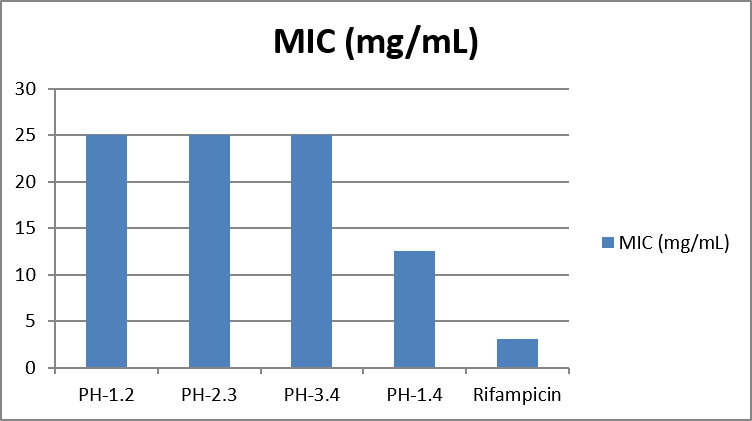
The present study successfully evaluated the in vitro anti-tubercular activity of binary Polyherbal formulations prepared from selected medicinal plants, namely Aegle marmelos, Moringa oleifera, Vitex negundo, and Alstonia scholaris. The formulations demonstrated measurable inhibitory activity against Mycobacterium tuberculosis, indicating that the combination of medicinal plant extracts can enhance antimycobacterial efficacy through the collective action of multiple phytoconstituents. Among the formulations tested, PH-1.4 demonstrated the highest inhibitory activity with a minimum inhibitory concentration (MIC) of 12.5 mg/mL, whereas PH-1.2, PH-2.3, and PH-3.4 showed MIC values of 25 mg/mL. As expected, the standard drug Rifampicin exhibited the greatest potency with an MIC of 3.125 mg/mL.
The study also suggests that binary Polyherbal formulations may provide synergistic therapeutic effects by combining antimicrobial, antioxidant, and immunomodulatory properties of the constituent plants. These findings support the traditional use of these medicinal plants and highlight their potential as complementary sources for the development of novel anti-tubercular agents.
In conclusion, the study demonstrates the binary Polyherbal formulations possess promising anti-tubercular activity, with PH-1.4 emerging as the most effective formulation among those evaluated. Although its activity was lower than that of Rifampicin, the results provide encouraging evidence for the therapeutic potential of plant-based combinations. Further studies involving phytochemical characterization of the active constituents, toxicity evaluation, and mechanism of action studies, formulation standardization, and in vivo investigations are required to validate their efficacy and safety before clinical application.
REFERENCES

K. Vijaya Sindhu, M. Santhosh, I. Supriya, D. Abhilasha, Development and In Vitro Screening of Binary Polyherbal Combinations from Four Medicinal Plants against Mycobacterium tuberculosis, Int. J. of Pharm. Sci., 2026, Vol 4, Issue 8, 1373-1390. https://doi.org/10.5281/zenodo.21840255
 10.5281/zenodo.21840255