
We use cookies to ensure our website works properly and to personalise your experience. Cookies policy
Shambhunath Institute of Pharmacy, Jhalwa Prayagraj.
The goal of the present study was to prepare and test Clidinium Bromide containing nanospheres for prolonged drug delivery via solvent evaporation technique. Nanospheres were formulated using a biodegradable polymer and their physicochemical properties were determined. The extent of drug content was satisfactory as confirmed by UV spectrophotometric analysis and formulations were compatible as confirmed by FTIR studies which showed no significant drug–polymer interaction. SEM images revealed the existence of discrete particles with smooth surface morphology and nearly spherical shape. The optimized formulation exhibited a particle size ranges from 192nm to 250nm, PDI was <0.3, zeta potential of ?27.2 ± 1.5 mV, and entrapment efficiency of 81.6 ± 2.1%. The in vitro drug release study indicated a controlled and sustained release profile with a release of ~78% in 8 hours. The results indicate that the nanospheres of Clidinium Bromide can be used as a potential sustained-release drug delivery system which has better therapeutic performance
There are two classes of nanoparticles: spherical nanospheres and spherical nanocapsules. The shape of both types of nanoparticles is uniform all the way through. Nanospheres are particles of a spherical shape whose diameter ranges from 10 to 200nm, which have different, superior properties depending on their size when compared with larger spheres made of the same material. The medication is often attached to, encapsulated in or integrated within a polymer matrix. The drug is physically distributed in the structure of the polymer matrix. Nanospheres can either be crystalline or amorphous, and they can help to stabilize the drug against chemical and enzyme degradation.[1] Nanospheres can be supplied specifically to the liver and phagocytically active cells thanks to the reticuloendothelial system. Nanospheres are small and can be injected intravenously without blocking capillaries or the needle, unlike many other colloidal systems which are used as diagnostic agents. Injections make only a small selection of the many applications of nanoparticles as a means of delivering medications and diagnostic imaging. However, the reticuloendothelial system swiftly eliminates them when given intravenously. To prepare monodisperse biodegradable nanospheres, amphiphilic copolymers have been designed. These nanospheres were found to have a longer half-life in circulation as well as less drug accumulation in the mice's liver.[2] The capacity of these colloidal particles to transport drugs has been directly related to their interaction with proteins and enzymes in various bodily fluids. Systems allowing for drug use or injection have a lot of benefits. Nanospheres can be used to release drugs at the exact right place in the body. The nanospheres can be used to develop a wide range of systems so that the patent life of effective and well-established pharmacophores can be extended.[3]
The chemical name for clidinium bromide (CLI) is 1-azoniabicyclo[2.2.2]octane, 3-[(hydroxydiphenylacetyl)oxy]. CLI has a strong antispasmodic and antisecretory activity in the gastrointestinal tract. [4] The molecular weight is 432.35 and the molecular structure of it is C 22 H 26 BrNO 3. It stops the muscarinic actions of acetylcholine at postganglionic parasympathetic neuroeffector sites. It is used to treat peptic ulcer disease and helps reduce stomach or abdominal cramps or spasms resulting from diverticulitis, irritable bowel syndrome and colicky abdominal pain.[5]
MATERIALS
Clidinium Bromide was obtained from Digital Vision, H.P. as a gift sample. Eudragit RS-100, Polyvinyl Alcohol, Ethyl Acetate and Ethanol was procured from college lab.
METHOD
Preparation of Clidinium Bromide loaded nanosphere:
The nanosphere was prepared by Emulsion solvent evaporation method in which Clidinium Bromide was dissolved in ethanol and Eudragit RS-100 was dissolved in ethyl acetate and stirred continuously using magnetic stirrer at 60o C till clear solution was obtained. After that both these mixture are mixed by using micropipette to form a primary emulsion, put the emulsion on probe sonicator for 15 minutes for the stable emulsion formation (Pulse mode 30-30 sec. on/off ). This emulsion was then added drop wise in the 25 ml of 2% PVA with a proper mixing. The solution obtained was then homogenised using a homogeniser for around 15-20 minutes. Once this was done, the solution was left to evaporate solvents for 4-6 hrs at room temp. Collect the solution by centrifuging the solution: 15000 rpm, 15 min, at room temperature; wash 2-3 times with distilled water and dry with freeze dryer. Then the nanospheres was collected and stored in a closed glass container.[6]
UV Spectroscopy of Clidinium Bromide:
Clidinium Bromide exhibits a characteristic maximum absorption value of UV spectrophotometric analysis (λmax) in a suitable solvent at around 212 nm. Beer-Lambert's Law can be used over an appropriate range of concentrations for a simple and accurate and reliable application of a UV method.[7]
Characterization of Clidinium Bromide loaded nanosphere:
Scanning electron microscopy (SEM): The form and the properties of the nanospheres produced were studied using scanning electron microscopy (SEM). The nanospheres are dispersed in water and spread out on the glass slide and then left to dry at room temperature (25°C) to obtain a thin layer. The size and shape was determined by scanning electron microscopy (SEM) and the results obtained.[8]
EE%: After washing the nanosphere with distilled water twice to remove the drugs that are absorbed to the surface, the nanosphere was centrifuged at 7000 rpm for 1 hour and the nanosphere and the supernatant were separated, the formulation was named as EE%. Absorbance of the entrapped drug was measured using UV visible spectrophotometer at 212nm.[9]
EE%=Total drug-Free drugTotal drug×100

Zeta Potential: It is used for measuring the electrical potential and surface charge properties of nanospheres. Both the dispersion media and the particle's composition have an impact on them. It is also used to measure the stability of the charge of particles and their aggregation. The zetasizer is used to determine it.[10]
Particle Size: Drug loading, stability, cellular uptake, and drug release behavior are influenced by particle size and so the size is an important parameter for the characterization of nanospheres. The commonly used method for measuring the hydrodynamic diameter of nanoparticles is dynamic light scattering (DLS). In general, the smaller and more evenly distributed nanospheres are more effective in therapeutic efficacy and bioavailability.[11]
PDI: A nanosphere formulation's homogeneity of particle size distribution is indicated by the polydispersity index (PDI). A low PDI value (< 0.3) will be regarded as narrow and good formulation and stability. Lower PDI means a more consistent performance in delivering medication, due to less aggregation.[11]
FT-IR: FT-IR analysis can be carried out by using FT-IR spectrophotometer to check the chemical integrity and possible chemical interaction between the drug and polymer.[12]
In-vitro release: An in vitro drug release study for drug loaded nanospheres can be done in vitro using the dialysis bag diffusion method. Using this method, a dialysis membrane tube filled with phosphate buffer solution is kept at 37°C while being shaken at a speed of 120 rpm.[13]
RESULT AND DISCUSSION
UV spectrometric Scan
λmax of Clidinium Bromide was obtained 212 nm under UV spectroscopy which is shown below:
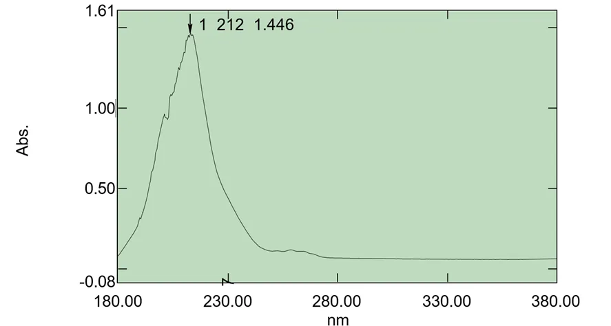
Figure: UV Spectroscopy Image of Clidinium Bromide
Calibration Curve of Clidinium Bromide
The absorbance data points were subjected to a linear regression analysis, which yielded a straight line to help determine the drug quantity using a linear equation. The result was a regression value of 0.99.
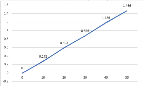
Figure: Calibration Curve of Clidinium Bromide
Table: Statistical data for calibration curve
|
S.No. |
Parameters |
Values |
|
1. |
λmax |
212nm |
|
2. |
Slope |
0.02956 |
|
3. |
R 2 |
0.99 |
Surface Morphology
Using scanning electron microscope surface morphology of the Clidinium Bromide nanosphere formulation was assessed. Using double-sided sticky tape, the sample was immediately placed onto the SEM sample holder, and scanning electron microscopy pictures were captured at 11mm x 300 SE magnifications at an acceleration voltage of 15 kV. The nanosphere's SEM picture is displayed in below figure.
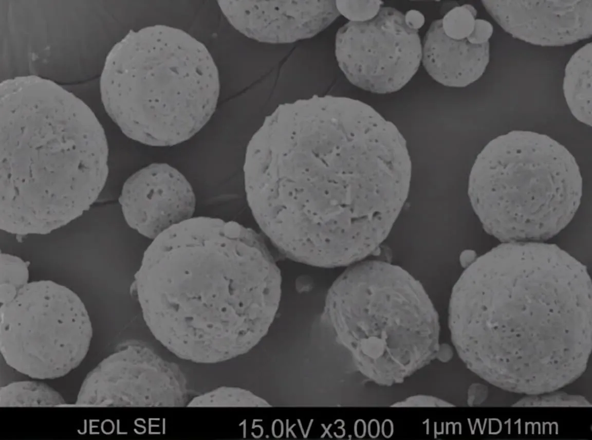
Figure: SEM of Clidinium Bromide Nanosphere
Percentage Yield
The reported percentage yield was obtained 84% which shows that the nanosphere contains a good amount of medicament
Entrapment Efficiency
The nanospheres had entrapment efficacy between 72.4 to 81.6. The highest drug entrapment efficacy was 81.6.
F2 selected as optimized batch
Particle size:
The Dynamic light scattering (DLS) method was used to analyse the mean particle size of nanospheres. These nanospheres minimum average diameter were reported to 192 nm & maximum average diameter to 250 nm.
Particle Size Analysis (DLS)
Table: Data of particle size of different batches
|
Batch |
Trial 1 (nm) |
Trial 2 (nm) |
Trial 3 (nm) |
Mean ± SD |
|
F1 |
205 |
212 |
213 |
210 ± 4.3 |
|
F2 |
238 |
247 |
250 |
245 ± 6.2 |
|
F3 |
192 |
201 |
201 |
198 ± 5.1 |
PDI:
Table: Data of Polydispersity Index
|
Batch |
PDI |
|
F1 |
0.22 |
|
F2 |
0.28 |
|
F3 |
0.19 |
Particle size increased with increase in polymer concentration. PDI <0.3 confirms uniform distribution.
Zeta Potential
The majority of the nanosphere particles in the formulation had this charge, as seen by the plot's peak at -27.2 mV, indicating a strong affinity between the particles.
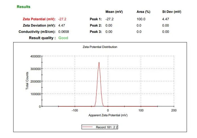
Figure: Zeta potential of Clidinium Bromide nanosphere
FTIR Specroscopy:
According to FTIR spectroscopy analysis, showed that the presence of characteristics functional group in the sample and there is no significant chances in characteristics peak of Cldinium Bromide, indicates the chemical stability and absence of drug-polymer interaction.The characteristics peak of Cldinium Bromide -OH/ -NH, aromatic, C-N are present. Peak 3459.66 cm-1 shows the presence of O-H & N-H stretching, 1634.01 cm-1 shows the aromatic C=C / N-H blending, 1384.28 cm-1 indicates C-N stretching/ CH blending & 520.62 cm-1 shows the aromatic ring deformation, these peaks confirms the compatability of Cldinium Bromide with PVA and Eudragit RS-100 and Cldinium Bromide is stable in nanosphere with these polymers.

Figure: FTIR of Cldinium Bromide loaded nanosphere
In vitro Drug Release Study
In an in-vitro dissolution test, Clidinium Bromide loaded nanosphere released the medication for upto 8 hrs The drug release increased in proportion to time, indicating that the medication will remain in body for a longer period of time and release continuously over time.
Medium: pH 6.8 Phosphate Buffer
Speed: 100 rpm
Temp: 37±0.5°C
Dissolution Data
Table: Statistical data of dissolution study
|
Time (Hrs) |
F1 (%) |
F2(%) |
F3(%) |
|
1 |
20 |
18 |
22 |
|
2 |
34 |
29 |
36 |
|
4 |
49 |
45 |
52 |
|
6 |
63 |
58 |
66 |
|
8 |
75 |
71 |
78 |
Table: Statistical table of F2 for % drug release
|
Time |
% Drug Release |
|
0 |
0 |
|
1 |
18 |
|
2 |
29 |
|
4 |
45 |
|
6 |
58 |
|
8 |
71 |
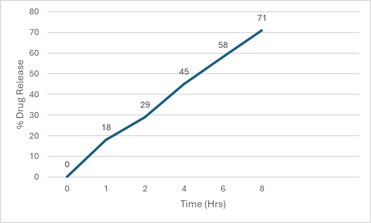
Figure: Graph of F2 drug release
CONCLUSION
Clidinium Bromide nanospheres were successfully developed by the solvent evaporation method and demonstrated desirable physicochemical characteristics, high drug entrapment, and sustained drug release. The optimized formulation exhibited good stability, uniform particle size, and effective controlled-release behavior. These findings suggest that Clidinium Bromide nanospheres have significant potential as a sustained-release drug delivery system for improved therapeutic performance and patient compliance.
REFERENCES


Mohammad Sarfraz, Sagar Bansal, Anurag Dwivedi, Sandeep Pandey, Development & Evaluation of Clidinium Bromide Nanospheres for Sustained Release, Int. J. of Pharm. Sci., 2026, Vol 4, Issue 7, 628-635, https://doi.org/10.5281/zenodo.21156380
 10.5281/zenodo.21156380