

We use cookies to ensure our website works properly and to personalise your experience. Cookies policy
Sarada Vilas College of Pharmacy Mysuru, Karnataka, India.
Dissolving microneedles (DMNs) have emerged as a promising approach for transdermal drug delivery, offering a minimally invasive, patient-friendly alternative to conventional injections and oral drug administration. These systems consist of microscopic needles prepared using biodegradable and biocompatible polymers that dissolve upon contact with the skin, releasing the incorporated drug without generating sharp medical waste. This review provides a comprehensive overview of the polymers used in DMN fabrication, including their properties, advantages, and limitations. It also discusses various fabrication technologies, such as micro moulding, 3D printing, and other advanced manufacturing techniques, along with their influence on needle structure, mechanical strength, drug loading, and dissolution behaviour. Furthermore, the therapeutic applications of DMNs in drug delivery, vaccine administration, chronic disease management, and the delivery of biological molecules are highlighted. Despite their considerable potential, challenges related to polymer safety, drug stability, manufacturing consistency, quality control, clinical evaluation, and regulatory approval remain important barriers to commercial translation. The review also explores emerging developments in smart polymer systems, personalized microneedle formulations, and scalable manufacturing approaches. Overall, dissolving microneedles represent an innovative drug delivery platform with the potential to improve treatment convenience, patient compliance, and therapeutic outcomes. Continued research, clinical validation, and clearer regulatory guidance will be essential for their successful transition from laboratory research to routine clinical applications.
1.1. Transdermal drug delivery: significance and therapeutic potential
The effectiveness of a medicine depends not only on its pharmacological properties but also on how safely, efficiently, and conveniently it can be delivered to the body. Although oral administration remains the preferred route for many medicines, it is not suitable for every therapeutic agent. Some drugs undergo extensive first-pass metabolism in the liver, degrade in the gastrointestinal tract, or show inconsistent absorption. Injectable formulations can overcome several of these limitations, particularly for peptides, proteins, vaccines, and other biological therapeutics. However, injections may cause pain, needle-related anxiety, and needlestick injuries, and they may require trained healthcare personnel. These challenges have encouraged the development of alternative drug delivery systems that improve treatment convenience without compromising therapeutic effectiveness. Transdermal drug delivery is one such approach because it uses the skin as an accessible route for administering selected medicines [1,2,3].
Transdermal drug delivery involves transporting a drug across the skin to achieve a systemic therapeutic effect. It differs from topical delivery, in which the primary objective is to treat a condition at or near the application site. Conventional transdermal patches have demonstrated clinical value for medicines such as nicotine, nitroglycerin, scopolamine, fentanyl, and certain hormonal agents. Depending on the drug and formulation, these systems can provide sustained drug release, reduce the frequency of administration, and help maintain relatively consistent drug concentrations in the bloodstream. They can also avoid gastrointestinal exposure and hepatic first-pass metabolism, making them useful for selected medicines with suitable physicochemical properties [1,2,3,4].
Another practical benefit is that a patch can generally be removed when treatment needs to be discontinued. However, removal does not always stop drug absorption immediately because some drug may remain in the formulation or skin. Transdermal delivery is also convenient for self-administration and avoids the equipment required for conventional injections. These features make it a valuable option for selected chronic conditions, pain management, smoking cessation, hormone replacement, and other therapeutic applications [2,4].
The therapeutic potential of transdermal delivery extends beyond conventional patches. Advances in biotechnology have increased interest in delivering peptides, proteins, vaccine antigens, and nucleic acid-based therapeutics. Many of these molecules are difficult to administer orally because of enzymatic degradation, poor gastrointestinal absorption, or limited permeability across biological membranes. Their delivery can also be complicated by sensitivity to temperature, moisture, and formulation conditions. The skin therefore represents an attractive alternative route, provided that an appropriate delivery system can overcome its protective barrier while preserving drug stability and patient safety [1,3,5].
However, the skin is designed to prevent the entry of potentially harmful substances and minimize water loss. This protective function also restricts the penetration of many therapeutic compounds. Consequently, the development of advanced transdermal systems requires a careful balance between increasing drug permeability and maintaining the skin's structural integrity. This challenge has stimulated research into chemical permeation enhancers, iontophoresis, sonophoresis, electroporation, and microneedle-based technologies [3,5].
1.2. Skin barrier function and limitations of conventional transdermal patches
The main obstacle to conventional transdermal drug delivery is the stratum corneum, the outermost layer of the epidermis. It consists of flattened, keratin-rich corneocytes surrounded by an organized extracellular lipid matrix. This arrangement is commonly described using the “brick-and-mortar” model, in which corneocytes form the bricks and intercellular lipids act as the mortar. Although relatively thin, this layer provides substantial resistance to the entry of foreign substances and helps protect the body from environmental exposure [5,6].
For a drug to cross intact skin, it must partition into the stratum corneum, diffuse through its complex structure, and subsequently move into the more hydrated viable epidermis and dermis. The extent of transport depends on several factors, including molecular size, lipophilicity, aqueous solubility, ionization, drug concentration, and the condition of the skin. Conventional passive transdermal systems are therefore most suitable for relatively potent drugs that can cross the skin efficiently and are required in comparatively small daily doses [1,3,6].
Large hydrophilic molecules, charged compounds, peptides, and proteins generally face greater difficulty crossing intact skin. Their limited ability to enter the lipid-rich pathways of the stratum corneum often results in insufficient drug transport. Increasing the drug concentration in a formulation does not necessarily resolve this problem because permeability, solubility, and the thermodynamic activity of the drug also influence absorption. As a result, many promising therapeutic agents cannot be delivered effectively through conventional patches without additional enhancement strategies [3,5,7].
Dose requirements create another limitation. A transdermal patch has a finite surface area, and the amount of drug delivered depends on skin permeability, drug potency, formulation characteristics, and application time. Medicines requiring relatively large doses may therefore be unsuitable for passive transdermal administration. Drug crystallization, inadequate solubility, poor partitioning into the skin, and difficulties in maintaining consistent release can further reduce delivery efficiency [1,3].
The performance of conventional patches may also be affected by sweating, friction, body movement, temperature, adhesion failure, and differences in individual skin characteristics. Some users experience irritation, allergic contact dermatitis, or local discomfort associated with the adhesive, excipients, or permeation enhancers. These factors do not diminish the clinical value of well-designed patches, but they demonstrate that passive transdermal delivery cannot meet the requirements of every drug or patient [2,4].
Several approaches have been investigated to improve penetration through the stratum corneum. Chemical permeation enhancers alter the barrier properties of the skin or improve drug partitioning. Iontophoresis uses a low electrical current to promote the transport of suitable molecules, while sonophoresis employs ultrasound-associated effects to increase permeability. Electroporation uses electrical pulses to create temporary changes in the skin barrier. Although these approaches can improve drug transport, their performance depends on the drug, formulation, operating conditions, and safety requirements. Some methods also require additional equipment or careful control of application conditions [3,5,7].
Microneedle technology offers an alternative strategy by creating microscopic pathways through the stratum corneum. Rather than relying entirely on passive diffusion or chemical modification, microneedles provide localized access through the skin barrier. This approach has expanded the possibilities for delivering compounds that are poorly absorbed from conventional patches, including selected macromolecules and biological therapeutics [5,7,8].
1.3. Microneedle technology: overcoming the skin barrier
Microneedles (MNs) are miniature needle-like structures, usually arranged in arrays, that penetrate the outer skin barrier while aiming to minimize disruption to deeper tissues. Their dimensions and geometry are designed to create microscopic pathways through the stratum corneum without the deeper penetration associated with conventional hypodermic needles. Microneedles have consequently attracted considerable interest as a minimally invasive approach to improving skin permeability and delivering compounds that cannot readily cross intact skin [7,8,9].
The principle of microneedle-mediated delivery is straightforward. When an array is applied to the skin, its projections penetrate the stratum corneum and create pathways through which a drug can enter the underlying tissue. Depending on the design, the drug may be applied after the pathways are formed, released from a surface coating, infused through hollow needles, transported from a separate reservoir, or incorporated into the needle material itself. Each approach offers a different balance of drug-loading capacity, release behavior, ease of administration, and manufacturing complexity [8,9,10].
Microneedles occupy an intermediate position between conventional patches and hypodermic injections. Unlike passive patches, they can bypass the principal skin barrier. Unlike conventional injection needles, they are designed to penetrate only a limited depth. Their small dimensions may reduce discomfort by limiting stimulation of deeper pain-sensitive structures, although the sensation experienced by users depends on needle geometry, insertion force, application technique, and individual skin characteristics. They should therefore be regarded as minimally invasive rather than universally painless or completely non-invasive [7,8,9].
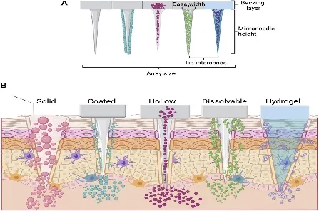
Microneedle systems are commonly classified as solid, coated, hollow, hydrogel-forming, or dissolving microneedles. Solid microneedles create microchannels through which a subsequently applied formulation can enter the skin. Coated microneedles carry the drug on their surfaces and release it during insertion. Hollow microneedles contain internal channels that permit liquid formulations to be delivered into the skin. Hydrogel-forming microneedles absorb interstitial fluid after insertion and swell, allowing drug molecules to diffuse through the hydrated structure from an associated reservoir. Dissolving microneedles incorporate the therapeutic agent into a soluble or biodegradable matrix that dissolves or degrades after insertion [8,9,10,11].
Each design has advantages and limitations. Solid microneedles may require a separate drug-application step, whereas coated systems are constrained by the available surface area and the efficiency of coating transfer. Hollow microneedles require reliable fluid flow and sufficient structural strength. Hydrogel-forming systems depend on swelling and diffusion through the hydrated network. Dissolving microneedles offer a different approach because the needle structure itself contains the drug and participates directly in its release [8,9,10,11].
The selection of an appropriate microneedle platform depends on the intended therapeutic agent, required dose, desired release profile, and method of administration. A device designed for a potent drug requiring a small dose may not be suitable for a vaccine or protein that requires a different payload or formulation environment. Mechanical strength, insertion consistency, storage stability, and ease of application must therefore be considered alongside drug-delivery performance [9,10,11].
1.4. Dissolving microneedles: a distinct approach to transdermal drug delivery
Dissolving microneedles (DMNs) are an important development in transdermal drug delivery because they combine skin penetration with the release of a drug-containing matrix. Unlike solid microneedles, which primarily create microchannels, and coated microneedles, which carry the drug on their external surfaces, dissolving microneedles incorporate the active pharmaceutical ingredient within the needle material. After insertion, interstitial fluid enters the matrix, allowing it to dissolve or disintegrate and release the incorporated drug into the skin [10,11,12].
The foundational study by Lee, Park, and Prausnitz demonstrated that dissolving microneedles could be fabricated from water-soluble materials, including carboxymethylcellulose and amylopectin. Their work showed that model therapeutic compounds could be incorporated into the needle matrix and released after insertion. The study also examined the preservation of protein activity, helping establish the feasibility of using dissolving microneedles for the delivery of biologically active substances [12].
The material selected for a dissolving microneedle formulation strongly influences its mechanical performance, dissolution behavior, drug compatibility, and storage stability. Commonly investigated materials include hyaluronic acid, polyvinylpyrrolidone, polyvinyl alcohol, carboxymethylcellulose, dextran, and selected biodegradable polymers. These materials differ in their molecular weight, viscosity, water affinity, mechanical strength, degradation behavior, and compatibility with active pharmaceutical ingredients. Material selection must therefore reflect the intended drug, required dose, desired release profile, and manufacturing conditions [11,12,13,14].
An effective polymer matrix must be strong enough to penetrate the skin without bending or fracturing prematurely. At the same time, it must dissolve at a suitable rate to release the incorporated drug. These requirements can conflict: increasing polymer concentration may improve mechanical strength but delay dissolution, whereas a highly water-soluble formulation may dissolve rapidly but provide insufficient resistance to deformation. Formulation development therefore requires careful optimization of polymer composition, needle geometry, drug distribution, and processing conditions [12,13,14].
Several fabrication techniques have been investigated, including micro molding, drawing-based methods, and other microfabrication approaches. Micro molding is widely studied because it allows polymer solutions containing the drug to be introduced into needle-shaped cavities. However, the quality of the finished array depends on factors such as polymer viscosity, mold filling, drying conditions, air-bubble removal, and the uniformity of drug distribution. These variables can affect needle dimensions, mechanical strength, insertion performance, and the amount of drug delivered [13,14].
One potential advantage of dissolving microneedles is the reduction of intact sharps waste. Because the drug-containing projections are designed to dissolve or degrade after insertion, they may not leave intact needle shafts requiring disposal in the same way as conventional needles or reusable solid microneedles. This feature could be useful in settings where access to trained personnel or specialized sharps-disposal facilities is limited. Nevertheless, the actual disposal and safety benefits depend on the formulation, backing structure, dissolution behavior, and completeness of material dissolution [11–14].
Dissolving microneedles have also been investigated for the delivery of vaccines, peptides, proteins, and other therapeutic agents that are difficult to administer through intact skin. The skin contains antigen-presenting cells and other immune-associated structures that may support responses to vaccines delivered into this tissue. Sullivan and colleagues demonstrated the potential of dissolving polymer microneedle patches for influenza vaccination, providing important experimental evidence for this application [15]. However, vaccine performance depends on antigen stability, dose, release characteristics, and the type of immune response required; delivery through the skin does not automatically guarantee superior immunogenicity.
Human studies have further strengthened interest in microneedle-based vaccination. In a randomized phase 1 trial, Rouphael and colleagues evaluated an inactivated influenza vaccine delivered through a microneedle patch, examining safety, immunogenicity, acceptability, and self-administration. The findings supported the feasibility of this approach in humans, although the results of a particular vaccine and device cannot be generalized automatically to every dissolving microneedle formulation or therapeutic indication [16].
The release characteristics of dissolving microneedles can be adjusted through formulation design. Rapidly dissolving matrices may be useful when prompt drug delivery is required, while selected polymer compositions and multilayer designs may be investigated for more complex release profiles. However, dissolution alone does not establish sustained release. Once the matrix dissolves, drug movement depends on the active ingredient's physicochemical properties, interactions with surrounding tissue, and the design of the overall formulation. Claims of prolonged delivery therefore require appropriate release, pharmacokinetic, and in vivo evidence [12,13,14].
Despite their potential, dissolving microneedles have important limitations. Their small dimensions restrict the amount of material that can be incorporated into an array, which can be problematic for drugs requiring high doses or having low potency. Mechanical performance may also change with polymer composition, drug loading, residual moisture, and environmental humidity. Incomplete insertion, partial dissolution, uneven drug distribution, and inconsistent application force can cause differences between the intended dose and the amount actually delivered [13,14,17].
Drug stability is another major concern. Proteins, peptides, vaccines, and nucleic acid therapeutics may be sensitive to temperature, moisture, pH, and the stresses associated with processing and drying. Incorporating these molecules into a polymer matrix must not compromise their quality or biological activity during manufacturing and storage. Formulations should therefore be evaluated not only for mechanical strength and dissolution but also for drug integrity, potency, and stability under realistic storage conditions [14,17].
Translation from laboratory prototypes to clinically useful products presents additional challenges. Commercial development requires reproducible needle dimensions, consistent drug content, suitable mechanical properties, predictable dissolution, and reliable performance across manufacturing batches. Appropriate packaging is particularly important for moisture-sensitive formulations, while sterilization or aseptic processing must preserve both the structure of the device and the quality of the incorporated drug. Manufacturing costs, quality-control requirements, and regulatory expectations must also be addressed before large-scale use becomes feasible [17,18].
Dissolving microneedles should consequently be viewed as one option within the wider microneedle technology landscape, rather than as a universally superior delivery system. Their integration of skin penetration, drug incorporation, and matrix dissolution may simplify administration and reduce intact sharps waste. However, these benefits must be balanced against restrictions in drug-loading capacity, formulation stability, mechanical performance, and dose reproducibility. The suitability of the platform depends on the therapeutic requirements and the evidence supporting its performance [10,11,12,13,14,18].
1.5. Research gap and rationale for the present review
Research on dissolving microneedles has expanded to include polymer selection, needle geometry, fabrication techniques, drug incorporation, mechanical characterization, dissolution behavior, and therapeutic applications. Reviews have described their potential in vaccine delivery, dermatological treatment, systemic drug administration, and other areas of medicine. Recent publications have also examined advances in fabrication, emerging clinical applications, and the challenges associated with translating laboratory research into practical products [9,11,20,23].
Despite this progress, important questions remain unresolved. Many published studies focus on demonstrating the feasibility of a particular formulation, improving one aspect of its performance, or reporting promising results in laboratory and preclinical models. Direct comparisons between studies can be difficult because researchers use different polymers, needle dimensions, model drugs, application procedures, skin models, and methods for assessing insertion, dissolution, and drug delivery. Consequently, improvements observed in one formulation cannot automatically be assumed to apply to other drugs or therapeutic applications [13,14,18,22].
One important knowledge gap concerns the relationship between material properties and actual delivery performance. Biocompatibility, solubility, and ease of fabrication are important when selecting a polymer, but they must be considered alongside mechanical strength, insertion efficiency, drug-loading capacity, dissolution time, and drug stability. A formulation that performs well during mechanical testing may still fail to deliver a sufficient dose in vivo. Similarly, rapid dissolution does not necessarily produce greater bioavailability, and high drug loading does not guarantee uniform delivery [12,13,14,18].
A second gap involves the relationship between formulation design and the properties of the drug being delivered. Small molecules, peptides, proteins, vaccines, and nucleic acid therapeutics differ in size, solubility, stability, dose requirements, and sensitivity to manufacturing conditions. A matrix suitable for a low-dose, potent drug may be inappropriate for a product requiring a larger payload. Likewise, a formulation that preserves the mechanical properties of a small-molecule system may behave differently when loaded with a protein or another sensitive therapeutic agent [14,17,18].
A third concern is the gap between proof-of-concept research and clinical applicability. Translation requires evidence of reproducible manufacturing, consistent dosing, acceptable local and systemic safety, adequate shelf life, user-friendly administration, and clinically meaningful therapeutic outcomes. Differences in skin thickness, hydration, application force, and individual physiology may influence delivery in humans. Moisture-sensitive materials, specialized packaging, production costs, and quality-assurance requirements can further affect the feasibility of large-scale manufacturing [18,19,22].
Recent reviews have further highlighted challenges associated with drug-loading capacity, dose consistency, regulatory expectations, sterilization, and manufacturing scale-up. These concerns indicate that progress should not be judged solely by the novelty of the materials or fabrication methods. Greater attention is needed to practical measures of performance, including the reproducibility of delivered dose, stability during storage, ease of use, safety, and the ability to manufacture the product consistently at a reasonable cost [18,19,22,25].
The present review therefore aims to critically examine dissolving microneedles as an advanced platform for transdermal drug delivery, focusing on their scientific basis, material composition, design and fabrication, drug-loading strategies, mechanical properties, dissolution behavior, and therapeutic applications. It also considers limitations involving dose capacity, insertion consistency, drug stability, manufacturing reproducibility, storage, safety, and clinical translation. By examining how these factors interact, the review seeks to distinguish established findings from emerging possibilities and identify research priorities that may improve the practical value of dissolving microneedle systems.
Ultimately, dissolving microneedles offer a promising approach to making selected medicines easier to administer while overcoming the permeability limitations of intact skin. Their successful development, however, depends on more than the ability of the needle matrix to dissolve after insertion. A clinically useful system must deliver an appropriate dose consistently, preserve drug quality and activity, possess adequate mechanical strength, remain stable during storage, and be manufactured reliably at an acceptable cost. An integrated assessment of these requirements is essential to guide future research and support the translation of dissolving microneedles from experimental formulations into clinically useful drug delivery systems.
2. Fundamentals of Dissolving Microneedles
Dissolving microneedles (DMNs) are a promising approach to transdermal drug delivery in which microscopic projections made from water-soluble or biodegradable materials penetrate the outer barrier of the skin and release an incorporated drug as the needle matrix dissolves. Unlike conventional hypodermic needles, DMNs are designed to deliver therapeutic substances with minimal tissue disruption. They also differ from solid, coated, hollow, and hydrogel-forming microneedles because the drug is generally incorporated throughout, or concentrated within selected regions of, the dissolving structure itself. This design supports drug delivery without leaving conventional rigid needle components behind after successful application [26-28].
The development of DMNs has attracted considerable interest because they can simplify drug administration, reduce dependence on conventional injections, and support the delivery of selected small-molecule drugs, peptides, proteins, and vaccine antigens. Their performance, however, depends on several interconnected factors, including polymer composition, needle geometry, mechanical strength, drug distribution, skin insertion, and dissolution rate. Understanding these factors is essential for designing a system that delivers a suitable dose safely and consistently [26,27].
2.1. Structure, Components, and Mechanism of Action
2.1.1. Basic structure of dissolving microneedles
A dissolving microneedle patch generally consists of an array of microscopic projections attached to a supporting base or backing layer. Each projection has a sharp tip, a tapered body or shaft, and a broader base that connects it to the backing layer. These features work together to help the microneedle enter the skin while maintaining sufficient structural strength during application.
The dimensions of DMNs vary according to their intended use, formulation, and manufacturing technique. Many designs use needles several hundred micrometres in length, although the dimensions and spacing must be selected for the target skin layer and therapeutic purpose. Needle length, tip sharpness, base diameter, aspect ratio, and array density influence insertion efficiency, mechanical stability, and the distribution of the delivered drug [26,28].
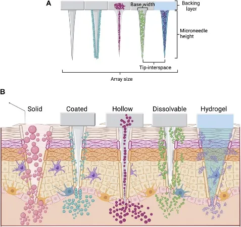
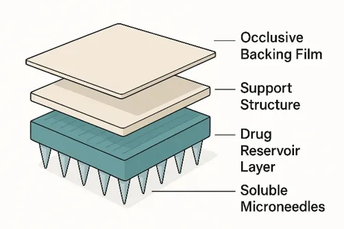
Figure 1: Basic Structure of a Dissolving Microneedle Patch
Schematic reference for illustrating the patch, needle array, skin surface, and drug-containing projections. Use a properly attributed image or create an original figure for publication.
The principal structural components and their functions are described below.
2.1.2. Components and materials used in dissolving microneedles
The selection of materials is a major factor in determining the performance of DMNs. An appropriate formulation must be strong enough to penetrate the skin, compatible with the incorporated drug, and capable of dissolving or degrading under suitable physiological conditions. The material should also maintain its physical integrity during storage and application [26,28].
Natural polymers. Hyaluronic acid, gelatin, chondroitin sulfate, dextran, and starch have been investigated for dissolving microneedle formulations. Hyaluronic acid is especially useful because of its water affinity and compatibility with several skin-related applications. Gelatin and starch can contribute to matrix formation, while dextran and chondroitin sulfate provide alternative formulation properties. Nevertheless, natural polymers can vary in molecular weight, composition, viscosity, and moisture sensitivity, making formulation consistency an important consideration [26,27].
Synthetic and semisynthetic polymers. Polyvinylpyrrolidone (PVP), polyvinyl alcohol (PVA), and carboxymethyl cellulose (CMC) are frequently studied because their physicochemical properties can be adjusted to suit different drug delivery requirements. PVP can facilitate water uptake and dissolution, whereas PVA may contribute to film formation and mechanical strength. CMC can support matrix formation and alter the rate at which the formulation hydrates. Their suitability depends on polymer grade, concentration, molecular weight, and the properties of the incorporated drug [26,28].
Sugars and other soluble materials. Materials such as maltose and trehalose may be used as matrix-forming or stabilizing components. Depending on the formulation, sugars can improve the solid-state properties of the patch or help protect sensitive biological substances during processing and storage. Their performance is influenced by humidity, crystallization, and the physical state of the finished formulation [27,29].
Active pharmaceutical ingredients. The API is the therapeutic component responsible for the intended pharmacological effect. DMNs have been investigated for delivering small-molecule drugs, peptides, proteins, and vaccine antigens. The drug's molecular size, water solubility, stability, required dose, and interaction with the polymer determine how it should be incorporated into the needle matrix. Heat-sensitive or moisture-sensitive drugs may require particular care during manufacturing and packaging [26,27].
Formulation additives. Depending on the drug and polymer, plasticizers, stabilizers, buffers, and other excipients may be incorporated to improve mechanical strength, maintain drug stability, or adjust dissolution behavior. However, each additional component must be assessed for compatibility, safety, and its influence on the final product [26,28].
2.1.3. Mechanism of action
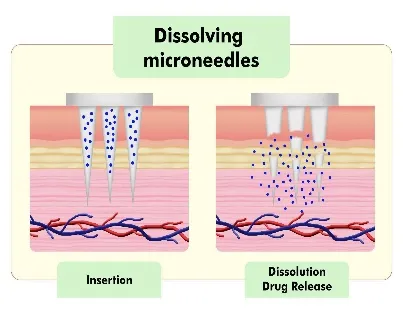
DMNs operate through a sequence of physical and formulation-related events. The basic principle is often described as a poke-and-release mechanism: the microneedles penetrate the skin, absorb local moisture, dissolve or degrade, and release the drug into the tissue.
Mechanism of action: step-by-step
The action of dissolving microneedles can be understood as a sequence of four stages.
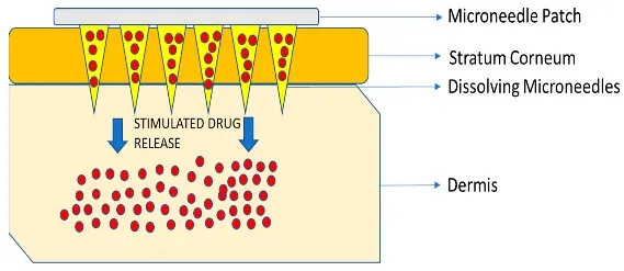
Stage 1 — Application and penetration
The patch is pressed against the skin. The pointed projections cross the stratum corneum, the outermost skin barrier, and enter the superficial skin layers.
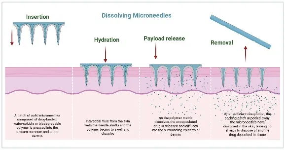
Stage 2 — Contact with skin fluid
Water from the surrounding tissue enters the soluble matrix. The material begins to hydrate, soften, swell, or dissolve, depending on its composition.
Stage 3 — Drug release
As the needle matrix dissolves or breaks apart, the incorporated drug becomes available in the skin tissue. The release rate depends on the formulation and the surrounding conditions.
Stage 4 — Patch removal and post-delivery
Once the intended dissolution period has been reached, the backing can be removed according to the product's instructions. The drug remains within the skin or moves into deeper tissues or circulation, depending on its properties.
Conceptual illustration of the mechanism described in references 26–29.
The process begins when the applied force overcomes the resistance of the skin surface. After insertion, tissue fluid interacts with the water-responsive matrix. As the matrix dissolves or breaks down, the drug is liberated and becomes available for transport within the surrounding tissue. Depending on the formulation, release may occur rapidly or over a longer period.
The drug does not necessarily enter the bloodstream immediately. Some formulations are intended to produce a local effect within the skin, whereas others are designed to allow systemic absorption. The final outcome depends on the depth of deposition, the drug's physicochemical properties, and its ability to move through the surrounding tissue [26,27,28].
2.2. Types of Microneedles and Their Classification
Microneedles can be classified according to their material composition, physical structure, fabrication approach, and method of drug delivery. The most widely used functional classification includes solid, coated, hollow, dissolving, and hydrogel-forming microneedles. Although these systems share the ability to bypass part of the skin's outer barrier, their drug-loading strategies and release mechanisms differ substantially [27,29,30].
2.2.1. Solid microneedles
Solid microneedles are manufactured from materials such as stainless steel, titanium, silicon, or suitable polymers. Their primary function is to create temporary microchannels in the skin. After the needles are removed, a drug-containing patch, cream, gel, or solution is applied to the treated area. This approach is often called the poke-and-patch method.
Solid microneedles are useful when skin pretreatment is sufficient to improve drug transport. However, they generally require a separate drug application step, and the effectiveness of delivery depends on the persistence of the microchannels and the characteristics of the subsequently applied formulation [29,30].
2.2.2. Coated microneedles
Coated microneedles have a drug-containing layer deposited on the surface of otherwise solid projections. When inserted into the skin, the coating dissolves or disperses, releasing the drug. This design can support rapid delivery and is suitable when the required dose can be accommodated within the coating.
A major limitation is the relatively restricted surface area available for drug loading. The coating must also remain sufficiently stable during storage and handling while dissolving efficiently after insertion. These requirements can make high-dose delivery and uniform coating challenging [27,29].
2.2.3. Hollow microneedles
Hollow microneedles contain a small internal channel through which a liquid formulation can be delivered. Drug administration may be driven by a syringe, pump, or applied pressure, depending on the device design.
Their principal advantage is the ability to deliver liquid formulations without relying on dissolution of the needle material. Nevertheless, fabrication is more complex because the internal lumen must be formed accurately and remain unobstructed. Leakage, blockage, insertion resistance, and the need for a suitable fluid-delivery system are important practical considerations [29,30].
2.2.4. Dissolving microneedles
Dissolving microneedles are produced from water-soluble or biodegradable matrices containing the drug. After insertion, the matrix dissolves or degrades, releasing the incorporated therapeutic substance. Because the drug can be distributed within the needle body rather than restricted to a surface coating, these systems may offer greater formulation flexibility.
DMNs can also reduce the risk of leaving rigid, contaminated needle waste after successful use. However, they must balance sufficient mechanical strength before insertion with suitable dissolution behavior after insertion. Incomplete insertion, inadequate hydration, or poor formulation stability can compromise their performance [26,27,28,29].
2.2.5. Hydrogel-forming microneedles
Hydrogel-forming microneedles absorb interstitial fluid and swell after insertion. Unlike conventional dissolving microneedles, their cross-linked structures are generally designed to remain intact rather than dissolve completely in the skin. The swollen microneedles can create pathways through which a drug moves from a separate reservoir into the tissue.
These systems may be useful when prolonged delivery is required. Their limitations include dependence on swelling behavior, the need for a compatible drug reservoir, and possible delays before sufficient fluid uptake and drug transport occur [29,30].
2.3 Drug incorporation and delivery
Drug incorporation and delivery
In DMNs, the active pharmaceutical ingredient is incorporated into the material that forms the needle structure. During application, the projections enter the skin and expose the drug-containing matrix to tissue moisture. Hydration initiates the dissolution or degradation process, allowing the drug to become available for transport within the skin.
The way in which the drug is incorporated influences its distribution and release. A drug dispersed uniformly throughout the polymer matrix may be released differently from one concentrated near the needle tips. Tip-loaded designs can direct a greater proportion of the drug towards the tissue during insertion, whereas drug distributed throughout the needle body may be released as progressively deeper or broader regions of the matrix dissolve. These outcomes depend on the geometry and formulation and should be verified experimentally [26,27,31].
Several mechanisms can contribute to drug release:
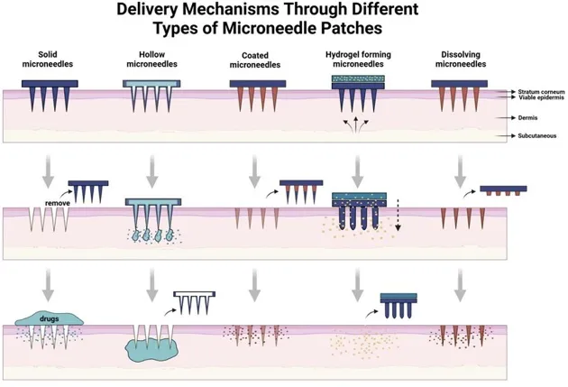
Figure 2: Drug Delivery Mechanism.
2.4. Advantages and Limitations Compared with Other Microneedle Systems
Dissolving microneedles offer a combination of minimally invasive skin penetration and drug release from a soluble or degradable matrix. However, their advantages must be considered alongside limitations related to mechanical strength, dose capacity, drug stability, manufacturing reproducibility, and storage. The most suitable microneedle system depends on the therapeutic objective, the drug's properties, and the required delivery profile [26,27,28,29,30].
2.4.1. Advantages of dissolving microneedles
1. Minimally invasive drug delivery
DMNs create microscopic pathways through the skin without the need for conventional hypodermic needles. Their small dimensions may reduce discomfort compared with conventional injections, although pain and tolerability depend on the design, insertion method, and application site.
2. Improved access across the skin barrier
The microneedles bypass part of the stratum corneum, allowing selected drugs to reach the epidermis or superficial dermis. This can improve delivery of substances that do not readily pass through intact skin when applied in conventional topical formulations.
3. Reduced sharps waste
Because the drug-containing projections are designed to dissolve or degrade, they can reduce the generation of rigid, contaminated needle waste. This feature may simplify disposal compared with systems that leave reusable or disposable solid needles after administration. Nevertheless, the complete patch and its packaging must still be disposed of appropriately.
4. Potential for self-administration
DMN patches may be easier to use than conventional injections when their application procedure is simple and reliable. This could support treatment adherence in selected settings. However, consistent dosing requires appropriate instructions, adequate insertion, and confirmation that the patch has been used correctly.
5. Flexibility in drug formulation
The drug can be incorporated into the needle matrix, allowing the formulation to be adapted for selected small-molecule drugs, peptides, proteins, and vaccine antigens. The actual suitability depends on drug stability, compatibility with the polymer, and the dose required.
6. Possibility of targeted deposition
The dimensions of the needle array and the distribution of the drug within the matrix can be adjusted to influence where the drug is initially deposited. This is particularly relevant for treatments intended to act locally within the skin.
7. Simplified delivery architecture
Unlike hollow microneedles, DMNs do not require an internal fluid channel or a separate pumping mechanism. Unlike coated microneedles, the drug need not be confined to a thin surface layer. These characteristics may simplify certain aspects of design and manufacture, although formulation and quality-control requirements remain substantial [26,27,28,29,30].
2.4.2. Limitations of dissolving microneedles
1. Limited drug-loading capacity
Although drug incorporation throughout the needle matrix may offer greater loading flexibility than surface coating, the total dose remains constrained by the size of the patch, the number of needles, the volume of the needle material, and the proportion of drug that can be incorporated without compromising mechanical performance. Consequently, DMNs may be unsuitable for drugs requiring large administration doses.
2. Mechanical strength requirements
The microneedles must remain intact during manufacture, packaging, handling, and insertion. Excessive drug loading or an unsuitable polymer composition may weaken the structure. If the projections bend, fracture, or fail to penetrate sufficiently, drug delivery can become incomplete or inconsistent.
3. Moisture sensitivity and storage stability
Many DMN formulations contain hydrophilic polymers or sugars. Exposure to humidity may cause moisture uptake, softening, deformation, or changes in dissolution behavior. Protective packaging and controlled storage conditions are therefore important, particularly for products intended for long-term storage.
4. Drug stability during manufacture
Some therapeutic substances, especially proteins and other biological molecules, can be affected by temperature, pH, moisture, drying, or interactions with excipients. The manufacturing process must preserve the drug's chemical integrity and biological activity.
5. Variability in dissolution and drug delivery
Differences in skin thickness, hydration, insertion force, application time, and matrix composition may influence the amount of drug released. Reliable clinical performance therefore requires standardized application procedures and suitable performance testing.
6. Difficulty in achieving prolonged release
Many water-soluble DMNs are designed for relatively rapid drug release. Although polymer composition and structural design can modify release behavior, prolonged delivery is not guaranteed by slow dissolution alone. Extended-release formulations require evidence that the drug remains available at the intended rate for the required period.
7. Manufacturing and regulatory challenges
Reproducible production requires control of needle geometry, drug content, mechanical properties, residual moisture, and dissolution performance. Scaling up from laboratory prototypes to commercial production may introduce variability. Regulatory assessment must also address material safety, drug stability, manufacturing quality, packaging, and clinical performance [26,28,30,33].
3. Polymer Selection for Dissolving Microneedles
Polymers play an important role in the development of dissolving microneedles because they form the main structure of the needles and help deliver the drug into the skin. The selected polymer should be safe, strong enough to enter the skin, and capable of dissolving after insertion. It should also be compatible with the drug and should not affect its stability or therapeutic activity. Both natural and synthetic polymers are used for preparing dissolving microneedles. The choice of polymer mainly depends on its mechanical strength, water solubility, drug-loading capacity, biocompatibility, and dissolution rate [38,39,40].
3.1. Essential Properties of Polymers Used in Dissolving Microneedles
The selection of a suitable polymer is very important because it directly affects the strength, safety, and drug-release behaviour of dissolving microneedles. The ideal polymer should have the following properties:
1. Biocompatibility: The polymer should be safe for the skin and should not cause irritation, inflammation, or unwanted allergic reactions after application.
2. Biodegradability: The polymer should dissolve or break down safely after entering the skin without leaving harmful residues.
3. Mechanical strength: The polymer should provide sufficient strength to the microneedles so that they can penetrate the outer skin layer without bending or breaking.
4. Water solubility: The polymer should dissolve when it comes into contact with the moisture present in the skin. This helps release the drug from the microneedle structure.
5. Drug compatibility: The polymer should not react with the drug or cause its degradation. It should maintain the stability and effectiveness of the incorporated drug.
6. Good film-forming ability: The polymer should easily form a uniform and smooth structure during the manufacturing process.
7. Drug-loading capacity: The polymer should be able to hold a sufficient amount of drug without affecting the shape and strength of the microneedles.
8. Stability: The polymer should maintain its physical and chemical properties during storage and should not absorb excessive moisture from the environment.
9. Controlled dissolution: The polymer should dissolve at a suitable rate to provide effective drug release according to the intended treatment.
Therefore, the ideal polymer should provide a balance between mechanical strength, safety, drug stability, and dissolution behaviour [38,39,40,41].
3.2. Natural Polymers
Natural polymers are obtained from natural sources such as plants, animals, and microorganisms. They are widely used in dissolving microneedles because of their good biocompatibility, biodegradability, and ability to interact with biological tissues. Many natural polymers are also water-soluble, which makes them suitable for drug delivery through the skin [38,39].
3.2.1. Hyaluronic Acid (HA)
Hyaluronic acid is a naturally occurring substance found in the human body, especially in the skin and connective tissues. It is widely used in dissolving microneedles because it is biocompatible, biodegradable, and capable of holding large amounts of water.
Role in dissolving microneedles:
Hyaluronic acid is particularly useful because it is naturally present in the skin and generally shows good compatibility with skin tissues. However, its mechanical strength and dissolution rate may vary depending on its molecular weight and concentration [40,41,42].
3.2.2. Chitosan
Chitosan is a natural polymer obtained from chitin, which is mainly found in the shells of crustaceans such as prawns and crabs. It is widely studied in pharmaceutical formulations because of its biodegradability, biocompatibility, and antimicrobial properties.
Role in dissolving microneedles:
Chitosan is a promising material for drug delivery, particularly when antimicrobial or wound-healing properties are required. However, it has limited solubility in neutral water, so suitable formulation conditions are needed during preparation [38,39].
3.2.3. Gelatin
Gelatin is a natural protein obtained from collagen, which is commonly derived from animal connective tissues. It is widely used in pharmaceutical formulations because of its biocompatibility, biodegradability, and good film-forming properties.
Role in dissolving microneedles:
Gelatin is a useful material for dissolving microneedles because it can form stable structures and is compatible with many biological substances. However, its mechanical strength and dissolution rate may need improvement through polymer blending or crosslinking [39,43].
3.2.4. Sodium Alginate
Sodium alginate is a natural polysaccharide obtained mainly from brown sea weed. It is widely used in pharmaceutical and biomedical applications because of its biocompatibility, water-absorbing ability, and gel-forming properties.
Role in dissolving microneedles:
Sodium alginate is especially useful because it can form a gel structure and help regulate drug release. However, its dissolution and drug-release behaviour depend on its concentration and degree of crosslinking [41,44].
3.2.5. Pullulan
Pullulan is a natural polysaccharide produced by the microorganism Aureo basidium pullulans. It is water-soluble, biodegradable, and has good film-forming properties. Due to these characteristics, it is considered a useful material for pharmaceutical drug delivery systems.
Role in dissolving microneedles:
Pullulan is a promising polymer because of its good film-forming ability and water solubility. However, its mechanical strength may need to be improved by combining it with other polymers [38,39].
3.2.6. Dextran
Dextran is a natural polysaccharide produced by certain microorganisms. It is commonly used in pharmaceutical formulations because of its water solubility, biocompatibility, and biodegradability.
Role in dissolving microneedles:
Dextran is useful for developing dissolving microneedles because it can dissolve in the skin environment and release the incorporated drug. Its mechanical strength can be improved by combining it with other suitable polymers [38,40].
3.3. Synthetic and Semi-Synthetic Polymers
Synthetic and semi-synthetic polymers are also widely used in dissolving microneedle formulations. Synthetic polymers are prepared through chemical processes, where as semi-synthetic polymers are obtained by chemically modifying naturally occurring materials.
These polymers are preferred because their properties, such as molecular weight, viscosity, solubility, and mechanical strength, can be adjusted according to the requirements of the formulation. They also help improve the uniformity, stability, and drug-release characteristics of microneedles [38,42].
3.3.1. Polyvinylpyrrolidone (PVP)
Polyvinylpyrrolidone (PVP) is a synthetic, water-soluble polymer commonly used in pharmaceutical formulations. It is known for its good film-forming ability, water solubility, and compatibility with different drugs.
Role in dissolving microneedles:
PVP is one of the commonly used polymers for dissolving microneedles because of its good solubility and ability to form strong microneedle structures. However, it can absorb moisture during storage, which may affect the stability and mechanical strength of the formulation [40,42].
3.3.2. Polyvinyl Alcohol (PVA)
Polyvinyl alcohol (PVA) is a synthetic, water-soluble polymer widely used in pharmaceutical and biomedical applications. It is known for its good film-forming ability, flexibility, and mechanical strength.
Role in dissolving microneedles:
PVA is a useful polymer because it provides a good balance between strength and flexibility. Its dissolution rate depends on its molecular weight, degree of hydrolysis, and concentration [40,42].
3.3.3. Polyethylene Glycol (PEG)
Polyethylene glycol (PEG) is a synthetic, hydrophilic polymer widely used in pharmaceutical formulations. It is available in different molecular weights and is known for its water solubility and biocompatibility.
Role in dissolving microneedles:
PEG is particularly useful as a supporting polymer or formulation additive. PEG-based cross linkable materials, such as polyethylene glycol diacrylate (PEGDA), can also be used to improve the structural strength and control the degradation behaviour of microneedle systems [40,42].
3.3.4. Carboxymethyl Cellulose (CMC)
Carboxymethyl cellulose (CMC) is a semi-synthetic polymer obtained by chemically modifying natural cellulose. It is water-soluble and widely used in pharmaceutical formulations because of its thickening, stabilizing, and film-forming properties.
Role in dissolving microneedles:
CMC is considered a useful material for dissolving microneedles because of its good film-forming ability and compatibility with other polymers. However, its concentration needs to be optimized to obtain sufficient mechanical strength and suitable dissolution behaviour [41,42].
3.3.5. Other Cellulose Derivatives
Apart from carboxymethyl cellulose, other cellulose derivatives are also used in pharmaceutical drug delivery systems. These include hydroxypropyl methylcellulose (HPMC), hydroxypropyl cellulose (HPC), and methylcellulose (MC).
These polymers are obtained by modifying natural cellulose to improve its solubility and formulation properties.
Role in dissolving microneedles:
Among these materials, HPMC and HPC are particularly useful because of their film-forming ability and compatibility with other pharmaceutical ingredients. Their concentration and molecular weight can be adjusted to obtain the desired strength and dissolution properties [38,42].
3.4. Polymer Blends and Composite Systems
Polymer blends and composite systems are prepared by combining two or more polymers to obtain better properties than those provided by a single polymer. Sometimes, one polymer may provide good mechanical strength but dissolve slowly, while another polymer may dissolve quickly but have poor strength. By combining these polymers, it is possible to improve the overall performance of dissolving microneedles.
Role of polymer blends and composite systems:
Examples of polymer combinations:
1. PVP + PVA: This combination helps improve mechanical strength, flexibility, and film-forming properties while maintaining suitable water solubility.
2. Hyaluronic acid + PVP: This combination can improve the structural properties of microneedles while maintaining good biocompatibility and drug-release characteristics.
3. Chitosan + Gelatin: This combination can improve film formation, structural stability, and biological compatibility.
4. Sodium alginate + HPMC: This combination can improve the structural properties and help modify the swelling and drug-release behaviour.
5. Dextran + PVA: This combination can be used to improve the mechanical and formulation properties of dissolving microneedles.
Overall, polymer blends and composite systems provide an effective approach to developing dissolving microneedles with improved strength, stability, and drug-release properties. The selection of suitable polymers and their concentrations is important to achieve the desired performance [38,40,42].
4. Fabrication Strategies for Dissolving Microneedles
Dissolving microneedles are tiny, needle-shaped structures made from water-soluble or biodegradable materials. They are designed to enter the upper layer of the skin and dissolve after insertion, releasing the drug into the skin. Different fabrication methods are used to prepare these microneedles with suitable shape, size, strength and drug-loading capacity.
The main aim of these techniques is to produce microneedles that are sharp enough to enter the skin, strong enough to avoid breaking during application, and capable of dissolving at the required rate. [44,45,46]
4.1. Overview of Microneedle Fabrication Techniques
Different methods are used to manufacture dissolving microneedles. The selection of a suitable method depends on the type of polymer, drug properties, production cost, required needle shape and large-scale manufacturing needs.
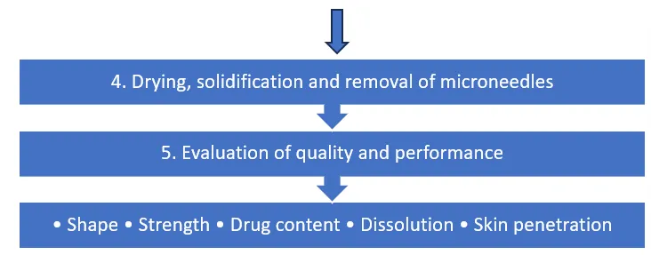
Flow chart:
Overview of Fabrication Techniques for Dissolving Microneedles
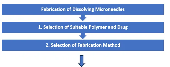
| 3. Selection of fabrication method | |
|---|---|
| a. Micromolding | f. Droplet-born air blowing |
| b. Centrifugation-assisted micromolding | g. 3D printing |
| c. Vacuum-assisted micromolding | h. Solvent casting and drying |
| d. Drawing lithography | i. Emerging technologies |
4.2. Micro molding Technique
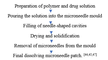
Micromolding is one of the most commonly used methods for preparing dissolving microneedles. In this technique, a liquid polymer solution containing the drug is poured into a mould containing tiny needle-shaped cavities. The solution fills the cavities and is allowed to dry. After drying, the solid microneedles are carefully removed from the mould.
Role: This technique helps to produce microneedles with a uniform shape, size and arrangement. It is suitable for preparing drug-loaded microneedle patches using polymers such as PVP, PVA and hyaluronic acid. It is also relatively simple and economical.
Flow chart:
4.3. Centrifugation-Assisted Micromolding
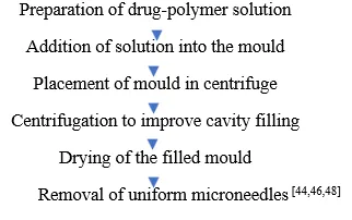
In this method, centrifugation is used to help the polymer solution enter all the tiny cavities of the mould. First, the drug and polymer solution is added to the mould. The mould is then placed in a centrifuge and rotated at a suitable speed. This process helps push the solution into the narrow needle-shaped spaces and reduces the amount of trapped air.
Role: The main role of centrifugation is to improve the filling of the mould cavities. It helps reduce empty spaces and air bubbles, resulting in more complete and uniform microneedle structures.
Flow chart:
4.5. Drawing Lithography
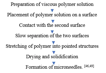
Drawing lithography is a technique in which a thick or viscous polymer solution is stretched to form needle-like structures. In this method, a small amount of polymer solution is placed between two surfaces. The surfaces are slowly separated, which stretches the polymer and forms pointed structures. The structures are then dried or solidified to obtain microneedles.
Role: This method is useful for producing microneedles without using a conventional mould. It allows control over the needle height and shape by adjusting the pulling distance and polymer properties. However, controlling the uniformity of all needles can be challenging.
Flow chart:
4.6. Droplet-Born Air Blowing (DAB) Technique
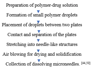
Droplet-born air blowing is a relatively simple method used to prepare dissolving microneedles. In this technique, small droplets of polymer solution are placed between two plates. The plates are brought together and then slowly separated. During separation, the polymer droplets are stretched into needle-like shapes. Air is blown over the stretched polymer to help it dry and solidify.
Role: The main role of this technique is to form microneedles without using a traditional needle-shaped mould. It is useful for preparing drug-loaded microneedles and can reduce the number of manufacturing steps. Insulin-loaded dissolving microneedles have also been prepared using this method.
Flow chart:
4.7. Three-Dimensional (3D) Printing Technologies
Three-dimensional printing is a modern technique used to prepare microneedles by building structures layer by layer using a computer-designed model. Different printing methods, such as stereolithography (SLA), digital light processing (DLP) and two-photon polymerization, can be used to produce microneedle structures or the moulds required for their preparation.
In this technique, a digital design is first created using computer software. The printer then produces the required structure using a suitable printing material. Depending on the technology, the printed structure may be used directly or as a master mould for casting dissolving polymers.
Role: 3D printing helps produce microneedles with customized shapes, sizes and arrangements. It is useful for developing complex designs and reducing the time needed to prepare prototypes. However, printing equipment can be expensive, and the resolution and material compatibility may limit its use in large-scale production.
Flow chart:
4.8. Solvent Casting and Drying Methods
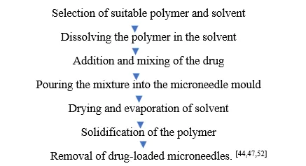
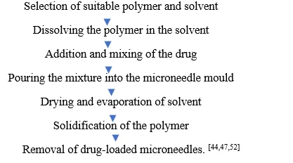
Solvent casting is a simple technique used for preparing dissolving microneedles. In this method, a suitable polymer is dissolved in a solvent, and the drug is mixed into the solution. The prepared mixture is poured into a microneedle mould and allowed to dry. During drying, the solvent evaporates, leaving behind solid polymer microneedles containing the drug.
Role: This method helps incorporate drugs into water-soluble or biodegradable polymer structures. It is relatively inexpensive and does not require highly complicated equipment. The drying process is important because it affects the strength, shape and final quality of the microneedles.
Flow chart:
4.9. Emerging Manufacturing Technologies
New manufacturing technologies are being developed to improve the quality, production speed and accuracy of dissolving microneedles. These methods aim to overcome the limitations of traditional moulding techniques.
Some important emerging techniques include:
5. Evaluation Parameters of Dissolving Microneedles
Evaluation of dissolving microneedles is an important step carried out after their preparation. Different tests are performed to check the physical appearance, shape, size, strength, drug content, skin penetration ability, dissolution and drug release properties of the prepared microneedle patches.
These tests help to determine whether the microneedles are properly formed, whether they can enter the skin without breaking, and whether they can dissolve and release the drug effectively. [55,56,57,58]
5.1. Physical Appearance and Visual Inspection
The prepared microneedle patches are first examined visually to check their overall appearance. The patch is observed for uniformity, colour, cracks, broken needles, air bubbles and other visible defects. The surface of the patch should be smooth, and the needles should be properly formed. [55,56]
5.2. Morphological Characterization
Morphological characterization is performed to study the shape and surface structure of the microneedles. Optical microscopy and scanning electron microscopy (SEM) are commonly used to observe the needle structure. The sharpness of the tips, needle arrangement, surface appearance and uniformity are examined. [55,56,57]
5.3. Measurement of Needle Height, Width and Tip Sharpness
The dimensions of the microneedles are measured using optical microscopy or scanning electron microscopy. Parameters such as needle height, base width, tip diameter and distance between individual needles are recorded. These measurements help to assess the uniformity of the microneedle array. [55,57,58]
5.4. Mechanical Strength Test
Mechanical strength testing is performed to determine whether the microneedles can withstand the pressure applied during skin insertion without breaking or bending. A texture analyser or universal testing machine is commonly used. The microneedle patch is subjected to compression, and the force required to deform or break the needles is measured. [56,57,58]
5.5. Skin Insertion and Penetration Test
This test is performed to check whether the microneedles can successfully enter the skin. Suitable skin models, such as porcine skin, animal skin or human skin samples obtained with appropriate ethical approval, may be used. After applying the microneedles, the skin is examined using microscopy or staining methods such as trypan blue staining to observe the insertion points. [55,56,57,58]
5.6. Dissolution Test
The dissolution test is carried out to determine how quickly the microneedles dissolve after entering the skin or coming into contact with a suitable dissolution medium. Changes in needle shape and size are observed at different time intervals using microscopy. The time required for the needles to dissolve is recorded. [55,56,58]
5.7. Drug Content Estimation
Drug content estimation is performed to determine the total amount of drug present in the prepared microneedle patch. A known quantity of the microneedle formulation is dissolved in a suitable solvent, and the drug concentration is measured using analytical techniques such as UV-visible spectrophotometry or high-performance liquid chromatography (HPLC). [55,57,58]
5.8. Drug Content Uniformity
Drug content uniformity is used to check whether the drug is distributed evenly throughout the microneedle patch or between different patches. Samples from different patches or different regions of the formulation are collected and analysed. The results are compared to identify variations in drug distribution. [55,57,59]
5.9. In-vitro Drug Release Study
The in-vitro drug release study is performed to determine the amount of drug released from the microneedle formulation over a particular period. Suitable dissolution apparatus or diffusion cells may be used with an appropriate release medium. Samples are collected at different time intervals and analysed to measure the amount of drug released. [55,57,58,65]
5.10. Skin Permeation Study
The skin permeation study is carried out to determine how much drug passes through the skin after applying the microneedle patch. Franz diffusion cells are commonly used for this purpose. A suitable skin sample is placed between the donor and receptor compartments, and the amount of drug reaching the receptor medium is measured at different time intervals. [55,56,57,62]
5.11. Water Content and Moisture Analysis
Dissolving microneedles are generally prepared using water-soluble or biodegradable polymers. Therefore, the amount of moisture present in the final patch is important. Moisture content can be measured using a moisture analyser, Karl Fischer titration or thermogravimetric analysis (TGA). [56,63,65]
5.12. Swelling and Water Uptake Study
The swelling study is performed to determine the ability of the polymer material to absorb water when it comes into contact with a suitable aqueous medium. The initial weight of the sample is recorded, followed by exposure to the medium for a specified period. The change in weight is then measured. [63,64]
5.13. Fourier Transform Infrared Spectroscopy (FTIR)
FTIR spectroscopy is used to study the chemical groups present in the drug, polymer and final microneedle formulation. The obtained spectra are compared to identify possible changes in characteristic peaks. [55,63,64]
5.14. Differential Scanning Calorimetry (DSC)
DSC is used to study the thermal properties of the drug, polymer and prepared microneedle formulation. It measures changes in heat flow when the sample is heated or cooled under controlled conditions. [55,63,64]
5.15. X-ray Diffraction (XRD)
XRD is used to determine the crystalline or amorphous nature of the drug and polymer formulation. The diffraction pattern of the pure drug is compared with that of the prepared microneedles to identify changes in the physical state of the drug. [55,63,64]
5.16. Stability Studies
Stability studies are conducted to determine whether the microneedle patch maintains its quality during storage. The patches are stored under suitable temperature and humidity conditions for a specified period. Physical appearance, moisture content, mechanical strength and drug content are examined at selected intervals. [55,56,63,65]
5.17. Skin Irritation and Biocompatibility Studies
These studies are performed to evaluate whether the microneedle formulation causes irritation or harmful effects when it comes into contact with the skin. Suitable in-vitro cell-based tests and, where scientifically and ethically justified, appropriate skin irritation studies may be performed. [56,63,64]
6. Therapeutic Applications of Dissolving Microneedles
Dissolving microneedles are a promising drug delivery system used to deliver different types of medicines through the skin. They are prepared using water-soluble or biodegradable polymers that dissolve after insertion into the skin and release the drug.
These microneedles are gaining attention because they are minimally invasive, easy to apply and generally cause less pain than conventional injections. They are being studied for the treatment of several diseases, including diabetes, cancer, skin disorders, infections and inflammatory conditions. They are also being explored for vaccine delivery, wound healing and cosmetic applications. [66,67,68]
6.1. Diabetes Treatment
Dissolving microneedles are being investigated as an alternative method for delivering insulin through the skin. Patients with diabetes often require regular insulin injections, which may cause pain and discomfort. Insulin-loaded dissolving microneedles can enter the skin and release insulin when the polymer material dissolves.
Researchers are also developing glucose-responsive microneedle patches that can respond to changes in blood glucose levels and release insulin accordingly. This approach may provide a more convenient method of insulin delivery in the future. [66,67,70,71]
6.2. Cancer Treatment
Dissolving microneedles are being studied for delivering anticancer drugs directly into the skin or tumour-associated tissues. They can carry different anticancer agents, nanoparticles and immunotherapeutic substances.
They are particularly being investigated for skin cancers such as melanoma. Some research has also explored their use in cancer immunotherapy, where therapeutic substances are delivered through the skin to stimulate the immune system against cancer cells. [67,72,73]
6.3. Vaccine Delivery
Dissolving microneedles are considered a promising method for vaccine delivery because they can deliver vaccine antigens directly into the skin, which contains many immune cells. After application, the microneedles dissolve and release the vaccine into the skin.
They have been investigated for vaccines against influenza and other infectious diseases. This method may reduce the fear of injections, avoid sharp needle waste and make vaccination easier and more convenient. [66,67,68,69]
6.4. Treatment of Skin Diseases
Dissolving microneedles are being explored for treating different skin conditions, such as psoriasis, acne, eczema and other inflammatory skin disorders. Drugs such as corticosteroids, anti-inflammatory agents and other therapeutic substances can be incorporated into the polymer matrix.
After insertion, the microneedles dissolve and release the drug into the affected skin area. This approach may improve local drug delivery compared with conventional creams and ointments. [66,67,68]
6.5. Wound Healing
Dissolving microneedles have shown promising results in wound healing research. They can deliver therapeutic substances such as growth factors, antibacterial agents and other wound-healing compounds directly into damaged skin.
Some formulations use biodegradable polymers such as hyaluronic acid and are designed to support tissue repair and skin regeneration. Research has also investigated simvastatin-loaded dissolving microneedles for improving wound healing. [67,74,75]
6.6. Hair Growth and Scalp Disorders
Dissolving microneedles are being investigated for delivering drugs into the scalp for the treatment of hair loss and other scalp-related problems. They can deliver substances such as valproic acid and other hair-growth-promoting agents into the skin around hair follicles.
Research has shown that valproic acid-loaded dissolving microneedles may support hair regrowth by influencing pathways involved in hair follicle development. This approach is being explored as an alternative to conventional topical hair treatments. [66,76]
6.7. Pain and Inflammation Management
Dissolving microneedles are also being studied for delivering pain-relieving and anti-inflammatory medicines through the skin. Different therapeutic agents can be incorporated into the polymer material and released after the needles dissolve.
They may be useful for the local treatment of inflammation, swelling and certain painful skin conditions. However, their effectiveness depends on the drug used and the condition being treated. [66,67,68]
6.8. Antibacterial and Antimicrobial Applications
Dissolving microneedles are being explored for delivering antibacterial and antimicrobial drugs into infected skin and wounds. These formulations can release the drug directly at the affected area after insertion.
Research is investigating their use in controlling bacterial growth, treating infected wounds and improving local antimicrobial drug delivery. [67,74,75]
6.9. Delivery of Proteins and Peptides
Proteins and peptides are important therapeutic substances used for treating various diseases. However, many of these molecules are difficult to administer orally because they may be broken down in the gastrointestinal tract.
Dissolving microneedles are being studied for delivering protein- and peptide-based medicines directly through the skin. Insulin is one of the important examples. Other biological molecules are also being investigated for transdermal delivery using suitable polymer formulations. [66,67,70,71]
6.10. Nucleic Acid and Gene-Based Therapy
Recent research has explored dissolving microneedles for delivering genetic materials such as DNA, messenger RNA (mRNA), microRNA (miRNA) and small interfering RNA (siRNA).
These materials are being investigated for applications in cancer treatment, vaccination and gene regulation. The genetic material can be incorporated into suitable polymer or carrier systems and released into the skin after the microneedles dissolve. Most of these applications are still under preclinical investigation. [67,72,73,74]
6.11. Cosmetic Applications and Skin Rejuvenation
Dissolving microneedles are also being explored in cosmetic and skincare applications. They can deliver ingredients such as hyaluronic acid, peptides and other skin-related substances into the skin.
Research has investigated their possible use in improving skin hydration, reducing the appearance of fine lines and supporting skin rejuvenation. They are also being studied for improving the delivery of cosmetic ingredients compared with ordinary topical application. [66,67,68]
7. Regulatory Challenges and Future Perspectives
7.1. Regulatory Challenges
Dissolving microneedles (DMNs) have gained considerable attention as a promising drug delivery system because they are minimally invasive, easy to use, and capable of delivering drugs through the skin without leaving sharp needle waste behind. Despite these advantages, their approval and commercial use still face several regulatory challenges. Since DMNs combine drug delivery technology with microneedle-based devices, their evaluation requires careful consideration of both pharmaceutical and medical device requirements. Regulatory authorities must ensure that these systems are safe, effective, reliable, and suitable for large-scale production.
One of the major challenges is the lack of specific regulatory guidelines designed exclusively for dissolving microneedle-based products. Existing regulations for conventional transdermal patches, injections, and medical devices may not fully address the unique characteristics of DMNs. Therefore, manufacturers may face difficulties in determining the appropriate regulatory pathway for product development and approval.[⁷⁷,⁷⁸]
Another important concern is the selection and safety of polymers used in DMN formulations. Polymers such as hyaluronic acid, polyvinyl alcohol, polyvinylpyrrolidone, and other biodegradable materials must be evaluated for their biocompatibility, toxicity, degradation behaviour, and possible skin irritation. Even when a polymer is generally considered safe, its suitability must be established for the intended route of administration, concentration, formulation, and duration of exposure.[⁷⁹]
Quality control and manufacturing consistency also present significant challenges. The mechanical strength, needle dimensions, drug content, dissolution rate, and drug release behaviour must remain consistent from one batch to another. Variations during fabrication, drying, mould filling, or storage can affect the performance of microneedles and may lead to inconsistent drug delivery. Therefore, suitable quality control methods and validated manufacturing procedures are essential to ensure product reliability.[⁸⁰,⁸¹]
Stability is another important factor that requires attention. Some drugs, particularly proteins, peptides, vaccines, and other biological molecules, may lose their activity during processing or storage because of temperature, humidity, or other environmental conditions. The polymer matrix must protect the incorporated drug while maintaining the physical and chemical stability of the microneedle system throughout its shelf life.[⁸²]
In addition, the performance of DMNs may vary depending on skin thickness, hydration, age, and individual skin conditions. Factors such as insertion depth, dissolution time, and the amount of drug released into the skin must be properly evaluated. Clinical studies are therefore necessary to establish the safety, effectiveness, and reproducibility of these systems in humans.[⁸³]
Overall, the development of clear regulatory standards, appropriate testing methods, and reliable manufacturing practices will be essential for the successful translation of dissolving microneedles from laboratory research into commercially available pharmaceutical products.
7.2. Future Perspectives
The future of dissolving microneedle technology looks promising, particularly with the growing interest in patient-friendly and minimally invasive drug delivery systems. Continuous improvements in polymer science, formulation development, and fabrication techniques are expected to make DMNs more effective and suitable for a wider range of therapeutic applications.
One important area of future development is the use of advanced biodegradable and biocompatible polymers. Researchers are exploring natural and synthetic polymers with improved mechanical strength, dissolution properties, and drug compatibility. The development of new polymer combinations may help overcome current limitations in drug loading and provide better control over drug release. In addition, smart and stimuli-responsive polymer systems may offer opportunities for controlled drug delivery in response to specific physiological conditions.[⁸⁴,⁸⁵]
Another promising direction is the improvement of manufacturing technologies. Although micromoulding is widely used, advanced techniques such as 3D printing, microfabrication, and automated production may help improve needle uniformity, reduce manufacturing errors, and support large-scale production. These technologies could also make it easier to customize microneedle shapes, sizes, and drug-loading capacities according to the requirements of different medicines.[⁸⁶]
The application of DMNs in vaccine delivery is another important area with considerable potential. Since these systems can deliver vaccines through the skin, they may help improve patient acceptance by reducing the discomfort associated with conventional injections. They may also support easier administration and potentially reduce dependence on trained personnel. However, further studies are needed to establish the stability, immune response, dose accuracy, and long-term effectiveness of microneedle-based vaccines.[⁸⁷]
DMNs may also find wider applications in the treatment of chronic diseases such as diabetes, cardiovascular disorders, hormonal conditions, and certain skin diseases. Their potential use for delivering peptides, proteins, and other biological drugs could provide alternatives to conventional administration methods. However, drug stability, loading capacity, and consistent delivery must be carefully addressed before these applications can be widely adopted.[⁸⁸]
Another important development will be the introduction of standardized quality control and evaluation methods. Future research should focus on establishing reliable procedures for testing mechanical strength, insertion efficiency, dissolution time, drug content uniformity, drug release, skin compatibility, and stability. These improvements will help generate reproducible results and provide stronger scientific evidence for regulatory assessment.[⁸⁹]
From a regulatory perspective, greater cooperation between researchers, pharmaceutical manufacturers, and regulatory authorities will be necessary. Clearer guidelines for product classification, polymer safety, manufacturing quality, clinical evaluation, and long-term stability would help reduce uncertainty during product development. International harmonization of testing procedures and quality standards could also make it easier to introduce DMN-based products into different markets.[⁹⁰]
In the coming years, the combination of advanced materials, innovative fabrication methods, improved quality control, and appropriate regulatory guidance may help transform dissolving microneedles from an emerging research technology into a practical drug delivery option. Although several scientific and regulatory challenges remain, continued research and clinical validation could establish DMNs as an important alternative to conventional drug administration systems.
REFERENCES

Harshith Kumar M. S., Kuldeep M. G., Venkatesh K., Hanumanthachar K. Joshi, Dissolving Microneedles as a Novel Drug Delivery System: Polymers, Fabrication Technologies, Application and Regulatory Perspective - a Comprehensive Review., Int. J. of Pharm. Sci., 2026, Vol 4, Issue 11, 1879-1908. https://doi.org/10.5281/zenodo.24155367
 10.5281/zenodo.24155367