
View Article

Abstract

Reactive oxygen species (ROS) are a common trigger for skin aging, which is caused by both intrinsic (chronological) processes and external causes including UV radiation and pollution. Excess ROS cause wrinkles, dryness, and loss of elasticity by damaging lipids, proteins, and DNA, upregulating matrix metalloproteinases (MMPs), and degrading dermal collagen and elastin [11–20]. The shoe flower or China rose, Hibiscus rosa-sinensis Linn. (Malvaceae), is a commonly available plant whose flowers and leaves include flavonoids, anthocyanins, phenolic compounds, mucilage, vitamins, and sterols [1-3]. Plant polyphenols are appealing topical antioxidants. In experiments, plant extracts exhibit antioxidant, anti-inflammatory, wound-healing, antibacterial, and hair-growth properties [7–10]. Water-rich, non-greasy, and esthetically pleasing, hydrogels can contain hydrophilic plant extracts and moisturize the skin [25–29]. The botany, phytochemistry, and antioxidant pharmacology of H. rosa-sinensis, the biology of skin aging, the principles of hydrogel production, and the assessment criteria pertinent to a leaf plus flower extract hydrogel are all summarized in this paper. The motivation for the current endeavor is supported by the fact that no study that particularly creates a combined leaf and flower extract hydrogel for anti-aging usage was found in the reviewed literature.
Keywords

Hibiscus rosa-sinensis, hydrogel, antioxidant, anti-aging, Carbopol, DPPH, topical delivery, herbal cosmetics

Introduction

× Popup Image

figure

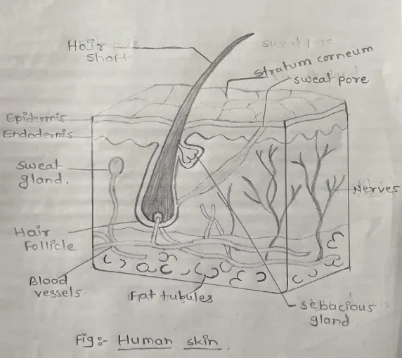
Figure 1: Human Skin

The skin, being the outermost layer of the body, is continuously exposed to oxidants from both internal metabolism and the environment. Skin has been utilized as a standard model to study the connection between oxidative stress and aging since Harman's free radical theory proposed that ROS buildup is a significant driver of aging [11,15]. Consumer interest in natural, plant-based cosmeceuticals has grown since they are believed to be safer and are often high in polyphenols that may enhance the skin's natural antioxidant defenses [14,21,24].

An successful topical antioxidant requires three things: a source of active compounds, a delivery strategy that enables them to reach the skin while preserving their stability, and a suitable evaluation to show their quality and performance. H. rosa-sinensis is inexpensive, readily accessible, and has a long history of traditional use in India [4-6]. Spreadable, cooling, and suitable for aqueous extracts are hydrogels composed of polymers such xanthan gum, Carbopol, or HPMC [25, 29]. This article's goal is to combine these three components and provide a logical method for creating and assessing a hydrogel made from Hibiscus leaf and flower extract.

2. Botany and Traditional Uses of Hibiscus rosa-sinensis

Originally from East Asia, H. rosa-sinensis is an evergreen flowering shrub in the Malvaceae family that is grown as a decorative and therapeutic plant in tropical and subtropical areas [1, 3]. Shoe flower, China rose, Japa, and Rudrapushpa are common names. Wounds, inflammation, fever, cough, diabetes, infections, hair loss, and stomach diseases have all been treated with various parts in traditional medicine [1,4-6]. Additionally, flowers are utilized as a natural colorant and in hair oils, which suggests a cosmetic function.
figure

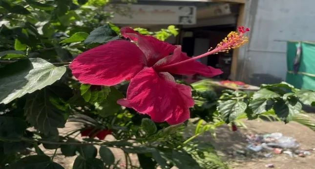
Figure 1: Hibiscus rosa-sinensis

2.1 Taxonomy and Morphology

Table 1: Taxonomical Classification

Rank Classification
Kingdom Plantae
Order Malvales
Family Malvaceae
Genus Hibiscus
Species Hibiscus rosa-sinensis L.

The plant is a woody shrub with branches that grows to a height of one to three meters. Its simple, alternating, oval leaves have toothed margins. The flowers have five petals, a conspicuous staminal column, and are big, showy, and funnel-shaped. Although varieties vary from white and yellow to pink and orange, red is the most common color [1,3]. The cultivar and color utilized in the project should always be noted because flower color affects pigment and antioxidant content [7].

3. Phytochemistry

There are bioactive substances in every section of the plant. Flavonoids, tannins, terpenoids, anthocyanins, saponins, cyclopeptide alkaloids, and vitamins are reported in reviews [1-3].

Table 2: Phytochemicals Present in Hibiscus rosa-sinensis

Plant part Reported constituents Relevance to anti-aging
Flower Ascorbic acid, thiamin, riboflavin, flavonols (quercetin-3-diglucoside, quercetin-3,7-diglucoside, kaempferol glycosides), anthocyanins (cyanidin-3,5-diglucoside, cyanidin-3-sophoroside-5-glucoside) and mucilage [2,3,7] UV absorption, metal chelation, radicalscavenging, film formation, and moisturizing mucilage
Leaf Alkaloids, phenolics, mucilage, sterols, glycosides, reducing sugars, and resin [1-3,7] Hydrating polysaccharides and additional phenolic antioxidants
Stem and root sterculic and malvalic acids, beta-sitosterol, taraxeryl acetate, saponins, and tannins [1,2]. Not utilized in the current formulation

Cultivar, flower color, season, solvent, and extraction method all affect the phenolic and anthocyanin content [7,35]. When the extract is put in a gel with a pH that is almost neutral, it matters because anthocyanins are sensitive to temperature and pH and break down at higher pH and heat [34]. It is therefore advised to standardize using total phenolic content (Folin-Ciocalteu) [39], total flavonoid content (aluminum chloride technique) [40], and total anthocyanin content (pH-differential method) [41].

3.1 Antioxidant chemistry of the main constituent classes

Table 3: Antioxidant Chemistry of the Main Constituent Classes

Class Why it scavenges radicals Practical point
Flavonols (quercetin and kaempferol glycosides) The radical that results from phenolic -OH groups donating an electron or hydrogen atom is stabilized by delocalization; quercetin's catechol group additionally chelates copper and iron [20,34]. Although they dissolve easily in water, glycosides are less able to penetrate skin than aglycones [32].
Anthocyanins (cyanidin glycosides) The resultant radical can be stabilized and given electrons by the conjugated phenolic ring system [34]. They are most stable in acidic conditions, and their color and stability vary with pH [34].
Phenolic acids and tannins Each molecule has several hydroxyl groups. Add astringency; some polymers and proteins may be bound.
Ascorbic acid and other vitamins reducing agents that support the regeneration of more antioxidants [14] Sensitive to heat and oxygen

3.2 Choice of solvent

What is extracted depends on the polarity of the solvent. Water yields higher mucilage and anthocyanin-rich fractions, but ethanol and hydroalcoholic combinations typically recover more phenolics and flavonoids [1,35]. An aqueous or hydroalcoholic extract (with the alcohol completely eliminated) is more useful than a non-polar one since the dried extract must disperse in water for a hydrogel. Comparing two solvents is a helpful experiment because hawthorn hydrogels generated with ethanolic extracts shown more antioxidant activity than aqueous ones in most experiments [50].

4. Antioxidant Activity of H. rosa-sinensis

Free radicals are scavenged by flower and leaf extracts, according to several in vitro investigations. examined extracts from hibiscus and cassia flowers and connected their phenolic content to antibacterial and antioxidant properties [7]. According to other research, ethanolic flower extracts suppress hydroxyl radicals and lipid peroxidation, flavonoids extracted from the petals exhibit antioxidant action, and a 70% ethanol leaf extract protects proteins and lipids from oxidative damage [1,2,3,7]. Additionally, reviews point out that aqueous extracts contain more mucilage and anthocyanins, but ethanol and methanol typically extract more phenolics than water [1,35].

The common in vitro methods used to quantify antioxidant capacity are summarised below.

Table 4: Antioxidant Activity of H. rosa-sinensis

Assay Principle Reference
DPPH Hydrogen/electron donation decolourises the purple DPPH radical (about 517 nm) [36]
ABTS Reduction of the pre-formed ABTS radical cation [37]
FRAP Reduction of Fe(III)-TPTZ to Fe(II) at low pH [38]
Total phenolics Folin-Ciocalteu reduction [39]
Total flavonoids AlCl3 complexation [40]

5. Skin Aging and Oxidative Stress

The term "skin aging exposome" refers to both the intrinsic (genetic, hormonal, and cellular senescence) and extrinsic (mostly caused by UV light, smoking, and pollution) components of skin aging [17,18]. ROS are produced by both mechanisms through photochemical processes, NADPH oxidases, and mitochondria [15]. In addition to decreasing collagen production, ROS stimulate signaling pathways (AP-1, NF-kB) that increase MMP-1, MMP-3, and MMP-9, which break down type I and III collagen and cause fine and coarse wrinkles, loss of firmness, and compromised barrier function [12,13]. Damage is exacerbated by lipid peroxidation, inflammation, and advanced glycation end products [13,15,16].

Topical antioxidants can mitigate UV-induced inflammation, neutralize ROS, and indirectly shield the extracellular matrix [14,19,20]. As indicators of anti-aging potential, natural products have been tested for antioxidant activity, anti-collagenase, and anti-elastase [21–23]. Recent studies emphasize that plant-derived compounds have anti-inflammatory, barrier-supporting, ROS-neutralizing, and MMP-inhibiting actions, and that delivery technique is just as crucial as the active ingredient [55].

5.1 Molecular pathways

Increased ROS from UV exposure and mitochondrial activity trigger transcription factors AP-1 and NF-kB as well as mitogen-activated protein kinase (MAPK) cascades. Collagen is produced more slowly and broken down more quickly as a result of AP-1's interference with TGF-beta/Smad signaling and upregulation of MMP-1, MMP-3, and MMP-9 [12,13]. The enzymatic defenses of the skin, such as glutathione peroxidase, catalase, and superoxide dismutase, also deteriorate with age [15,19].

Table 5: Molecular Pathways

Aging change Visible sign Where an antioxidant gel could help
ROS rise, lipid peroxidation Dullness, roughness Scavenging at the skin surface [14,20]
MMP activation, collagen loss Wrinkles, sagging Indirect, by lowering ROS-driven MMP signalling [12,13]
Chronic low-grade inflammation Redness, uneven tone Anti-inflammatory polyphenols [20,55]
Water loss from the skin Dryness, flaking Water content of the gel, humectants, mucilage [1,25]
UV damage Photoaging, pigmentation UV-absorbing polyphenols, as an adjunct only and not a sunscreen replacement [20,53]

6. Anti-Aging Relevance of Hibiscus Constituents

  • Antioxident and photoprotective effect: Photoprotective and antioxidant properties. In hairless mice, topical preparations containing flavonoids like quercetin decreased UVB-induced oxidative damage [33]. UV absorbance in the 270–300 nm region was seen in a gel containing H. rosa-sinensis and other floral extracts, which the authors connected to polyphenols [53].
  • Skin Penetration: Glycosylation reduces cutaneous absorption, although flavonoid aglycones penetrate skin more readily than glycosides [32]. A major portion of the action of hibiscus flavonoids and anthocyanins is anticipated in the stratum corneum and upper epidermis, which is suitable for surface antioxidant protection because they are mostly glycosides and surpass the 500 Dalton guideline for easy passive skin entrance [31].
  • Wound healing and tissue repair: Rats' wound healing was enhanced by an H. rosa-sinensis extract [9], indicating support for dermal repair.
  • Hair growth: In experimental models, leaf and flower extracts stimulated hair growth [8], suggesting their application in cosmetic products.
  • Antimicrobial activity Antimicrobial activity: Human pathogenic bacteria were effectively combated by flower extracts [10], which could aid in product preservation but cannot take the place of a preservative.
  • Mucilage: Mucilaginous polysaccharides have the ability to increase viscosity, slide, and hydration [1].

The "anti-aging" claim for the formulation is best framed as antioxidant-based unless additional assays are included because direct experimental evidence on collagenase, elastase, or MMP inhibition by H. rosa-sinensis are sparse in comparison to its antioxidant findings [21–23].

6.1 Mechanistic hypothesis and evidence status

Table 6: Mechanistic Hypothesis and Evidence Status

Proposed action Likely constituents Evidence status
Direct ROS scavenging Anthocyanins, flavonols, phenolics Reported for H. rosa-sinensis extracts in vitro [1,3,7]
Metal chelation Catechol-type flavonoids such as quercetin General flavonoid chemistry [34]; not tested for this plant in the sources reviewed
Protection from UV-induced oxidation Flavonols Shown for quercetin formulations [33]; UV absorbance of a Hibiscus gel reported [53]
Collagenase and elastase inhibition Polyphenols, tannins Suggested by studies of other plants [21-23]; to be tested for Hibiscus
Hydration and barrier support Mucilage, humectants, gel water Plausible [1,25]; can be measured by corneometry

This table keeps the review correct by separating reported data for H. rosa-sinensis from extrapolated data. It also highlights areas where your experiments can yield additional information.

7. Hydrogels as Topical Carriers

Large volumes of water can be stored in three-dimensional hydrophilic polymer networks called hydrogels [25, 26]. They provide a cooling effect, hydration, ease of washing, patient acceptance, minimal discomfort, and compatibility with water-soluble extracts when applied topically [27, 28]. Poor lipophilic chemical solubilization and the possibility of microbiological growth necessitate the use of a preservative.

Table 7: Hydrogels as Topical Carriers

Component Examples Function
Gelling agent Carbopol 934/940, HPMC, xanthan gum, sodium CMC, chitosan Forms the network; controls viscosity and release [25,28,29]
Neutraliser Triethanolamine, NaOH Ionises Carbopol to give a clear gel [29]
Humectant / penetration aid Glycerin, propylene glycol Moisturising; retains water; helps spreadability
Preservative Methylparaben, propylparaben Prevents microbial growth

After neutralization to a pH of roughly 6-7, carbopol, a cross-linked polyacrylic acid, swells and thickens; its rheology is dependent on this pH [29]. The final gel pH is often regulated near 5-6.5 to prevent irritation and keep anthocyanins reasonably stable because the skin's surface is rather acidic [34].

7.1 Choice of gelling agent

Table 8: Gelling Agents

Polymer Key properties Cautions
Carbopol 934 / 940 (carbomer) Clear gel, high viscosity at low concentration, good spreadability after neutralisation [29] Electrolytes and acidic extract components can thin the gel; needs pH adjustment
HPMC Non-ionic, stable over a wide pH range, film-forming Slow to hydrate; needs hot-water dispersion
Xanthan gum Shear-thinning, tolerant of salts Can feel stringy; supports microbial growth
Sodium CMC Low cost, anionic Viscosity falls at low pH and with salts
Chitosan Cationic, mucoadhesive, mildly antimicrobial Needs dilute acid; may interact with anionic extract components

skin sensation and rheology [25, 28, 48, 51]. Trial batches should determine whether the hibiscus extract reduces the viscosity of Carbopol at the selected pH because it contains mucilage and acidic pigments.

8. Formulation Approach for a Leaf and Flower Extract Hydrogel

8.1 Collection and authentication

Fresh flowers and leaves are gathered, verified by a botanist, cleaned, shade-dried, and ground into powder. Standard procedures are used to determine pharmacognostic standards like foreign matter, ash values, and extractive values [47].

8.2 Extraction

Ethanol, hydroalcoholic, or aqueous solvents are frequently used in maceration or Soxhlet extraction, and contemporary methods (aided by microwaves or ultrasound) can increase yield and reduce time [35]. The dry extract is kept in a cool, dark, airtight container after the solvent is evaporated under low pressure. For both leaf and flower extracts, preliminary phytochemical testing and TPC/TFC estimation should be documented [39, 40].

8.3 Preparation of the hydrogel

1. Add the gelling agent (such as Carbopol 940) to a portion of the water and let it swell for a few hours.

2. In the leftover water or glycol, dissolve the humectant and preservative.

3. Gently swirl the leaf and floral extracts into the swollen polymer after dissolving them in a tiny amount of the aqueous solution.

4. Make up to weight and use triethanolamine to adjust the pH to the appropriate level.

Illustrative starting ranges (to be optimised by trial batches; they are not fixed recommendations):

Table 9: Ingredients for Hydrogel Preparation

Ingredient Typical range explored
Leaf + flower extract 0.5-5% w/w (several levels, for example 1, 2 and 3%, with a blank)
Carbopol 940 0.5-1.5% w/w (about 1% gave optimal consistency in a hawthorn hydrogel [50]; 1.5% was used in a Centella-Moringa emulgel [52])
Glycerin / propylene glycol 5-15% w/w
Triethanolamine q.s. to pH 5.5-6.5
Methylparaben 0.1-0.2% w/w

8.4 Suggested batch plan

A simple design compares each extract alone with the combination, plus a blank.

Table 10: Batch Plan

Batch Leaf extract Flower extract Purpose
F0 none none Blank gel (control)
F1 1% none Leaf only
F2 none 1% Flower only
F3 0.5% 0.5% Combination at 1% total
F4 1% 1% Combination at 2% total
F5 1.5% 1.5% Combination at 3% total

The percentages serve as examples; following a trial run, determine the final levels based on color, pH, and DPPH readings. A reasonable check for synergy is provided by comparing F1, F2, and F3 (equal total extract).

8.5 Common formulation problems

Table 11: Arising Formulation Problems

Problem Probable cause Possible remedy
Lumps (fish-eyes) Carbopol added too fast Sprinkle slowly into a vortex and let it swell overnight
Thin gel after adding extract Electrolytes or acids lower viscosity Re-adjust pH; raise polymer level; add HPMC or xanthan gum
Colour fades or browns Anthocyanin breakdown by pH, light or heat Keep pH near 5.5; use opaque packaging; consider a chelator or antioxidant [34]
Turbidity or sediment Insoluble extract fraction Filter the extract; remove all alcohol from hydroalcoholic extract
Microbial growth Aqueous base plus plant material Use a preservative; run a microbial limit test

9. Evaluation of the Hydrogel

Table 12: Evaluation Parameters of the Hydrogels

Parameter Method / purpose Notes
Organoleptic  Colour, odour, appearance, homogeneity  Anthocyanin colour change can signal degradation [34] 
pH  Digital pH meter (1% dispersion)  Target near skin pH; a herbal hydrogel stayed at 5.5-6.5 over 90 days [50] 
Viscosity and rheology  Brookfield viscometer  Shear-thinning flow is desirable [29] 
Spreadability and extrudability  Parallel-plate method; tube extrusion  Patient acceptance and dose uniformity [51] 
Extract content  Marker such as TPC or TFC  Assures uniformity [39,40] 
Antioxidant activity of the finished gel  DPPH, ABTS, FRAP versus extract and blank gel  Confirms the extract remains active after formulation [36-38,50] 
In vitro release / permeation  Franz diffusion cell with a synthetic or animal membrane  Release kinetics fitted to Higuchi and Korsmeyer-Peppas models [44-46] 
Skin irritation  Primary irritation index (Draize-type), or human volunteers with ethical approval  Safety for topical use [42,49] 
Stability  ICH Q1A(R2) conditions, for example 40 C / 75% RH, plus room temperature and refrigerated  Observe pH, viscosity, colour and antioxidant activity [43] 
Microbial limit  Total viable count  Quality control 

9.1 Method notes

  • Antioxidant activity of the gel: A weighed quantity of gel should be dispersed in the solvent, centrifuged or filtered, and the supernatant should be reacted with DPPH. The absorbance at approximately 517 nm should then be measured to determine the percentage of inhibition. Compare the ascorbic acid, simple extract, and blank gel [36,50].
  • In vitro diffusion: The gel is held on a membrane above a receptor medium (such as phosphate buffer pH 7.4) in a Franz cell that is stirred and maintained at 32–37 C. Periodically, samples are taken out and tested, such as for total phenolics, and the volume is replenished [46]. Higuchi, Korsmeyer-Peppas, zero-order, and first-order models are fitted to cumulative release against time [44, 45].
  • Irritation: The Institutional Ethics Committee must approve the use of a patch test in healthy people or the Draize-type grading of erythema and edema (0–4) in animals [42,49].
  • Stability: For the duration specified in the protocol, store samples at room temperature, in a refrigerator, and at 40 +/- 2 C/75 +/- 5% RH. At predetermined intervals, compare pH, viscosity, color, and antioxidant activity [43]. Antioxidant tests, pH, rheology, spreadability, and volunteer acceptability studies have all been used in the literature to evaluate anti-aging hydrogels made from different plants. A Gac fruit hydrogel was evaluated for physical characteristics, DPPH activity, and skin irritation in volunteers [49]; an Oenanthe javanica hydrogel (0.8% extract) maintained its physical appearance and won volunteer preference for its smoothness [48]; Spreadability, antioxidant, and antibacterial activity of gels and emulgels based on centella were assessed [51,52]; and a hawthorn hydrogel maintained antioxidant activity comparable to the extract; in the majority of tests, ethanolic extracts outperformed aqueous ones [50]. A template for assessing a hibiscus hydrogel is provided by these studies.

Table 13: An Overview of Published Herbal Gel and Hydrogel Research

Plant Base Evaluations reported
Oenanthe javanica [48] Carbopol, HPMC, xanthan gum, glycerin pH, appearance, rheology, volunteer smoothness rating
Gac fruit [49] Carbopol 940 Physical properties, DPPH, skin irritation in volunteers
Hawthorn [50] Carbopol 940 (1%) pH over 90 days, TBARS, beta-carotene bleaching, DPPH, reducing power
Centella asiatica madecassoside [51] Carbopol 940, HPMC Spreadability, rheology, antimicrobial and antioxidant activity
Centella plus Moringa [52] Carbopol emulgel with stearic acid Organoleptic, physical quality, DPPH

11. Challenges and Future Perspectives

  • Standardized extracts, preferably with HPLC fingerprinting, are necessary due to batch-to-batch fluctuation in plant material.
  • Antioxidants, chelators, acidity buffering, light-protective packaging, or encapsulation (liposomes, nanoparticles) may be required for anthocyanin instability [34].
  • Glycosides have low skin penetration; penetration enhancers or nano-carriers may be investigated [32].
  • Mechanistic tests (collagenase, elastase, tyrosinase, MMP inhibition) and, lastly, in vivo or clinical data on hydration, elasticity, and wrinkles are required for anti-aging claims [21–23].
  • Since combined extracts have been shown to work better than single extracts in other herbal anti-aging medicines, a comparison of leaf extract, flower extract, and the combination (potential synergy) would bring innovation [52].

CONCLUSION

Hydrogels are an appropriate, patient-friendly carrier for the abundant, readily available polyphenols with proven antioxidant action found in H. rosa-sinensis leaves and flowers. A standardized leaf and flower extract hydrogel makes sense as an anti-aging option because oxidative stress is closely associated with skin aging. A good project should standardize the extracts, optimize the gel base, assess stability, irritation, and physicochemical characteristics, and show antioxidant activity in the finished product. Since there is now insufficient direct evidence for integrated leaf and flower hydrogels, this is a promising subject for future research.

REFERENCES

  1. Amtaghri S, Qabouche A, Slaoui M, Eddouks M. A comprehensive overview of Hibiscus rosa-sinensis L.: its ethnobotanical uses, phytochemistry, therapeutic uses, pharmacological activities, and toxicology. Endocr Metab Immune Disord Drug Targets. 2023. doi:10.2174/1871530323666230522113405
  2. Missoum A. An update review on Hibiscus rosa sinensis phytochemistry and medicinal uses. J Ayurvedic Herb Med. 2018;4 (3):135-146.
  3. Al-Snafi AE. Chemical constituents and pharmacological effects of Hibiscus rosa-sinensis - a review. IOSR J Pharm. 2018;8 (7):101-119.
  4. Jadhav VM, Thorat RM, Kadam VJ, Sathe NS. Traditional medicinal uses of Hibiscus rosa-sinensis. J Pharm Res. 2009;2 (8):1220-1222.
  5. Khristi V, Patel VH. Therapeutic potential of Hibiscus rosa sinensis: a review. Int J Nutr Pharmacol Neurol Dis. 2016;6 (2):105-109.
  6. Kumar A, Singh A. Review on Hibiscus rosa sinensis. Int J Res Pharm Biomed Sci. 2012;3 (2):534-538.
  7. Mak YW, Chuah LO, Ahmad R, Bhat R. Antioxidant and antibacterial activities of hibiscus (Hibiscus rosa-sinensis L.) and Cassia (Senna bicapsularis L.) flower extracts. J King Saud Univ Sci. 2013;25 (4):275-282.
  8. Adhirajan N, Ravi Kumar T, Shanmugasundaram N, Babu M. In vivo and in vitro evaluation of hair growth potential of Hibiscus rosa-sinensis Linn. J Ethnopharmacol. 2003;88 (2-3):235-239.
  9. Bhaskar A, Nithya V. Evaluation of the wound-healing activity of Hibiscus rosa sinensis L (Malvaceae) in Wistar albino rats. Indian J Pharmacol. 2012;44 (6):694-698.
  10. Ruban P, Gajalakshmi K. In vitro antibacterial activity of Hibiscus rosa-sinensis flower extract against human pathogens. Asian Pac J Trop Biomed. 2012;2 (5):399-403.
  11. Harman D. Aging: a theory based on free radical and radiation chemistry. J Gerontol. 1956;11 (3):298-300.
  12. Fisher GJ, Kang S, Varani J, et al. Mechanisms of photoaging and chronological skin aging. Arch Dermatol. 2002;138 (11):1462-1470.
  13. Pillai S, Oresajo C, Hayward J. Ultraviolet radiation and skin aging: roles of reactive oxygen species, inflammation and protease activation, and strategies for prevention of inflammation-induced matrix degradation. Int J Cosmet Sci. 2005;27 (1):17-34.
  14. Masaki H. Role of antioxidants in the skin: anti-aging effects. J Dermatol Sci. 2010;58 (2):85-90.
  15. Rinnerthaler M, Bischof J, Streubel MK, Trost A, Richter K. Oxidative stress in aging human skin. Biomolecules. 2015;5 (2):545-589.
  16. Papaccio F, D'Arino A, Caputo S, Bellei B. Focus on the contribution of oxidative stress in skin aging. Antioxidants. 2022;11 (6):1121.
  17. Krutmann J, Bouloc A, Sore G, Bernard BA, Passeron T. The skin aging exposome. J Dermatol Sci. 2017;85 (3):152-161.
  18. Tobin DJ. Introduction to skin aging. J Tissue Viability. 2017;26 (1):37-46.
  19. Zhang S, Duan E. Fighting against skin aging: the way from bench to bedside. Cell Transplant. 2018;27 (5):729-738.
  20. Dunaway S, Odin R, Zhou L, Ji L, Zhang Y, Kadekaro AL. Natural antioxidants: multiple mechanisms to protect skin from solar radiation. Front Pharmacol. 2018;9:392. doi:10.3389/fphar.2018.00392
  21. Mukherjee PK, Maity N, Nema NK, Sarkar BK. Bioactive compounds from natural resources against skin aging. Phytomedicine. 2011;19 (1):64-73.
  22. Thring TSA, Hili P, Naughton DP. Anti-collagenase, anti-elastase and anti-oxidant activities of extracts from 21 plants. BMC Complement Altern Med. 2009;9:27.
  23. Ndlovu G, Fouche G, Tselanyane M, Cordier W, Steenkamp V. In vitro determination of the anti-aging potential of four southern African medicinal plants. BMC Complement Altern Med. 2013;13:304.
  24. Dureja H, Kaushik D, Gupta M, Kumar V, Lather V. Cosmeceuticals: an emerging concept. Indian J Pharmacol. 2005;37 (3):155-159.
  25. Ahmed EM. Hydrogel: preparation, characterization, and applications: a review. J Adv Res. 2015;6 (2):105-121.
  26. Hoffman AS. Hydrogels for biomedical applications. Adv Drug Deliv Rev. 2002;54 (1):3-12.
  27. Calo E, Khutoryanskiy VV. Biomedical applications of hydrogels: a review of patents and commercial products. Eur Polym J. 2015;65:252-267.
  28. Peppas NA, Bures P, Leobandung W, Ichikawa H. Hydrogels in pharmaceutical formulations. Eur J Pharm Biopharm. 2000;50 (1):27-46.
  29. Islam MT, Rodriguez-Hornedo N, Ciotti S, Ackermann C. Rheological characterization of topical carbomer gels neutralized to different pH. Pharm Res. 2004;21 (7):1192-1199.
  30. Prausnitz MR, Langer R. Transdermal drug delivery. Nat Biotechnol. 2008;26 (11):1261-1268.
  31. Bos JD, Meinardi MM. The 500 Dalton rule for the skin penetration of chemical compounds and drugs. Exp Dermatol. 2000;9 (3):165-169.
  32. Chuang SY, Lin YK, Lin CF, et al. Elucidating the skin delivery of aglycone and glycoside flavonoids: how the structures affect cutaneous absorption. Nutrients. 2017;9 (12):1304.
  33. Casagrande R, Georgetti SR, Verri WA Jr, et al. Protective effect of topical formulations containing quercetin against UVB-induced oxidative stress in hairless mice. J Photochem Photobiol B. 2006;84 (1):21-27.
  34. Castaneda-Ovando A, Pacheco-Hernandez ML, Paez-Hernandez ME, Rodriguez JA, Galan-Vidal CA. Chemical studies of anthocyanins: a review. Food Chem. 2009;113 (4):859-871.
  35. Azmir J, Zaidul ISM, Rahman MM, et al. Techniques for extraction of bioactive compounds from plant materials: a review. J Food Eng. 2013;117 (4):426-436.
  36. Brand-Williams W, Cuvelier ME, Berset C. Use of a free radical method to evaluate antioxidant activity. LWT Food Sci Technol. 1995;28 (1):25-30.
  37. Re R, Pellegrini N, Proteggente A, et al. Antioxidant activity applying an improved ABTS radical cation decolorization assay. Free Radic Biol Med. 1999;26 (9-10):1231-1237.
  38. Benzie IFF, Strain JJ. The ferric reducing ability of plasma (FRAP) as a measure of "antioxidant power": the FRAP assay. Anal Biochem. 1996;239 (1):70-76.
  39. Singleton VL, Rossi JA. Colorimetry of total phenolics with phosphomolybdic-phosphotungstic acid reagents. Am J Enol Vitic. 1965;16 (3):144-158.
  40. Chang CC, Yang MH, Wen HM, Chern JC. Estimation of total flavonoid content in propolis by two complementary colorimetric methods. J Food Drug Anal. 2002;10 (3):178-182.
  41. Giusti MM, Wrolstad RE. Characterization and measurement of anthocyanins by UV-visible spectroscopy. Curr Protoc Food Anal Chem. 2001;F1.2.1-F1.2.13.
  42. Draize JH, Woodard G, Calvery HO. Methods for the study of irritation and toxicity of substances applied topically to the skin and mucous membranes. J Pharmacol Exp Ther. 1944;82 (3):377-390.
  43. ICH Harmonised Tripartite Guideline. Stability testing of new drug substances and products Q1A (R2). 2003.
  44. Korsmeyer RW, Gurny R, Doelker E, Buri P, Peppas NA. Mechanisms of solute release from porous hydrophilic polymers. Int J Pharm. 1983;15 (1):25-35.
  45. Higuchi T. Mechanism of sustained-action medication: theoretical analysis of rate of release of solid drugs dispersed in solid matrices. J Pharm Sci. 1963;52:1145-1149.
  46. Franz TJ. Percutaneous absorption: on the relevance of in vitro data. J Invest Dermatol. 1975;64 (3):190-195.
  47. Kokate CK. Practical Pharmacognosy. 4th ed. New Delhi: Vallabh Prakashan; 1994.
  48. Formulation of hydrogel containing Oenanthe javanica extract for topical use: extraction, formulation and characterization. Asian J Med Health Sci (UniKL). Available from: https://ejournal.unikl.edu.my/index.php/ajmhs/article/view/910
  49. Development and evaluation of a hydrogel containing Momordica cochinchinensis Spreng extract for topical applications. Braz J Pharm Sci. Available from: https://journals.usp.br/bjps/article/download/205307/194913/629760
  50. Development of paraben-free hydrogel based on plant extracts for topical application (hawthorn, Crataegus monogyna; conference paper). Instituto Politecnico de Braganca repository. Available from: https://bibliotecadigital.ipb.pt/entities/publication/d7adde3c-2f7f-4d52-9084-1ffe97396932
  51. Physicochemical assessment and evaluation of antibacterial and antioxidant activity of madecassoside gels. Trop J Nat Prod Res. Available from: https://tjnpr.org/index.php/home/article/download/7338/6459/17191
  52. Formulation and antioxidant activity test of Centella asiatica herba extract and Moringa oleifera leaves extract as an anti-aging emulgel. Trop J Nat Prod Res. Available from: https://tjnpr.org/index.php/home/article/download/3700/4121/7367
  53. Formulation of gel and its UV protective study of some medicinal flowers (Hibiscus rosa-sinensis, Rosa damascena, Spathodea campanulata). Asian J Pharm Anal. Available from: https://mail.i-scholar.in/index.php/Ajpa/article/view/135689
  54. Hibiscus rosa-sinensis flower extract (INCI: Hibiscus Rosa-Sinensis Flower Extract). SpecialChem Cosmetics Ingredient Database. Available from: https://www.specialchem.com/cosmetics/inci-ingredients/hibiscus-rosa-sinensis-flower-extract
  55. The natural defense: anti-aging potential of plant-derived substances and technological solutions against photoaging. PubMed Central (PMC12386611). Available from: https://www.ncbi.nlm.nih.gov/pmc/articles/PMC12386611/

Reference

  1. Amtaghri S, Qabouche A, Slaoui M, Eddouks M. A comprehensive overview of Hibiscus rosa-sinensis L.: its ethnobotanical uses, phytochemistry, therapeutic uses, pharmacological activities, and toxicology. Endocr Metab Immune Disord Drug Targets. 2023. doi:10.2174/1871530323666230522113405
  2. Missoum A. An update review on Hibiscus rosa sinensis phytochemistry and medicinal uses. J Ayurvedic Herb Med. 2018;4 (3):135-146.
  3. Al-Snafi AE. Chemical constituents and pharmacological effects of Hibiscus rosa-sinensis - a review. IOSR J Pharm. 2018;8 (7):101-119.
  4. Jadhav VM, Thorat RM, Kadam VJ, Sathe NS. Traditional medicinal uses of Hibiscus rosa-sinensis. J Pharm Res. 2009;2 (8):1220-1222.
  5. Khristi V, Patel VH. Therapeutic potential of Hibiscus rosa sinensis: a review. Int J Nutr Pharmacol Neurol Dis. 2016;6 (2):105-109.
  6. Kumar A, Singh A. Review on Hibiscus rosa sinensis. Int J Res Pharm Biomed Sci. 2012;3 (2):534-538.
  7. Mak YW, Chuah LO, Ahmad R, Bhat R. Antioxidant and antibacterial activities of hibiscus (Hibiscus rosa-sinensis L.) and Cassia (Senna bicapsularis L.) flower extracts. J King Saud Univ Sci. 2013;25 (4):275-282.
  8. Adhirajan N, Ravi Kumar T, Shanmugasundaram N, Babu M. In vivo and in vitro evaluation of hair growth potential of Hibiscus rosa-sinensis Linn. J Ethnopharmacol. 2003;88 (2-3):235-239.
  9. Bhaskar A, Nithya V. Evaluation of the wound-healing activity of Hibiscus rosa sinensis L (Malvaceae) in Wistar albino rats. Indian J Pharmacol. 2012;44 (6):694-698.
  10. Ruban P, Gajalakshmi K. In vitro antibacterial activity of Hibiscus rosa-sinensis flower extract against human pathogens. Asian Pac J Trop Biomed. 2012;2 (5):399-403.
  11. Harman D. Aging: a theory based on free radical and radiation chemistry. J Gerontol. 1956;11 (3):298-300.
  12. Fisher GJ, Kang S, Varani J, et al. Mechanisms of photoaging and chronological skin aging. Arch Dermatol. 2002;138 (11):1462-1470.
  13. Pillai S, Oresajo C, Hayward J. Ultraviolet radiation and skin aging: roles of reactive oxygen species, inflammation and protease activation, and strategies for prevention of inflammation-induced matrix degradation. Int J Cosmet Sci. 2005;27 (1):17-34.
  14. Masaki H. Role of antioxidants in the skin: anti-aging effects. J Dermatol Sci. 2010;58 (2):85-90.
  15. Rinnerthaler M, Bischof J, Streubel MK, Trost A, Richter K. Oxidative stress in aging human skin. Biomolecules. 2015;5 (2):545-589.
  16. Papaccio F, D'Arino A, Caputo S, Bellei B. Focus on the contribution of oxidative stress in skin aging. Antioxidants. 2022;11 (6):1121.
  17. Krutmann J, Bouloc A, Sore G, Bernard BA, Passeron T. The skin aging exposome. J Dermatol Sci. 2017;85 (3):152-161.
  18. Tobin DJ. Introduction to skin aging. J Tissue Viability. 2017;26 (1):37-46.
  19. Zhang S, Duan E. Fighting against skin aging: the way from bench to bedside. Cell Transplant. 2018;27 (5):729-738.
  20. Dunaway S, Odin R, Zhou L, Ji L, Zhang Y, Kadekaro AL. Natural antioxidants: multiple mechanisms to protect skin from solar radiation. Front Pharmacol. 2018;9:392. doi:10.3389/fphar.2018.00392
  21. Mukherjee PK, Maity N, Nema NK, Sarkar BK. Bioactive compounds from natural resources against skin aging. Phytomedicine. 2011;19 (1):64-73.
  22. Thring TSA, Hili P, Naughton DP. Anti-collagenase, anti-elastase and anti-oxidant activities of extracts from 21 plants. BMC Complement Altern Med. 2009;9:27.
  23. Ndlovu G, Fouche G, Tselanyane M, Cordier W, Steenkamp V. In vitro determination of the anti-aging potential of four southern African medicinal plants. BMC Complement Altern Med. 2013;13:304.
  24. Dureja H, Kaushik D, Gupta M, Kumar V, Lather V. Cosmeceuticals: an emerging concept. Indian J Pharmacol. 2005;37 (3):155-159.
  25. Ahmed EM. Hydrogel: preparation, characterization, and applications: a review. J Adv Res. 2015;6 (2):105-121.
  26. Hoffman AS. Hydrogels for biomedical applications. Adv Drug Deliv Rev. 2002;54 (1):3-12.
  27. Calo E, Khutoryanskiy VV. Biomedical applications of hydrogels: a review of patents and commercial products. Eur Polym J. 2015;65:252-267.
  28. Peppas NA, Bures P, Leobandung W, Ichikawa H. Hydrogels in pharmaceutical formulations. Eur J Pharm Biopharm. 2000;50 (1):27-46.
  29. Islam MT, Rodriguez-Hornedo N, Ciotti S, Ackermann C. Rheological characterization of topical carbomer gels neutralized to different pH. Pharm Res. 2004;21 (7):1192-1199.
  30. Prausnitz MR, Langer R. Transdermal drug delivery. Nat Biotechnol. 2008;26 (11):1261-1268.
  31. Bos JD, Meinardi MM. The 500 Dalton rule for the skin penetration of chemical compounds and drugs. Exp Dermatol. 2000;9 (3):165-169.
  32. Chuang SY, Lin YK, Lin CF, et al. Elucidating the skin delivery of aglycone and glycoside flavonoids: how the structures affect cutaneous absorption. Nutrients. 2017;9 (12):1304.
  33. Casagrande R, Georgetti SR, Verri WA Jr, et al. Protective effect of topical formulations containing quercetin against UVB-induced oxidative stress in hairless mice. J Photochem Photobiol B. 2006;84 (1):21-27.
  34. Castaneda-Ovando A, Pacheco-Hernandez ML, Paez-Hernandez ME, Rodriguez JA, Galan-Vidal CA. Chemical studies of anthocyanins: a review. Food Chem. 2009;113 (4):859-871.
  35. Azmir J, Zaidul ISM, Rahman MM, et al. Techniques for extraction of bioactive compounds from plant materials: a review. J Food Eng. 2013;117 (4):426-436.
  36. Brand-Williams W, Cuvelier ME, Berset C. Use of a free radical method to evaluate antioxidant activity. LWT Food Sci Technol. 1995;28 (1):25-30.
  37. Re R, Pellegrini N, Proteggente A, et al. Antioxidant activity applying an improved ABTS radical cation decolorization assay. Free Radic Biol Med. 1999;26 (9-10):1231-1237.
  38. Benzie IFF, Strain JJ. The ferric reducing ability of plasma (FRAP) as a measure of "antioxidant power": the FRAP assay. Anal Biochem. 1996;239 (1):70-76.
  39. Singleton VL, Rossi JA. Colorimetry of total phenolics with phosphomolybdic-phosphotungstic acid reagents. Am J Enol Vitic. 1965;16 (3):144-158.
  40. Chang CC, Yang MH, Wen HM, Chern JC. Estimation of total flavonoid content in propolis by two complementary colorimetric methods. J Food Drug Anal. 2002;10 (3):178-182.
  41. Giusti MM, Wrolstad RE. Characterization and measurement of anthocyanins by UV-visible spectroscopy. Curr Protoc Food Anal Chem. 2001;F1.2.1-F1.2.13.
  42. Draize JH, Woodard G, Calvery HO. Methods for the study of irritation and toxicity of substances applied topically to the skin and mucous membranes. J Pharmacol Exp Ther. 1944;82 (3):377-390.
  43. ICH Harmonised Tripartite Guideline. Stability testing of new drug substances and products Q1A (R2). 2003.
  44. Korsmeyer RW, Gurny R, Doelker E, Buri P, Peppas NA. Mechanisms of solute release from porous hydrophilic polymers. Int J Pharm. 1983;15 (1):25-35.
  45. Higuchi T. Mechanism of sustained-action medication: theoretical analysis of rate of release of solid drugs dispersed in solid matrices. J Pharm Sci. 1963;52:1145-1149.
  46. Franz TJ. Percutaneous absorption: on the relevance of in vitro data. J Invest Dermatol. 1975;64 (3):190-195.
  47. Kokate CK. Practical Pharmacognosy. 4th ed. New Delhi: Vallabh Prakashan; 1994.
  48. Formulation of hydrogel containing Oenanthe javanica extract for topical use: extraction, formulation and characterization. Asian J Med Health Sci (UniKL). Available from: https://ejournal.unikl.edu.my/index.php/ajmhs/article/view/910
  49. Development and evaluation of a hydrogel containing Momordica cochinchinensis Spreng extract for topical applications. Braz J Pharm Sci. Available from: https://journals.usp.br/bjps/article/download/205307/194913/629760
  50. Development of paraben-free hydrogel based on plant extracts for topical application (hawthorn, Crataegus monogyna; conference paper). Instituto Politecnico de Braganca repository. Available from: https://bibliotecadigital.ipb.pt/entities/publication/d7adde3c-2f7f-4d52-9084-1ffe97396932
  51. Physicochemical assessment and evaluation of antibacterial and antioxidant activity of madecassoside gels. Trop J Nat Prod Res. Available from: https://tjnpr.org/index.php/home/article/download/7338/6459/17191
  52. Formulation and antioxidant activity test of Centella asiatica herba extract and Moringa oleifera leaves extract as an anti-aging emulgel. Trop J Nat Prod Res. Available from: https://tjnpr.org/index.php/home/article/download/3700/4121/7367
  53. Formulation of gel and its UV protective study of some medicinal flowers (Hibiscus rosa-sinensis, Rosa damascena, Spathodea campanulata). Asian J Pharm Anal. Available from: https://mail.i-scholar.in/index.php/Ajpa/article/view/135689
  54. Hibiscus rosa-sinensis flower extract (INCI: Hibiscus Rosa-Sinensis Flower Extract). SpecialChem Cosmetics Ingredient Database. Available from: https://www.specialchem.com/cosmetics/inci-ingredients/hibiscus-rosa-sinensis-flower-extract
  55. The natural defense: anti-aging potential of plant-derived substances and technological solutions against photoaging. PubMed Central (PMC12386611). Available from: https://www.ncbi.nlm.nih.gov/pmc/articles/PMC12386611/

Photo
Zeenat Shaikh
Corresponding author

Department of Pharmacy, SND Institute of Pharmacy, Babhulgaon, Yeola, Maharashtra, India.

Photo
Bharat Pawar
Co-author

Department of Pharmacy, SND Institute of Pharmacy, Babhulgaon, Yeola, Maharashtra, India.

Photo
Pratik More
Co-author

Department of Pharmacy, SND Institute of Pharmacy, Babhulgaon, Yeola, Maharashtra, India.

Photo
Sayali Sonawane
Co-author

Department of Pharmacy, SND Institute of Pharmacy, Babhulgaon, Yeola, Maharashtra, India.

Zeenat Shaikh, Bharat Pawar, Pratik More, Sayali Sonawane, Formulation and Evaluation of Hibiscus Rosa-Sinensis Leaf and Flower Extract Hydrogel for Antioxidant & Anti-Aging Potential: A Review, Int. J. of Pharm. Sci., 2026, Vol 4, Issue 11, 1909-1920. https://doi.org/10.5281/zenodo.24155466

More related articles
Recent Advances in Standardization and Quality Con...
Datta More, Sanjay Bais, Yuvraj Arjun...
Evaluation of Analytical Quality Standards in the ...
Yogini A. Kawale, Upadesh Lade, Kunal Zode, Revati Donode, Priyan...
Beyond the Breakouts: Exploring the Pathophysiolog...
Aditya, Praveen Kumar, Pranav Priyadarshani, Nikita...
More related articles
Evaluation of Analytical Quality Standards in the Indian Pharmacopoeia: A Compar...
Yogini A. Kawale, Upadesh Lade, Kunal Zode, Revati Donode, Priyanka Lilhare...