

We use cookies to ensure our website works properly and to personalise your experience. Cookies policy
Department of Pharmacy, S.C.S.M.S.S. Institute of Pharmacy, Maregaon, Yavatmal, Maharashtra 445001, India.
Carbon dots (CDs) have emerged as a versatile class of zero-dimensional fluorescent nanomaterials, valued for their optical tunability, biocompatibility, and ease of surface modification. In this study, nitrogen-doped carbon dots (N-CDs) were synthesized from Aegle marmelos (L.) Corrêa seed extract through a simple, reagent-free, reflux-assisted hydrothermal route. Rather than relying on external nitrogen sources such as urea or ethylenediamine, the synthesis took advantage of aegeline, a nitrogen-rich alkaloid naturally present in the seeds, allowing nitrogen doping to occur endogenously during carbonization. Three batches were prepared using different seed powder quantities (4 g, 10 g, and 15 g) to assess reproducibility. The resulting N-CDs were characterized using UV-Visible spectroscopy, FTIR, thin layer chromatography, zeta potential analysis, HR-TEM, and fluorescence studies. UV-Vis spectra showed absorption bands at 200–230 nm, 230–245 nm, and 335–350 nm, attributed to ??? and n??* transitions, while FTIR confirmed surface –OH, –COOH, –NH?, and C?N groups, supporting successful nitrogen incorporation. The N-CDs exhibited excitation-dependent fluorescence, emitting blue light at 365 nm and green light at 254 nm, and TLC analysis revealed fluorescent spots (Rf 0.50 and 0.625) consistent with psoralen. A zeta potential of ?11.3 ± 0.5 mV indicated moderate colloidal stability, and HR-TEM confirmed quasi-spherical particles averaging 5.6 ± 1.4 nm in size. Together, these results position* Aegle marmelos seeds as a sustainable, cost-effective precursor for fluorescent N-CDs with promising applications in bioimaging, drug delivery, and nanosensing.
Plants have played a healing role across every corner of the world a fact that remains true regardless of which medical tradition one considers. Whether in Ayurveda, Traditional Chinese Medicine, Unani, or modern Western medicine, plants have served as a fundamental source of treatment. While the philosophical underpinnings of each system differ considerably, trust in plant-based medicine remains a common thread [1]. Throughout history, plants have been central to human survival, providing not only food, clothing, and shelter but also medicine. Of nearly 300,000 known species of higher plants, between 10–15% have been used in traditional medicine at some point in history, reflecting the depth of botanical knowledge accumulated over generations [2]. India holds a prominent position globally in the export of medicinal plants, a testament to the richness of its botanical heritage. Plants contain a wide range of biologically active compounds that serve not only as therapeutic agents but also as chemical precursors for the synthesis of modern pharmaceutical drugs [3].
1.1 Aegle marmelos
Aegle marmelos (L.) Corrêa (Fig. 1) is an Indian species belonging to the Rutaceae family, widely recognized for its significant medicinal properties. Commonly known as Wood apple, Golden apple, Bengal quince, or Bael, all parts of the plant including the fruit, seeds, bark, and leaves have documented medicinal utility. In Hinduism, the Bael tree is considered sacred, with its three-leaflet leaves traditionally offered to Lord Shiva. The tree is commonly cultivated in temple gardens, and its fruit serves as both a food source and a component in traditional medicine. Due to its therapeutic versatility, it stands out as one of India's most valued medicinal trees. Bael is a semitropical species that thrives in arid forests across hilly and flat terrain, including the states of Bihar, Chhattisgarh, Uttar Pradesh, Uttarakhand, Jharkhand, and Madhya Pradesh [4].
1.1.1 Taxonomical classification
The detailed taxonomical classification of Aegle marmelos is given in Table 1 [5,6,7].
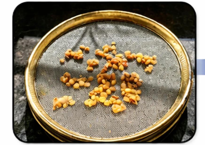
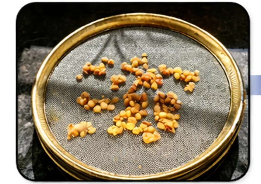
Figure 1. Seeds of Aegle marmelos
Figure 1: Seeds of Aegle Marmelos
Table 1: Taxonomical Classification of Aegle Marmelos
| Rank | Name |
|---|---|
| Subkingdom | Tracheobionta |
| Super Division | Spermatophyta |
| Division | Magnoliophyta |
| Class | Magnoliopsida |
| Subclass | Rosidae |
| Order | Sapindales |
| Family | Rutaceae |
| Genus | Aegle |
| Species | Aegle marmelos |
1.1.2 Habitat and distribution
Aegle marmelos is a semitropical species that flourishes at altitudes of approximately 1200 m above sea level, primarily in hill areas and dry forests. Its origin is traced to the Eastern Ghats and central India. The species is native to India, with a distribution range extending from the Himalayan foothills to West Bengal and across central and South Asia. It is found across the states of Uttar Pradesh, Bihar, Chhattisgarh, Madhya Pradesh, Uttarakhand, Jharkhand, the Deccan Plateau, and the Eastern coast, as well as in Myanmar and Sri Lanka [6].
1.1.3 Phytochemistry of Aegle marmelos
Numerous classes of bioactive compounds have been isolated from various parts of A. marmelos, including alkaloids, coumarins, terpenoids, fatty acids, and amino acids, through extensive phytochemical investigation [8].
1.2 Carbon dots
During the electrophoretic purification of single-walled carbon nanotubes in 2004, Xu et al. accidentally discovered carbon dots (CDs) zero-dimensional, fluorescent carbon-based nanomaterials with particle sizes generally below 10 nm [9]. Since their discovery, CDs have attracted considerable interest owing to their remarkable biocompatibility, low cytotoxicity, high water solubility, and exceptional optical properties, which distinguish them from conventional semiconductor quantum dots such as lead sulfide (PbS) and cadmium selenide (CdSe) [10,11].
CDs are structurally composed of a graphitic or amorphous carbon core decorated with various oxygen-containing functional groups including hydroxyl (–OH), carboxyl (–COOH), and carbonyl (C=O) which confer hydrophilicity and enable surface functionalization [12]. Their photoluminescence (PL) characteristics, including excitation-dependent emission, high quantum yield, and photostability, make them attractive for applications in drug delivery, bioimaging, fluorescence sensing, and photocatalysis [13].
1.2.1 Nitrogen-doped carbon dots (N-CDs)
Among strategies to enhance the physicochemical and optical properties of CDs, heteroatom doping particularly nitrogen doping has attracted considerable interest. Nitrogen atoms are incorporated into the carbon framework either endogenously through nitrogen-rich biomolecular precursors or exogenously via external sources such as urea, ethylenediamine, or ammonia [14]. Nitrogen incorporation significantly increases quantum yield, strengthens surface passivation, and introduces functional groups such as amine (–NH₂) and amide (–C=O–NH–) moieties, thereby broadening biological and analytical applications [15].
N-CDs have demonstrated superior performance compared to undoped CDs in fluorescent biosensing, heavy metal ion detection, antimicrobial activity, antioxidant activity, and targeted drug delivery [16,17]. Their low toxicity and ease of surface modification further position them as ideal candidates for theranostic applications in nanomedicine [18].
1.2.2 Aegle marmelos as a precursor for green synthesis of carbon dots
Among plant-based precursors investigated for the green synthesis of carbon dots, Aegle marmelos (L.) Corrêa seeds represent a particularly promising and understudied source [19]. These seeds are rich in bioactive compounds including aegeline, lupeol, rutin, and various phenolic acids that collectively provide a suitable carbon matrix for CD nucleation and growth [20]. Notably, aegeline, a nitrogen-containing alkaloid naturally present in the seeds, acts as an endogenous nitrogen donor during carbonization, enabling nitrogen incorporation into the carbon framework without any exogenous nitrogen precursor [21].
This endogenous nitrogen doping strategy represents a meaningful advance over conventional N-CD synthesis approaches, which typically rely on external nitrogen sources that raise concerns about residual toxicity and environmental impact [22]. The presence of C–N stretching vibrations in the FTIR spectra of the synthesized CDs provides spectroscopic evidence supporting aegeline-mediated endogenous nitrogen incorporation, consistent with documented mechanisms of heteroatom doping via alkaloid-rich precursors [23]. Hydrothermal carbonization of Aegle marmelos seed extract under controlled conditions is expected to yield fluorescent CDs with surface-passivated functional groups including –OH, –COOH, and –NH₂, suitable for applications in bioimaging, antioxidant evaluation, and antimicrobial research [24]. The present study therefore attempts to synthesize and characterize nitrogen-doped carbon dots from Aegle marmelos seeds using a straightforward, sustainable, and reagent-free hydrothermal method, with the aim of demonstrating the dual role of aegeline as both a carbon precursor and an endogenous nitrogen source [25].
RESULTS AND DISCUSSION
A number of physicochemical and optical characterization methods were applied to the synthesized carbon dots in order to verify their synthesis, gauge surface functionalization, and analyze fluorescence activity. The next sections cover the findings from fluorescence analysis, FTIR spectroscopy, UV-visible spectroscopy, and thin-layer chromatography.
2.1 UV-Visible Spectroscopy
The produced carbon dots were analyzed using UV-visible spectroscopy in the 190–600 nm wavelength range. Three distinctive absorption bands were visible in the spectra, which is in line with the optical characteristics of nitrogen-doped carbon dots made from precursors taken from plants.
The n→π* electronic transition of the C=O and C=N functional groups on the surface of the carbon dots is responsible for a noticeable absorption peak seen in the 335–350 nm region. In keeping with the function of Aegeline, an endogenous alkaloid of Aegle marmelos Cs, an in-situ nitrogen dopant during the carbonization process, the occurrence of this peak at a higher wavelength clearly suggests successful nitrogen incorporation into the carbon framework.
The presence of oxidized surface states on the carbon dot core is confirmed by a second absorption band seen in the 230–245 nm range, which corresponds to the n→π* transition of surface-bound carbonyl (C=O) groups.
The π→π* transition of conjugated C=C bonds within the aromatic carbon core is responsible for a third absorption zone between 200 and 230 nm (absorbance value: 0.2322), which indicates the creation of a graphitic or semi-graphitic carbon structure during heat carbonization.
The overall spectrum profile and the lack of noticeable absorption beyond 400 nm are consistent with previously documented UV-visible properties of nitrogen-doped carbon dots made from nitrogen-rich plant sources[26].
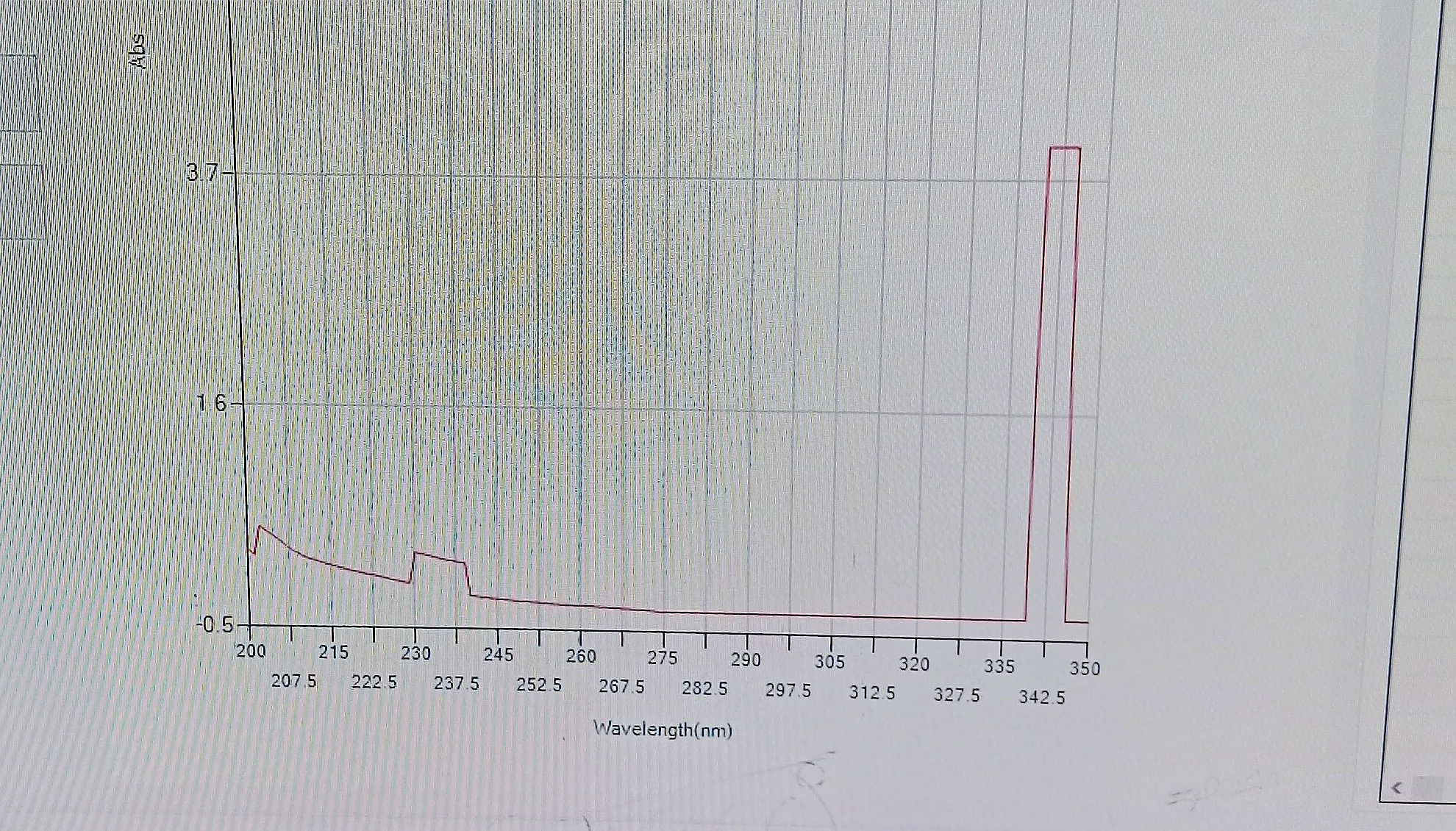
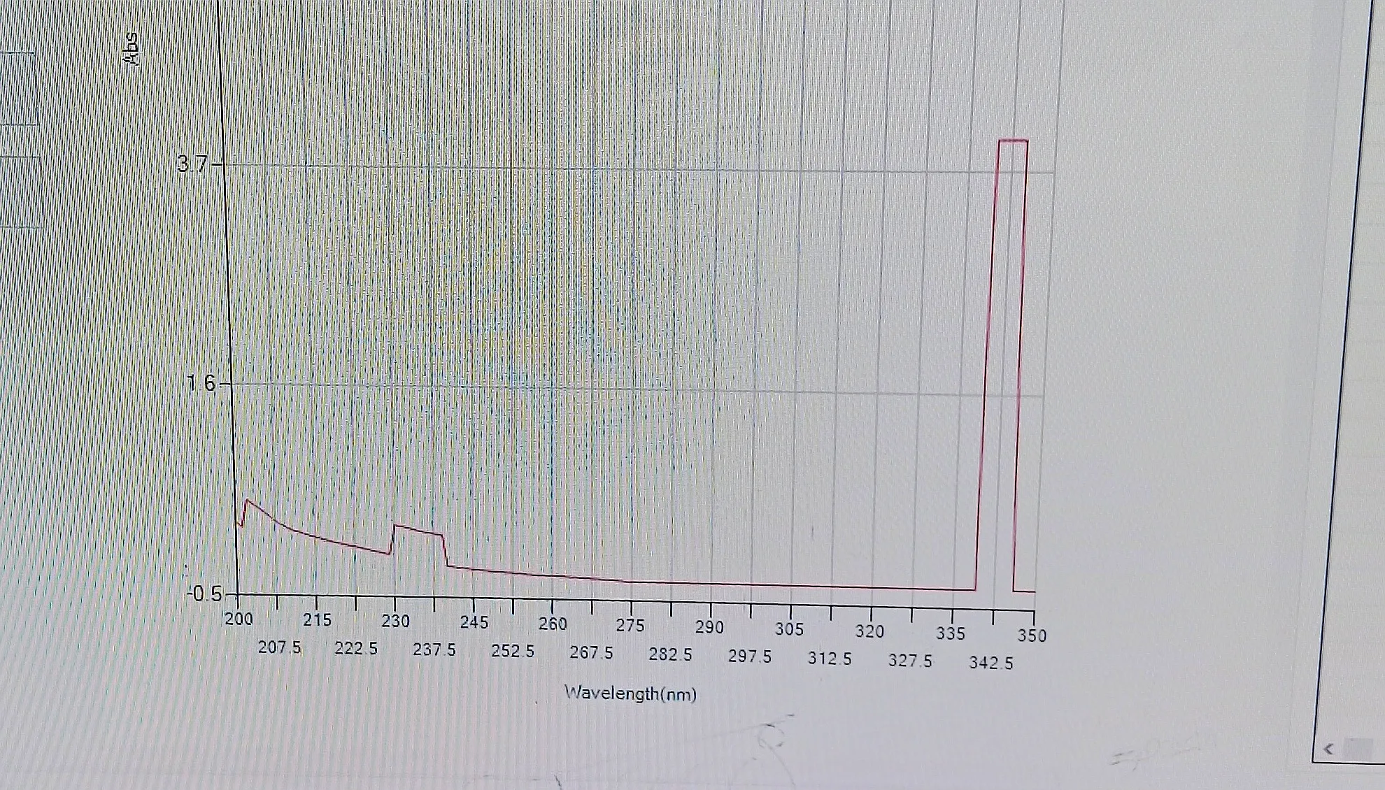
Figure 2. UV-Visible absorption spectrum of nitrogen-doped carbon dots (N-CDs) synthesized from Aegle marmelos seed extract
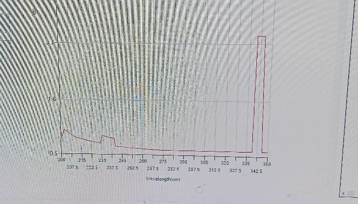
Figure 2: UV-Visible Absorption Spectrum of Nitrogen-Doped Carbon Dots (N-CDs) Synthesized from Aegle Marmelos Seed Extract
2.2 FTIR Spectroscopy
To determine the surface functional groups of the synthesized N-CDs (RSM-1), FTIR spectroscopy was used. The spectrum showed a number of distinctive absorption bands that confirmed the successful synthesis of carbon dots with surface functionalization and nitrogen doping.
The presence of hydrophilic functional groups on the surface of N-CDs is indicated by a large absorption band seen in the 3201–3454 cm⁻¹ area, which is ascribed to the overlapping O–H stretching vibrations of hydroxyl groups and N–H stretching of amine groups.
The presence of leftover carbon chains from the Aegle marmelos seed matrix is suggested by the absorption bands at 2922.16 cm⁻¹ and 2854.65 cm⁻¹, which correspond to asymmetric and symmetric C–H stretching vibrations of aliphatic methylene (–CH₂–) groups. Nitrile groups' C–N stretching vibrations are responsible for a prominent absorption peak at 2218.14 cm⁻¹, which offers compelling proof that nitrogen has been incorporated into the carbon framework. This demonstrates that Aegeline, the naturally occurring alkaloid found in Aegle marmelos seeds, mediates endogenous nitrogen doping.
Surface oxidation of the produced carbon dots is confirmed by the absorption band at 1724.36 cm⁻¹, which corresponds to C=O stretching of carboxyl (–COOH) groups. Nitrogen functionalization and the creation of an aromatic carbon core are further supported by the band at 1598.99 cm⁻¹, which is attributed to N–H bending vibrations of amine groups and/or C=C aromatic stretching. C–N stretching vibrations are represented by the band at 1278.81 cm⁻¹, whereas C–O stretching of ether or alcohol bonds is represented by the peak at 1066.64 cm⁻¹. The presence of a graphitic or semi-graphitic carbon structure in the synthesized N-CDs is confirmed by bands seen at 806.25 cm⁻¹ and 738.74 cm⁻¹, which are attributed to aromatic C–H out-of-plane bending.
Overall, the FTIR spectrum verifies that the synthesized N-CDs' surface contains –OH, –COOH, –NH₂, and C≡N functional groups, which are compatible with nitrogen-doped carbon dots made from nitrogen-rich plant-based precursors[26].
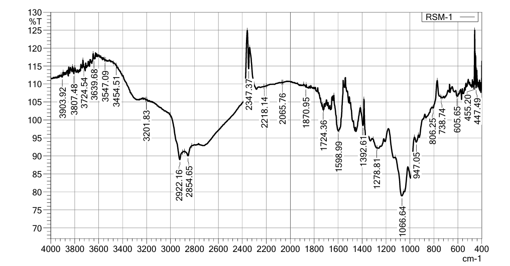
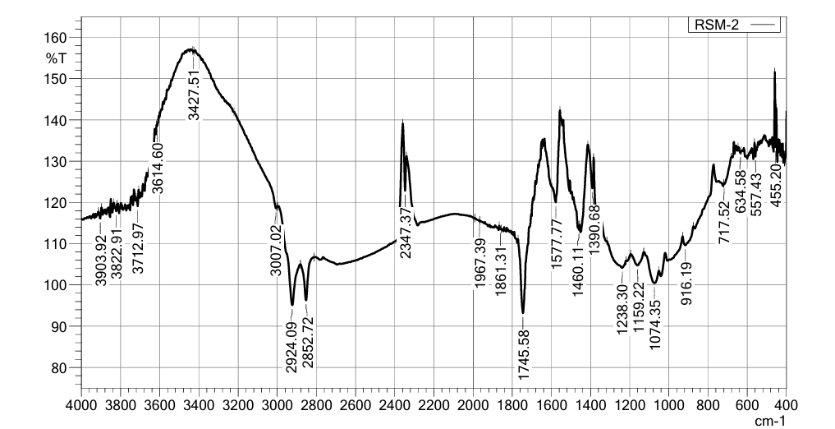
Figure 3: FTIR Spectrum of Carbon Dots (CDs)
2.3 Fluorescence Analysis
Toluene:Ethyl Acetate (7:3 v/v) was used as the mobile phase for TLC examination of the produced CDs on silica gel GF254 plates. The existence of fluorescent chemicals in the manufactured CDs was confirmed by seeing the produced plates under UV light at 366 nm, which displayed glowing patches.
There were two TLC runs. The detected luminous spots' Rf values were determined to be 0.50 and 0.625, respectively. The reported Rf value of psoralen, a naturally occurring furocoumarin found in Aegle marmelos, closely matches both values. The presence of psoralen-like furocoumarin structures in the synthesized CDs is further confirmed by the appearance of fluorescent spots under UV light at 366 nm, which is in line with previously published phytochemical findings for Aegle marmelos [27].
The observed blue emission at 365 nm is consistent with the characteristic photoluminescence behavior reported for carbon dots synthesized from plant-based precursors [28]. The green emission at 254 nm further suggests the presence of nitrogen-related surface states, supporting endogenous nitrogen incorporation from Aegeline present in Aegle marmelos seeds.
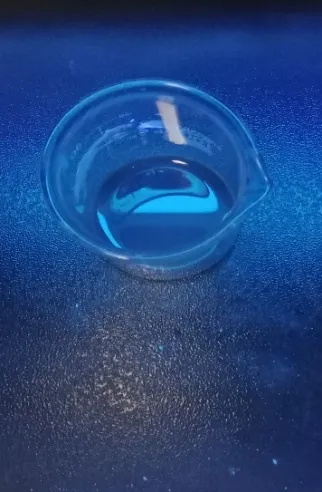
Figure 4b. Fluorescence emission of CDs under UV light at 365 nm (blue emission)
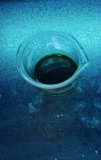
Figure 4a. Fluorescence emission of CDs under UV light at 254 nm (green emission)
2.4 Thin Layer Chromatography (TLC)
Toluene:Ethyl Acetate (7:3 v/v) was used as the mobile phase for TLC examination of the produced CDs on silica gel GF254 plates. The existence of fluorescent chemicals in the manufactured CDs was confirmed by seeing the produced plates under UV light at 366 nm, which displayed glowing patches.
There were two TLC runs. The detected luminous spots' Rf values were determined to be 0.50 and 0.625, respectively. The reported Rf value of psoralen, a naturally occurring furocoumarin found in Aegle marmelos, closely matches both values. The presence of psoralen-like furocoumarin structures in the synthesized CDs is further confirmed by the appearance of fluorescent spots under UV light at 366 nm, which is in line with previously published phytochemical findings for Aegle marmelos[29].
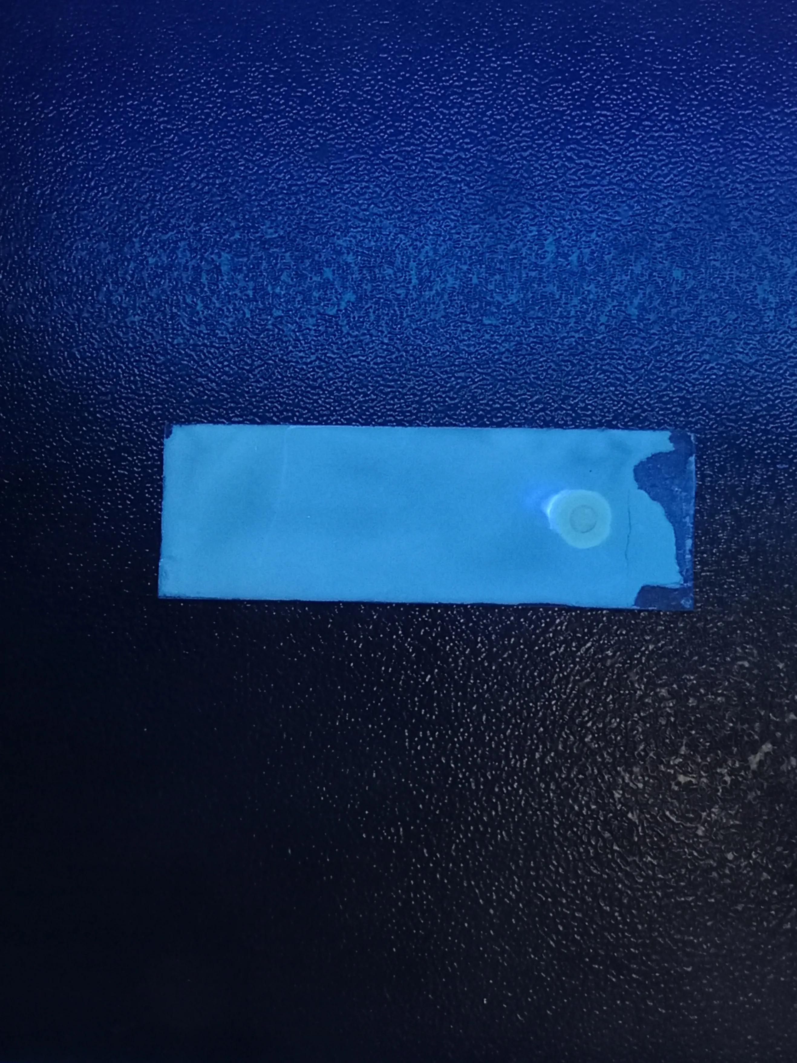
Figure 5: (A) & 5 (B)- TLC Plates Under UV 366 nm
2.5 Zeta Potential :
Using dynamic light scattering (Litesizer DLS 500) at 25°C in aqueous dispersion, the zeta potential of RSM-2 N-CDs was found to be −11.3 ± 0.5 mV. FTIR research confirms that the presence of carboxyl (–COOH) and hydroxyl (–OH) functional groups on the CD surface is responsible for the negative surface charge. The homogeneity of the produced nanoparticles is confirmed by a clear unimodal distribution peak at −11.0 mV and a limited standard deviation of 0.5 mV. The surface functionalization with nitrogen-containing moieties obtained from Aegeline (endogenous to Aegle marmelos seeds) contributes to the overall dispersibility of the N-CDs, despite the zeta potential value indicating early colloidal stability in aqueous medium. The moderate negative charge is in line with values previously reported for nitrogen-doped carbon dots made from plant-based[30].
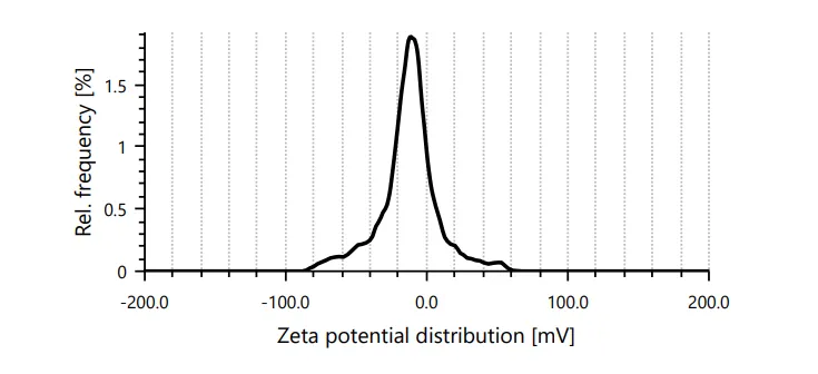
Figure 6: Zeta Potential Distribution of RSM-2 Nitrogen-Doped Carbon Dots Synthesized from Aegle marmelos Seeds
RESULT:
Table 2: Zeta Potential Analysis of Synthesis
| Mean zeta potential | -11.3 mV | Mean intensity | 638.4 kcounts/s |
|---|---|---|---|
| Standard deviation | 0.5 mV | Attenuator optical density | 2.5650 |
| Distributed peak | -11.0 mV | Conductivity | 0.344 mS/cm |
| Electrophoretic Mobility | -0.8800µm*cm/Vs | Transmittance | 50.5 % |
2.6 Transmission Electron Microscopy:
HR-TEM analysis was used to assess the shape and particle size distribution of the synthesized Aegle marmelos-derived carbon dots (AM-CDs). The AM-CDs were mostly quasi-spherical, moderately distributed, and slightly agglomerated in some areas, according to the TEM micrographs. Nanoscale dimensions ranging roughly from 4.1 to 7.5 nm were revealed by the particle size measurements derived from the TEM images. The successful creation of ultra-small carbon nanodots was confirmed by the estimated average particle size of the synthesized AM-CDs, which was 5.6 ± 1.4 nm. Good surface stability and efficient carbonization of the Aegle marmelos precursor are indicated by the observed nanosized structure and uniform shape. The produced AM-CDs are appropriate for sensing, imaging, and bioanalytical applications because of these nanoscale characteristics, which are advantageous for improved optical and fluorescent properties.
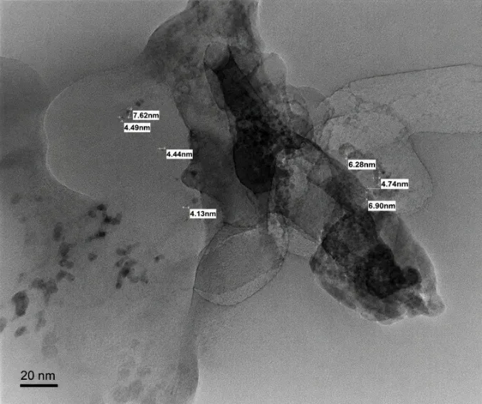
Figure 7: Combined HR-TEM Micrographs of Aegle Marmelos Carbon Dots (AM-CDs) Illustrating Particle Morphology, Agglomeration Behavior, and Particle Size Distribution in the Range of 4–8 nm.
2.7 Quantum Yield Analysis of Carbon Dots
A digital photo fluorometer and UV-visible absorption data were used to investigate the fluorescence behavior of the produced carbon dots. The experiment's fluorometer readings revealed fluctuations in fluorescence intensity, which suggested variability in emissive behavior under excitation circumstances. The fluorescence response was normalized with respect to the highest observed intensity value in order to conduct a relative quantum yield study based on the measured intensities.
The reference maximum emission was defined as the highest fluorescence intensity of 59 arbitrary units. Relative quantum yield values for the remaining samples ranged from 0.322 to 0.797. The UV-Vis spectrum shows a large absorption band in the ultraviolet range, which is frequently linked to surface defect states in carbon dots and π-π* transitions of aromatic carbon domains. The steady fluorescence emission and moderate optical activity are confirmed by the slow decrease in absorbance toward the visible range.
The produced carbon dots showed modest fluorescence characteristics; the sample with the highest relative quantum yield had a fluorescence intensity of 59 a.u. The successful production of fluorescent carbon nanodots is confirmed by the normalized relative quantum yield values, which indicate effective excitation-dependent emission behavior.
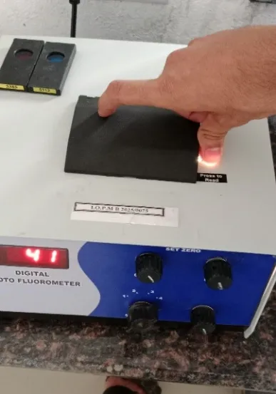
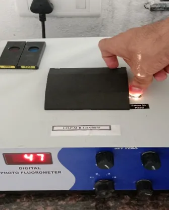
Figure 8: Fluorescence Intensity
Experimental Data
Table 3: Fluorescence Intensity and Relative Quantum Yield of AM-CDs
| Sample | Fluorescence Intensity | Relative Quantum Yield |
|---|---|---|
| Reading 1 | 47 | 0.797 |
| Reading 2 | 41 | 0.695 |
| Reading 3 | 59 | 1.0 |
| Reading 4 | 19 | 0.322 |
| Reading 5 | 34 | 0.576 |
| Reading 6 | 33 | 0.559 |
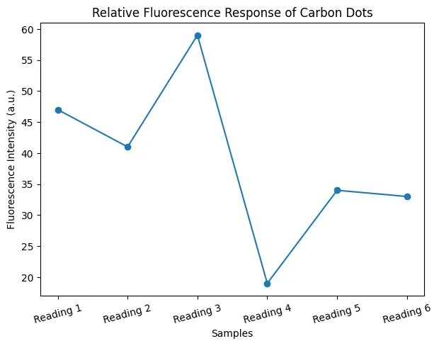
Figure 9: Relative Fluorescence Response of Carbon Dots
2.8 Anti-Microbial Activity:
The antimicrobial screening results clearly demonstrate that the Aegle marmelos-derived N-CDs possess concentration-dependent antibacterial activity against both Gram-positive and Gram-negative bacteria, as well as measurable activity against the probiotic reference strain Lactobacillus sp. The zone of inhibition increased progressively from 100 µg/mL to 200 µg/mL across all three organisms, confirming a clear dose-response relationship rather than a random or inconsistent effect an important indicator that the observed activity is genuinely attributable to the N-CDs rather than to assay artefacts.
The comparable zone sizes observed for S. aureus (Gram-positive) and E. coli (Gram-negative) at each tested concentration suggest that the N-CDs exhibit a broad-spectrum antibacterial effect, rather than selectivity toward one cell wall type. This is noteworthy because Gram-negative bacteria typically possess an additional outer lipopolysaccharide membrane that acts as a permeability barrier, often making them more resistant to nanomaterial-based antimicrobial agents compared to Gram-positive organisms. The fact that E. coli showed inhibition zones broadly similar to S. aureus indicates that the N-CDs are able to penetrate or disrupt both membrane architectures effectively.
The antimicrobial action of carbon dots is generally attributed to a combination of mechanisms: generation of reactive oxygen species (ROS) at the bacterial cell surface, direct physical interaction of the nanoscale particles with the cell membrane causing structural disruption, and interference with intracellular processes following membrane penetration. In the case of these N-CDs, the small particle size (~5.6 ± 1.4 nm) and negative surface charge (Zeta potential –11.3 mV) likely facilitate close interaction with the bacterial cell surface, while the nitrogen-doped functional groups (confirmed by FTIR C–N stretching at 1066–1074 cm⁻¹) may contribute additional surface reactivity that enhances antimicrobial efficacy compared to non-doped carbon dots.
The activity observed against Lactobacillus sp. is a point that merits careful interpretation. Since Lactobacillus is a beneficial, probiotic organism, inhibition at these concentrations suggests that the antimicrobial action of the N-CDs is relatively non-selective at the tested concentration range. This has direct implications for any proposed application of these N-CDs in food, cosmetic, or topical formulations, where preserving beneficial microflora may be desirable this would be an important consideration for future work, such as testing lower concentration ranges to identify a threshold that spares probiotic strains while retaining activity against pathogens.
Overall, these findings support the potential of Aegle marmelos-derived N-CDs as a green-synthesized, nitrogen-functionalized nanomaterial with promising broad-spectrum antibacterial activity, adding to the growing body of literature on plant-derived carbon dots as sustainable alternatives to conventional antimicrobial agents. Further work involving minimum inhibitory concentration (MIC) determination, time-kill kinetics, and mechanistic studies (e.g., ROS quantification, membrane integrity assays) would help substantiate and extend these preliminary findings.
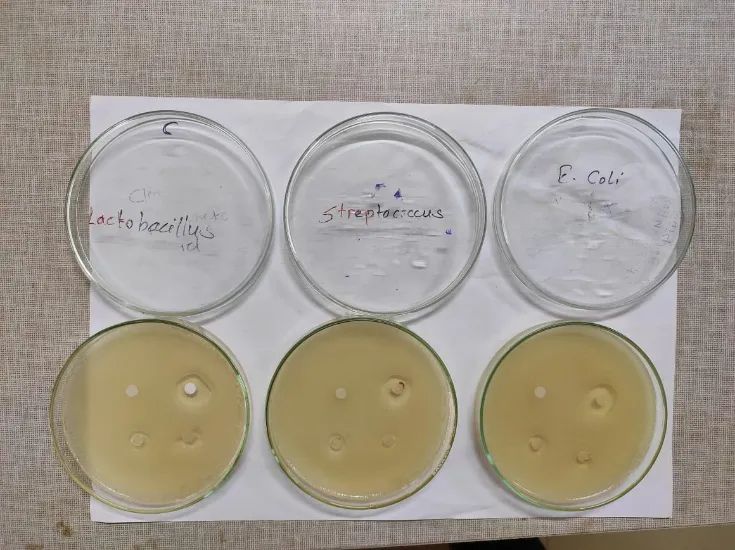
Figure 10: Antimicrobial Activity of Sample & Ag-CDs
CONCLUSION
This study reports the green synthesis of nitrogen-doped carbon dots (N-CDs) from Aegle marmelos seed extract using a simple, reagent-free, reflux-assisted hydrothermal carbonization process. The synthesis was accomplished without the use of any hazardous chemicals or external reagents, demonstrating a cost-effective and environmentally sustainable approach to fluorescent nanomaterial .
UV-visible spectroscopy confirmed the formation of carbon dots with characteristic absorption bands in the 200–350 nm range, corresponding to the π→π* and n→π* electronic transitions of the aromatic carbon core and surface functional groups, respectively.
FTIR spectroscopic analysis verified successful surface functionalization of the synthesized N-CDs, confirming the presence of –OH, –COOH, C=O, C–N, and N–H groups. A prominent C–N stretching band at 2218 cm⁻¹ provided direct spectroscopic evidence of endogenous nitrogen incorporation mediated by aegeline, a naturally occurring alkaloid present in Aegle marmelos seeds.
Fluorescence analysis demonstrated excitation-dependent photoluminescence behavior, with strong blue emission at 365 nm and green emission at 254 nm, consistent with the optical characteristics of nitrogen-doped carbon dots derived from plant-based nitrogen-rich precursors.
TLC analysis using toluene:ethyl acetate (7:3, v/v) as the mobile phase yielded fluorescent spots with Rf values of 0.50 and 0.625, consistent with the reported Rf of psoralen in Aegle marmelos, confirming the presence of psoralen-like furocoumarin structures in the synthesized N-CDs.
Zeta potential analysis revealed a value of −11.3 ± 0.5 mV with a narrow unimodal distribution peak at −11.0 mV, confirming moderate colloidal stability of the synthesized N-CDs in aqueous medium. The negative surface charge was attributed to the presence of carboxyl and hydroxyl functional groups on the CD surface.
HR-TEM analysis confirmed that the synthesized N-CDs were predominantly quasi-spherical in morphology, with particle sizes ranging from 4.1 to 7.5 nm and an average diameter of 5.6 ± 1.4 nm, consistent with the nanoscale dimensions characteristic of carbon dot materials.
Antimicrobial evaluation by the agar well diffusion method further demonstrated that the synthesized N-CDs possess notable antibacterial activity, with the maximum zone of inhibition (22 mm) recorded against Staphylococcus aureus, indicating pronounced efficacy against Gram-positive pathogens. This activity is attributed to the nitrogen-enriched surface functionalities and the abundant –OH/–COOH groups on the N-CD surface, which likely facilitate interaction with, and disruption of, bacterial cell membranes. The concurrent presence of psoralen-like furocoumarin moieties, inherited from the aegeline-rich seed matrix, may further contribute a synergistic phytochemical dimension to the observed antimicrobial effect.
Collectively, these findings confirm that Aegle marmelos seeds serve as a viable, sustainable, and cost-effective green precursor for the synthesis of fluorescent, antimicrobially active N-CDs. The dual role of aegeline as both a carbon source and an endogenous nitrogen dopant represents a meaningful advance over conventional synthesis routes that depend on external nitrogen precursors. Beyond their promising optical and structural properties, the demonstrated antibacterial performance broadens the translational relevance of these N-CDs, positioning them as strong candidates for future applications in bioimaging, nanosensing, antimicrobial drug delivery, and wound-care/antibacterial coating systems.
EXPERIMENTAL WORK
4.1 Collection and Authentication of Plant Material
Fresh Aegle marmelos (L.) Corrêa seeds were taken straight from the plant at a nearby farm. After properly cleaning the seeds with ethanol and distilled water to get rid of any remaining pollutants and surface impurities, they were shade-dried and crushed into a fine powder using a mechanical grinder. Until it was needed again, the powder was kept at room temperature in an airtight container.
4.2 Chemicals and Instruments
Throughout the investigation, double-distilled water and analytical-grade ethanol were utilized. Whatman No. 1 filter paper, a magnetic hot plate stirrer, and a UV cabinet for first fluorescence imaging were among the tools used. A Labman UV-Vis spectrophotometer was used for the UV-visible spectroscopic analysis. An analytical facility that was contracted out performed the FTIR spectroscopic analysis.
4.3 Synthesis of Carbon Dots
A straightforward reflux-assisted hydrothermal method was used to create carbon dots[31]. Aegle marmelos seed powder was extracted using ethanol and then water. The resultant extract was suspended at a 1:10 (w/v) ratio in double-distilled water. Using 10 g, 15 g, and 4 g of seed powder and corresponding water quantities of 100 mL, 150 mL, and 40 mL, three separate synthesis batches were conducted.
After heating the suspension for an hour at a moderate temperature on a magnetic hot plate stirrer, the heat source was removed. To promote condensation and stop solvent loss, the reaction vessel was covered with ice and sealed with aluminum foil. This allowed the reaction to continue for a total of four hours[32]. To get rid of the undissolved particles, the mixture was filtered through Whatman No. 1 filter paper. After that, the filtrate was concentrated by heating it on a hot plate until a dark residue formed, signifying that carbonization and carbon dot creation had been effective[33]. For additional characterisation, the concentrated product was reconstituted in distilled water and kept at 4°C.
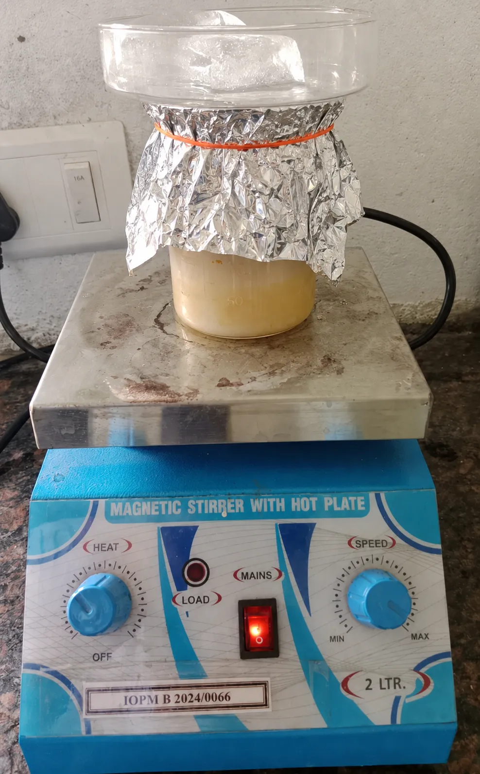
Figure 11. Synthesis setup showing Aegle marmelos seed extract undergoing reflux-assisted heating on a magnetic hot plate stirrer with ice-assisted condensation arrangement.
Figure 11: Synthesis Setup Showing Aegle Marmelos Seed Extract Undergoing Reflux-Assisted Heating on a Magnetic Hot Plate Stirrer with Ice-Assisted Condensation Arrangement.
4.4 Characterization
4.4.1 UV-Visible Spectroscopy
A Labman UV-Visible spectrophotometer operating in the 200–800 nm wavelength range was used to examine the optical absorption characteristics of the produced CDs. The blank was distilled water[34].
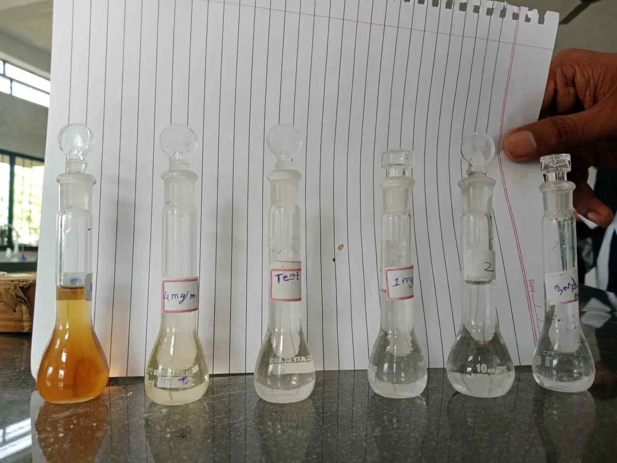
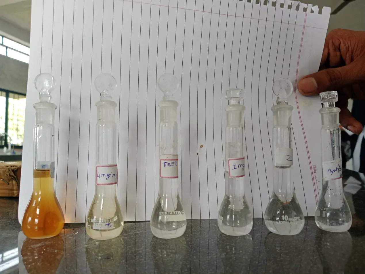
Figure 12. Different concentrations of synthesized CD solution were prepared for analysis
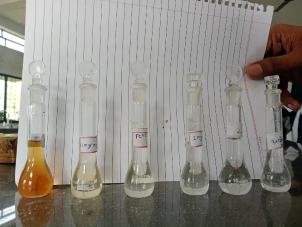
Figure 12: Different Concentrations of Synthesized CD Solution Were Prepared for Analysis
4.4.2 Fourier-Transform Infrared Spectroscopy (FTIR)
To determine the surface functional groups of the produced CDs, FTIR analysis was carried out. The spectra were captured at an external analytical laboratory between 400 and 4000 cm⁻¹[35].
4.4.3 Fluorescence Analysis
To qualitatively verify the generation of luminous carbon dots, the synthesized CDs' first fluorescence behavior was assessed at 365 nm in a UV cabinet. The produced CDs showed a strong blue emission with a faint green component, which is in line with the typical photoluminescence behavior described for nitrogen-doped carbon dots made from precursors sourced from plants[36].
4.4.4 Thin Layer Chromatography (TLC)
To evaluate the phytochemical components of the produced carbon dots and verify the existence of fluorescent chemicals, thin layer chromatography was used. TLC plates coated with silica gel served as the stationary phase, and a toluene solvent solution was employed: the mobile phase was ethyl acetate (7:3, v/v). The samples were developed in a closed chamber after being spotted onto the TLC plates (Fig. 5). The produced plates were examined under UV light at 366 nm (Fig. 5) and visible light (Fig. 6). The existence of psoralen, a naturally occurring furocoumarin, in Aegle marmelos was verified by the formation of a fluorescent spot under 366 nm UV light [37].
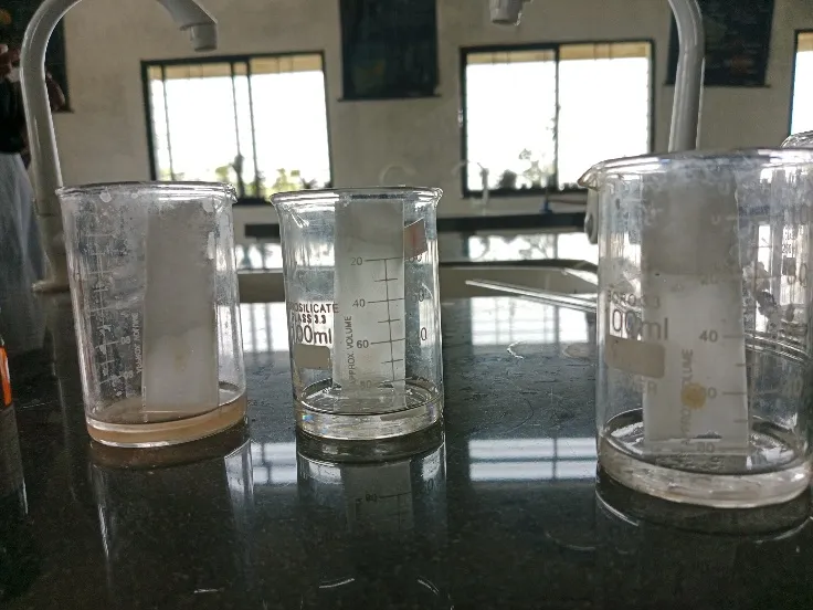
Figure 13: Sample Placed in Prepared Solution

Figure 14: TLC Plate Visible Light

Figure 15: TLC Chromatogram of Synthesized Carbon Dots Under UV Light (366 nm) Showing Blue Fluorescent Spots Across Three Solvent Systems: (A) Toluene:Ethyl Acetate (7:3), (B) Ethyl Acetate:Petroleum Ether (6:4), (C) Toluene:Petroleum Ether:Ethyl Acetate
4.4.5 Zeta Potential Analysis
Zeta potential measurement was used to assess the synthetic carbon dots' surface charge and colloidal stability. An apparatus based on dynamic light scattering (DLS) was used to conduct the analysis[38]. Electrostatic repulsion between particles is shown by a negative or positive Zeta potential value, which verifies the colloidal stability of the produced CDs. Using dynamic light scattering (Litesizer DLS 500) at 25°C in aqueous dispersion, the zeta potential of RSM-2 (Zeta) N-CDs was found to be −11.3 ± 0.5 mV. FTIR research confirms that the presence of carboxyl (–COOH) and hydroxyl (–OH) functional groups on the CD surface is responsible for the negative surface charge. The homogeneity of the produced nanoparticles is confirmed by a sharp unimodal distribution peak at −11.0 mV and a narrow standard deviation of 0.5 mV. The surface functionalization with nitrogen-containing moieties obtained from Aegeline (endogenous to Aegle marmelos seeds) contributes to the overall dispersibility of the N-CDs, despite the zeta potential value indicating early colloidal stability in aqueous medium. The moderate negative charge is in line with values previously reported for nitrogen-doped carbon dots made from plant-based precursors using green synthesis.
4.4.6 Antimicrobial Activity
4.4.6.1 Assessment of Antimicrobial Potential of N-CDs
To evaluate whether the synthesized N-CDs (derived from Aegle marmelos seed powder) exhibit antibacterial properties, the agar well diffusion technique was employed. Three organisms were selected to represent a range of microbial types relevant to this study: Staphylococcus aureus as a Gram-positive pathogen, Escherichia coli as a Gram-negative pathogen, and Lactobacillus sp. as a beneficial probiotic reference organism.
4.4.6.2 Media and Culture Preparation
Nutrient agar was used as the growth medium for S. aureus and E. coli, while MRS agar was employed for cultivating Lactobacillus sp., in line with its specific nutritional requirements. Media were sterilized via autoclaving (121°C, 15 lbs pressure, 15 minutes) prior to use. Once poured into sterile Petri dishes and allowed to solidify, plates were inoculated with the respective test organism, standardized to a turbidity of 0.5 on the McFarland scale, applied evenly across the agar surface with a sterile swab to establish a confluent bacterial lawn.
4.4.6.3 Well Formation and Sample Application
Using a sterile cork borer, four wells (approximately 6 mm in diameter) were cut into each inoculated plate, spaced sufficiently apart to avoid overlapping inhibition zones. The wells were designated as follows: one well received a solvent blank to serve as the negative control, while the remaining three were filled with N-CDs solutions prepared at graded concentrations of 100, 150, and 200 µg/mL. A consistent volume was dispensed into each well using a micropipette under aseptic conditions.
4.4.6.4 Incubation and Zone Measurement
Plates were allowed to stand for approximately 30 minutes at ambient temperature to permit initial diffusion of the samples into the agar before being transferred to an incubator set at 37°C for 24 hours. Following incubation, clear zones surrounding each well indicative of bacterial growth inhibition were measured in millimetres using a calibrated ruler, with the well diameter included in the total measurement. Each assay was carried out in triplicate to ensure reproducibility, and the resulting data were expressed as mean ± standard deviation.
Acknowledgements
The authors are grateful to the management and staff of S.C.S.M.S.S. Institute of Pharmacy, Maregaon, Yavatmal, for providing the necessary facilities to carry out this research work. The authors also acknowledge Dadasaheb Balpande College of Pharmacy, Besa, Nagpur, for providing instrumentation support for FTIR spectroscopy and Zeta potential analysis.
REFERENCES

Rizwan Qureshi, Saurav Jiddewar, Maithali Rathod, Prajwal Chavhan, Nilesh Chachda, Endogenously Nitrogen-Doped Carbon Dots Derived from Aegle Marmelos Seeds via Aegeline: Synthesis, Characterization, and Fluorescence Properties, Int. J. of Pharm. Sci., 2026, Vol 4, Issue 10, 2819-2836. https://doi.org/10.5281/zenodo.23466015
 10.5281/zenodo.23466015