

We use cookies to ensure our website works properly and to personalise your experience. Cookies policy
1PG, Research Scholler at University Institute of Pharmacy, Oriental University, Indore, MP.
2Professor at University Institute of Pharmacy, Oriental University, Indore, MP.
3Principal and Dean at University Institute of Pharmacy, Oriental University, Indore, MP.
4Principal and Professor at Oriental Collage of Pharmacy and Research, Oriental University, Indore, MP.
Myocardial infarction is associated with oxidative stress, myocardial cellular injury, and impairment of endogenous antioxidant defense. The present study was undertaken to evaluate the cardioprotective activity of hydroalcoholic root extract of Phyllanthus niruri against isoproterenol-induced myocardial infarction in Wistar rats. Animals were divided into normal control, isoproterenol control, propranolol-treated, and P. niruri extract-treated groups (200 and 400 mg/kg). Myocardial injury was assessed using serum cardiac biomarkers and tissue oxidative stress parameters. Isoproterenol administration markedly increased CK-MB from 116.24 ± 4.08 to 304.52 ± 8.96 U/L, LDH from 228.36 ± 6.14 to 536.48 ± 13.25 U/L, and AST from 82.46 ± 3.05 to 201.35 ± 5.84 U/L. Treatment with P. niruri at 400 mg/kg reduced CK-MB, LDH, and AST to 158.42 ± 5.12, 284.53 ± 9.34, and 106.28 ± 3.92 U/L, respectively. Isoproterenol also increased MDA from 1.82 ± 0.07 to 6.14 ± 0.18 nmol/mg protein, while decreasing SOD, CAT, and GSH levels. The 400 mg/kg extract restored these parameters to 10.84 ± 0.35 U/mg protein, 50.18 ± 1.62 U/mg protein, and 7.36 ± 0.28 µg/mg tissue, respectively, and reduced MDA to 2.56 ± 0.10 nmol/mg protein. Histopathological examination showed improved myocardial architecture with reduced necrosis, inflammation, and edema. These findings suggest that P. niruri root extract exhibits cardioprotective activity, possibly through reduction of oxidative stress and enhancement of endogenous antioxidant defense.
1.1Cardiovascular Diseases
Cardiovascular diseases (CVDs) are among the major causes of morbidity and mortality worldwide. The term cardiovascular diseases includes a group of disorders affecting the heart and blood vessels, with coronary artery disease, myocardial infarction, hypertension, and heart failure being some of the commonly encountered conditions.[1] The development of cardiovascular disorders is associated with several risk factors, including hypertension, dyslipidemia, diabetes mellitus, obesity, physical inactivity, unhealthy dietary habits, smoking, and increasing age. These factors may act individually or together to produce structural and functional changes in the cardiovascular system. [2]
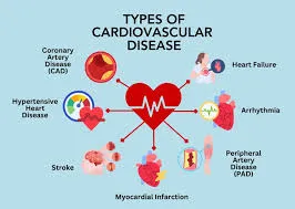
Figure 1: Cardiovascular Diseases (CVDs)
Among the different cardiovascular conditions, coronary artery disease is particularly important because it can reduce the blood supply to the myocardium. Prolonged impairment of coronary blood flow may result in myocardial ischemia and subsequent myocardial infarction. [3] Damage to cardiac tissue can affect myocardial contractility and may ultimately lead to complications such as arrhythmia, ventricular dysfunction, heart failure, and, in severe cases, death. Therefore, identification of effective approaches for prevention and management of myocardial injury remains an important area of cardiovascular research. [4]
1.2 Myocardial Infarction
Myocardial infarction (MI) is an acute condition characterized by myocardial injury resulting from an inadequate supply of oxygen to cardiac tissue. The imbalance between myocardial oxygen demand and oxygen supply can cause cellular dysfunction and, when sufficiently prolonged, irreversible damage to cardiomyocytes. The severity of myocardial injury depends on factors such as the duration and extent of ischemia and the ability of the myocardium to maintain adequate oxygenation. [5]
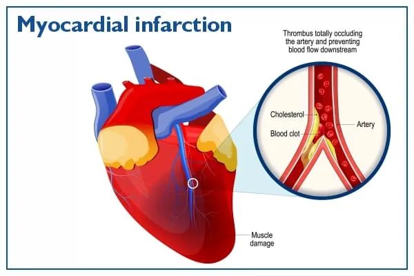
Figure 2: Myocardial Infarction (MI)
Oxidative stress plays an important role in the development of myocardial injury. Excessive generation of reactive oxygen species (ROS) can cause oxidation of cellular lipids, proteins, and other important biomolecules. Increased oxidative damage can disturb the normal structure and function of cardiomyocytes and may promote inflammatory responses and cellular death. Thus, oxidative stress is considered one of the important mechanisms involved in myocardial damage. [6]
Biochemical markers are commonly used to assess myocardial injury experimentally. Enzymes such as creatine kinase-MB (CK-MB), lactate dehydrogenase (LDH), aspartate aminotransferase (AST), and alanine aminotransferase (ALT) may show altered activity following cardiac tissue injury. [7] An increase in these markers reflects leakage of intracellular components into the circulation as a consequence of myocardial cell damage. Their estimation therefore provides useful biochemical evidence for evaluating the extent of cardiac injury and the protective effect of test treatments [8].
1.3 Experimental Model of Myocardial Infarction
Experimental animal models are widely used to investigate the mechanisms involved in myocardial injury and to evaluate potential cardioprotective agents. Such models provide an opportunity to study biochemical, physiological, and histopathological changes associated with cardiac damage under controlled experimental conditions. [9]
Isoproterenol (ISO), a synthetic β-adrenergic agonist, is commonly used to produce experimental myocardial injury in laboratory animals. Administration of a suitable dose of isoproterenol produces characteristic biochemical and structural changes resembling several features of myocardial infarction. The model is therefore useful for screening compounds and plant extracts with potential cardioprotective activity. [10]
The cardiotoxic effect of isoproterenol is associated with excessive β-adrenergic stimulation of the myocardium. Increased cardiac workload and oxygen demand, together with enhanced generation of reactive oxygen species, contribute to oxidative damage of myocardial tissue. Isoproterenol-induced oxidative stress can promote lipid peroxidation, damage cellular membranes, alter antioxidant defense mechanisms, and result in leakage of cardiac enzymes. These changes can be evaluated through biochemical parameters and histopathological examination of cardiac tissue. [11]
1.4 Oxidative Stress in Myocardial Injury
Oxidative stress occurs when the production of reactive oxygen species exceeds the capacity of the endogenous antioxidant defense system to neutralize them. The myocardium is particularly susceptible to oxidative damage because of its high metabolic activity and continuous requirement for oxygen. Excessive ROS generation can adversely affect membrane lipids, proteins, enzymes, and cellular structures. [12]
One of the important consequences of increased ROS generation is lipid peroxidation. Oxidation of membrane lipids can alter membrane integrity and permeability, resulting in cellular dysfunction and leakage of intracellular components. Malondialdehyde (MDA) is commonly used as an indicator of lipid peroxidation and provides an indirect measure of oxidative damage to cellular membranes. [13]
The body possesses several enzymatic and non-enzymatic antioxidant mechanisms to maintain redox balance. Superoxide dismutase (SOD) converts superoxide radicals into hydrogen peroxide, while catalase (CAT) helps in the decomposition of hydrogen peroxide into water and oxygen. [14] Reduced glutathione (GSH) is an important endogenous antioxidant that participates in the neutralization of reactive species and maintenance of cellular redox status. During myocardial injury, excessive oxidative stress may reduce the activity or availability of these antioxidant defenses. Therefore, evaluation of SOD, CAT, GSH, and MDA can provide useful information regarding oxidative changes associated with myocardial damage and the possible antioxidant effect of a test extract. [15]
1.5 Herbal Cardioprotection
Medicinal plants have traditionally been used for the management of several cardiovascular disorders and continue to attract interest as potential sources of pharmacologically active compounds. Plant-derived preparations contain diverse groups of phytoconstituents, including phenolic compounds, flavonoids, tannins, alkaloids, terpenoids, and other secondary metabolites. Several of these compounds have been reported to possess antioxidant, anti-inflammatory, and membrane-protective properties. [16]
Oxidative stress and inflammation are closely associated with the progression of myocardial injury. Consequently, plant extracts capable of reducing oxidative damage and supporting endogenous antioxidant defenses may have potential cardioprotective effects. Phenolic and flavonoid constituents are of particular interest because of their ability to interact with reactive oxygen species and contribute to the overall antioxidant activity of plant extracts.
The cardioprotective potential of medicinal plants can be evaluated using experimental models of myocardial injury. Changes in cardiac biomarkers, antioxidant enzymes, lipid peroxidation products, and myocardial histology provide useful evidence for determining whether a plant extract can attenuate cardiac damage. [17]
1.6 Phyllanthus niruri
Phyllanthus niruri is a medicinal plant belonging to the family Phyllanthaceae and has been traditionally used in various systems of herbal medicine. Different parts of the plant have been investigated for a range of pharmacological activities, including antioxidant, hepatoprotective, anti-inflammatory, and other biological effects. The plant contains several classes of secondary metabolites that may contribute to its pharmacological properties. [18]
Table 1: Botanical Profile of Phyllanthus niruri
| Parameter | Description |
|---|---|
| Botanical Name | Phyllanthus niruri Linn. |
| Family | Phyllanthaceae |
| Common Name | Bhui Amla |
| Habit | Annual herb |
| Distribution | Tropical regions |
| Part Used | Root/whole plant |
| Major Constituents | Flavonoids, lignans, phenolics |
| Major Uses | Liver, kidney and inflammatory disorders |
The antioxidant potential reported for P. niruri provides a rationale for investigating its possible protective effect against conditions in which oxidative stress plays an important role. Phytochemical constituents such as phenolic compounds, flavonoids, lignans, tannins, and other bioactive constituents have been associated with various biological activities of the plant. However, the pharmacological activity of a plant extract may vary according to the plant part, extraction solvent, geographical source, and method of preparation. [19]
In the present study, the root of P. niruri was selected for preparation of a hydroalcoholic extract. The use of a hydroalcoholic solvent system allows extraction of a broad range of polar and moderately polar phytoconstituents. Considering the reported antioxidant properties of P. niruri and the important role of oxidative stress in myocardial injury, investigation of its root extract in an experimental model of myocardial infarction is scientifically relevant. [20]
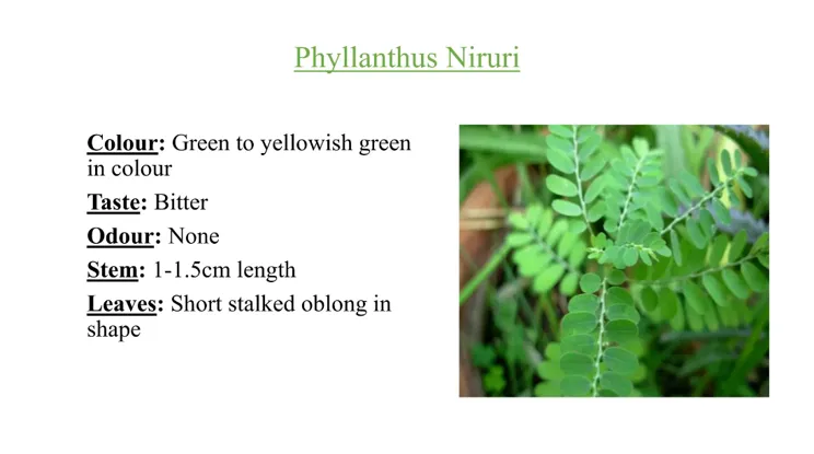
Figure 3: Phyllanthus niruri
1.7 Research Gap
Although Phyllanthus niruri has been investigated for several pharmacological activities, comparatively limited experimental information is available regarding the cardioprotective potential of its root extract in myocardial infarction. In particular, evidence concerning the effect of a hydroalcoholic root extract against isoproterenol-induced myocardial injury remains limited. Further experimental investigation is therefore required to determine whether the root extract can attenuate biochemical and oxidative changes associated with myocardial damage.
The present study was undertaken to evaluate the cardioprotective potential of the hydroalcoholic root extract of Phyllanthus niruri against isoproterenol-induced myocardial infarction in Wistar rats. The study focuses on biochemical markers of myocardial injury, antioxidant defense parameters, lipid peroxidation, and histopathological changes to obtain a comprehensive assessment of the protective effect of the extract.
MATERIALS AND METHODS
2.1 Plant Material
Roots of Phyllanthus niruri were collected from [indore], authenticated by [botanist], washed, shade-dried, powdered, and stored in an airtight container until extraction.
2.2 Instruments and Apparatus
Soxhlet apparatus, rotary evaporator, electronic balance, centrifuge, UV–Visible spectrophotometer, homogenizer, pH meter, microscope, microtome, water bath, refrigerator/freezer, micropipettes, and standard laboratory glassware were used.
RESULTS AND DISCUSSION
3.1 Effect of Phyllanthus niruri Root Extract on Cardiac Biomarkers
The cardioprotective effect of the hydroalcoholic root extract of Phyllanthus niruri was evaluated by estimating serum CK-MB, LDH, and AST levels. The administration of isoproterenol produced a marked elevation in all three cardiac biomarkers compared with the normal control group, indicating the development of myocardial injury.
Table 2: Effect of Phyllanthus niruri Root Extract on Cardiac Biomarkers
| Group | CK-MB (U/L) | LDH (U/L) | AST (U/L) |
|---|---|---|---|
| Normal Control | 116.24 ± 4.08 | 228.36 ± 6.14 | 82.46 ± 3.05 |
| Disease Control (ISO) | 304.52 ± 8.96 | 536.48 ± 13.25 | 201.35 ± 5.84 |
| Propranolol + ISO | 146.38 ± 5.24 | 268.42 ± 8.72 | 98.15 ± 4.22 |
| PN 200 mg/kg + ISO | 204.18 ± 6.34 | 356.27 ± 10.84 | 132.46 ± 4.98 |
| PN 400 mg/kg + ISO | 158.42 ± 5.12 | 284.53 ± 9.34 | 106.28 ± 3.92 |
RESULTS
The CK-MB level increased from 116.24 ± 4.08 U/L in the normal control group to 304.52 ± 8.96 U/L following isoproterenol administration. Treatment with P. niruri at 200 mg/kg reduced CK-MB to 204.18 ± 6.34 U/L, whereas the 400 mg/kg dose reduced it to 158.42 ± 5.12 U/L.
Similarly, LDH increased from 228.36 ± 6.14 U/L in the normal control group to 536.48 ± 13.25 U/L in the disease control group. Treatment with 200 and 400 mg/kg of the extract reduced LDH levels to 356.27 ± 10.84 and 284.53 ± 9.34 U/L, respectively.
AST also increased considerably after isoproterenol administration, from 82.46 ± 3.05 U/L to 201.35 ± 5.84 U/L. Treatment with the extract reduced AST to 132.46 ± 4.98 U/L at 200 mg/kg and 106.28 ± 3.92 U/L at 400 mg/kg.\
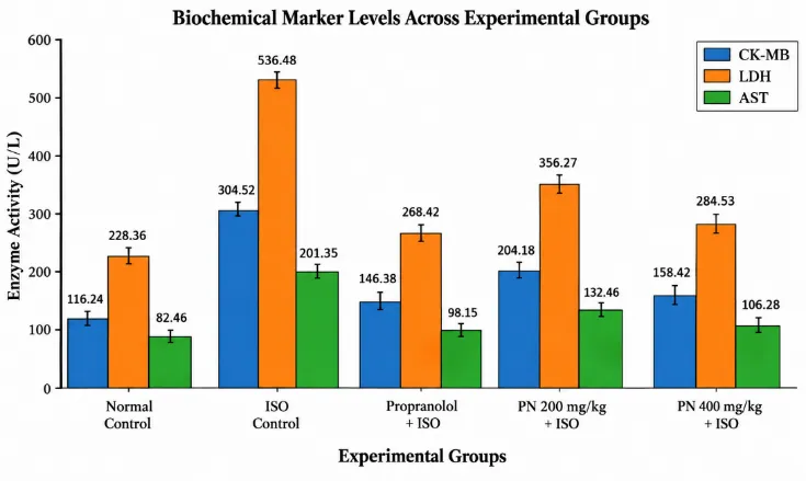
Figure 4: Effect of Phyllanthus Niruri Root Extract on Cardiac Biomarkers
DISCUSSION
The marked elevation of CK-MB, LDH, and AST in the disease control group indicates myocardial cellular injury and leakage of intracellular enzymes into the circulation. Pretreatment with P. niruri reduced these enzyme levels in a dose-dependent manner. Compared with the disease control group, the 400 mg/kg dose produced approximately 47.98% reduction in CK-MB, 46.96% reduction in LDH, and 47.22% reduction in AST. The higher dose therefore produced a greater reduction in cardiac enzyme leakage and showed values approaching those observed with propranolol.
3.2 Effect on Oxidative Stress and Antioxidant Parameters
Oxidative stress was evaluated by estimating MDA as a marker of lipid peroxidation and SOD, CAT, and GSH as indicators of endogenous antioxidant defense.
Table 3: Effect of Phyllanthus niruri Root Extract on Oxidative Stress Markers
| Group | MDA (nmol/mg protein) | SOD (U/mg protein) | CAT (U/mg protein) | GSH (µg/mg tissue) |
|---|---|---|---|---|
| Normal Control | 1.82 ± 0.07 | 12.64 ± 0.44 | 59.36 ± 1.92 | 8.58 ± 0.34 |
| Disease Control (ISO) | 6.14 ± 0.18 | 4.12 ± 0.18 | 22.48 ± 0.88 | 2.96 ± 0.12 |
| Propranolol + ISO | 2.18 ± 0.09 | 11.42 ± 0.36 | 53.26 ± 1.74 | 7.86 ± 0.26 |
| PN 200 mg/kg + ISO | 3.86 ± 0.13 | 8.24 ± 0.28 | 40.36 ± 1.32 | 5.94 ± 0.24 |
| PN 400 mg/kg + ISO | 2.56 ± 0.10 | 10.84 ± 0.35 | 50.18 ± 1.62 | 7.36 ± 0.28 |
RESULTS
Isoproterenol administration markedly increased MDA from 1.82 ± 0.07 to 6.14 ± 0.18 nmol/mg protein, indicating increased lipid peroxidation. Treatment with P. niruri reduced MDA to 3.86 ± 0.13 and 2.56 ± 0.10 nmol/mg protein at 200 and 400 mg/kg, respectively.
SOD activity decreased from 12.64 ± 0.44 U/mg protein in normal animals to 4.12 ± 0.18 U/mg protein after isoproterenol administration. Treatment with the extract increased SOD activity to 8.24 ± 0.28 U/mg protein at 200 mg/kg and 10.84 ± 0.35 U/mg protein at 400 mg/kg.
CAT activity decreased from 59.36 ± 1.92 to 22.48 ± 0.88 U/mg protein in the disease control group. The extract increased CAT activity to 40.36 ± 1.32 and 50.18 ± 1.62 U/mg protein at 200 and 400 mg/kg, respectively.
Similarly, GSH decreased from 8.58 ± 0.34 to 2.96 ± 0.12 µg/mg tissue following isoproterenol administration. Treatment with the extract increased GSH levels to 5.94 ± 0.24 and 7.36 ± 0.28 µg/mg tissue at 200 and 400 mg/kg, respectively.
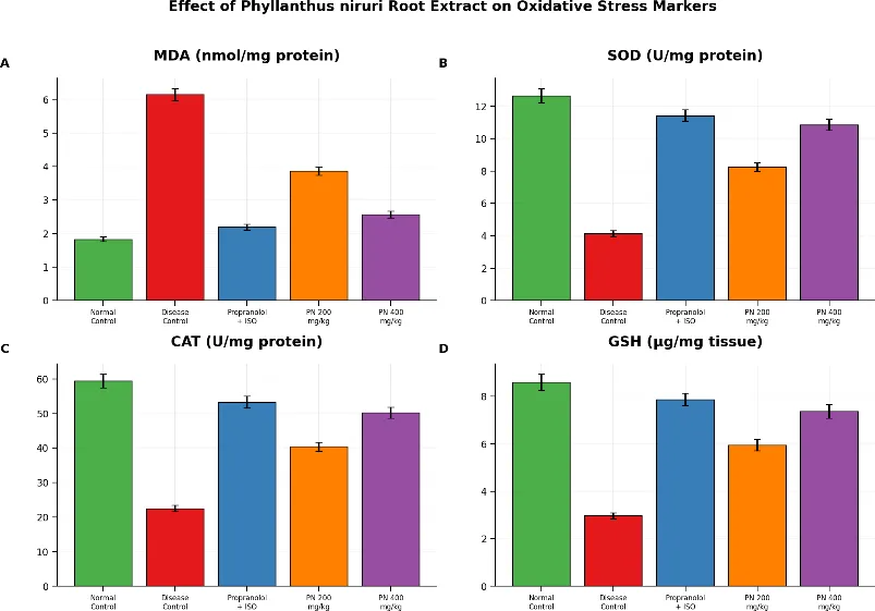
Figure 5: Effect on Oxidative Stress and Antioxidant Parameters
DISCUSSION
The increased MDA level in the isoproterenol-treated group indicates enhanced lipid peroxidation and oxidative damage to myocardial membranes. Treatment with P. niruri markedly reduced MDA, with the 400 mg/kg dose producing approximately 58.31% reduction compared with the disease control group.
The reduction in SOD, CAT, and GSH following isoproterenol administration indicates depletion of endogenous antioxidant defense. Treatment with P. niruri restored these parameters in a dose-dependent manner. At 400 mg/kg, SOD, CAT, and GSH reached 10.84 ± 0.35 U/mg protein, 50.18 ± 1.62 U/mg protein, and 7.36 ± 0.28 µg/mg tissue, respectively. These findings indicate that the extract was able to attenuate oxidative stress and improve the antioxidant status of myocardial tissue. The effect may be related to antioxidant phytoconstituents such as flavonoids, tannins, and phenolic compounds reported in the extract.
3.3 Histopathological Evaluation of Cardiac Tissue
Histopathological examination was performed to determine structural alterations in the myocardium following isoproterenol administration and treatment with P. niruri root extract.
Table 4: Histopathological Findings
| Group | Myocardial Structure | Necrosis | Inflammation | Edema |
|---|---|---|---|---|
| Normal Control | Normal architecture | Absent | Absent | Absent |
| Disease Control (ISO) | Severe myocardial damage | Severe | Severe infiltration | Severe |
| Propranolol + ISO | Near-normal structure | Mild | Mild | Mild |
| PN 200 mg/kg + ISO | Moderate protection | Moderate | Moderate | Mild |
| PN 400 mg/kg + ISO | Well-preserved structure | Mild/minimal | Mild | Minimal |
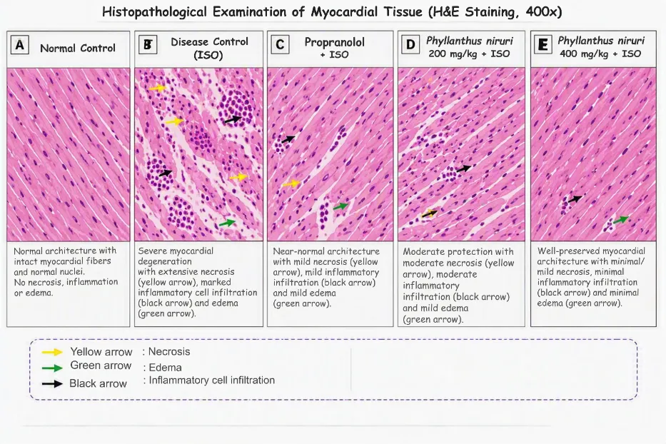
Figure 6: Histopathological Findings
RESULTS AND DISCUSSION
The normal control group showed normal myocardial architecture with intact cardiac muscle fibers and no apparent necrosis, inflammation, or edema. In contrast, the isoproterenol-treated disease control group showed severe myocardial damage accompanied by marked necrosis, inflammatory cell infiltration, edema, and degeneration of cardiac tissue.
Treatment with P. niruri root extract produced noticeable improvement in myocardial architecture. The 200 mg/kg dose showed moderate protection with a reduction in tissue damage, whereas the 400 mg/kg dose produced better preservation of myocardial structure with only mild or minimal necrosis, inflammation, and edema. The findings were broadly consistent with the biochemical results, particularly the reduction in cardiac biomarkers and MDA together with restoration of SOD, CAT, and GSH.
CONCLUSION
The present study demonstrated the cardioprotective potential of the hydroalcoholic root extract of Phyllanthus niruri against isoproterenol-induced myocardial injury in Wistar rats. Isoproterenol markedly increased CK-MB from 116.24 ± 4.08 to 304.52 ± 8.96 U/L and LDH from 228.36 ± 6.14 to 536.48 ± 13.25 U/L. Treatment with P. niruri at 400 mg/kg reduced CK-MB to 158.42 ± 5.12 U/L and LDH to 284.53 ± 9.34 U/L. The extract also reduced MDA from 6.14 ± 0.18 to 2.56 ± 0.10 nmol/mg protein and improved SOD, CAT, and GSH levels. Histopathological examination further showed improved myocardial architecture at the higher dose. Overall, the findings indicate that P. niruri root extract possesses promising cardioprotective activity, possibly through attenuation of oxidative stress and lipid peroxidation.
4. FUTURE SCOPE
Further studies should focus on identification and quantification of the active phytoconstituents responsible for the cardioprotective activity. Dose-response studies, molecular investigations involving oxidative stress and inflammatory pathways, and isolation of individual bioactive compounds may provide better understanding of the mechanism. Long-term toxicity, pharmacokinetic studies, and further validated animal studies are also required before considering potential clinical application.
REFERENCES


Mukesh Rathod, Dr. Jeevan Patel, Dr. Sudha Vengurlekar, Dr. Sachin Kumar Jain, Pharmacological Evaluation of Cardioprotective Activity of Phyllanthus Niruri Root Extract in Isoproterenol-Induced Myocardial Infarction in Experimental Rats, Int. J. of Pharm. Sci., 2026, Vol 4, Issue 10, 2862-2870. https://doi.org/10.5281/zenodo.23466326
 10.5281/zenodo.23466326