

We use cookies to ensure our website works properly and to personalise your experience. Cookies policy
1Research Scholar Sant Gahira Guru Vishwavidyalaya, Ambikapur, Dist. Sarguja (C.G.) & Govt. S.P.M. College –Sitapur, Surguja (C.G.) INDIA 497111.
2Department of Biotechnology, Government E. Raghavendra Rao PG Science College, Bilaspur, Chhattisgarh, India.
3Assistant Professor and Head, Department of Chemistry and Research Centre, Govt. S.P.M. College –Sitapur , Surguja (C.G.) INDIA 497111.
Senna surattensis is a medicinal plant belonging to the family Fabaceae, traditionally recognized for its potential therapeutic properties. The present study aims to investigate the anti-inflammatory and antimicrobial properties of Senna surattensis methanolic flower extract to explore its potential as a source of bioactive compounds. The anti-inflammatory potential of the extract can be evaluated using suitable in vitro assays, while its antimicrobial activity can be assessed against selected bacterial and fungal strains. The antibacterial activity is investigated against Staphylococcus aureus, Escherichia coli, Pseudomonas aeruginosa, and Bacillus subtilis. The antifungal activity is evaluated against Trichoderma asperellum and Candida parapsilosis. Antimicrobial efficacy can be assessed by measuring the zones of inhibition, while the minimum inhibitory concentration (MIC) can be determined to estimate the lowest concentration required to inhibit visible microbial growth. Anti-inflammatory activity was assessed by heat-induced albumin denaturation assay. The extract showed dose-dependent inhibition from 15.20% at 25 µg/mL to 65.44% at 200 µg/mL, while diclofenac sodium exhibited 24.94% to 82.78% inhibition. The activity is attributed to flavonoids and phenolics that stabilize albumin via hydrogen and hydrophobic interactions. Antibacterial activity was tested by agar well diffusion against S. aureus, B. subtilis, E. coli and P. aeruginosa. At 300 µg/mL, maximumzone of inhibition was observed for S. aureus (6.2 mm), followed by B. subtilis (5.6 mm), E. coli (5.4 mm) and P. aeruginosa (5.2 mm), with MIC values ranging from 61-170 µg/mL. In the present study, B. subtilis was found to be most
Medicinal plants have been used for centuries as an important source of therapeutic agents, and they continue to receive considerable scientific attention because of their diverse phytochemical composition and biological activities. The increasing prevalence of microbial infections and antimicrobial resistance, together with the adverse effects associated with prolonged use of some synthetic drugs, has increased interest in identifying safer and naturally derived therapeutic agents (Newman & Cragg, 2020)[1]. In this context, Senna surattensis (Burm.f.) H.S. Irwin & Barneby, also reported in earlier literature as Cassia surattensis, is a medicinally important plant belonging to the family Fabaceae. Different parts of the plant have been investigated for their phytochemical constituents and biological activities, suggesting that it may serve as a useful source of natural bioactive compounds (Fatima et al., 2012)[2].
Phytochemical studies have shown that S. surattensis contains several classes of secondary metabolites, including phenolic compounds, flavonoids, tannins, saponins and other constituents that may contribute to its pharmacological properties (Odebiyi et.al. 1978) [3]. Phenolic compounds and flavonoids are particularly important because of their ability to scavenge reactive oxygen species and reduce oxidative damage. Oxidative stress is closely associated with inflammation because excessive production of reactive oxygen species can activate inflammatory signaling pathways and promote tissue damage (Reuter et al., 2010) [4]. Therefore, the antioxidant potential of medicinal plants may provide an important basis for investigating their possible anti-inflammatory effects.
Experimental studies have provided evidence supporting the biological potential of S. surattensis. Uthaya Kumar et al. (2016)[5] reported that the seed extract of Cassia surattensis exhibited significant antioxidant and hepatoprotective effects in a paracetamol-induced toxicity model in mice. The treatment improved antioxidant status and reduced biochemical indicators associated with oxidative damage, which was attributed partly to the presence of polyphenolic constituents in the extract (Uthaya Kumar et al., 2016)[5]. Although antioxidant activity does not by itself establish direct anti-inflammatory activity, the close relationship between oxidative stress and inflammatory responses makes these findings relevant for further investigation of the anti-inflammatory potential of the plant (Reuter et al., 2010) [4].
The antimicrobial activity of S. surattensis is another important aspect of its pharmacological potential. Plant-derived phenolics, flavonoids, tannins and related compounds can affect microorganisms through several mechanisms, including alteration of cell membrane integrity, inhibition of microbial enzymes and interference with essential cellular processes (Cowan, 1999) [6]. Studies on Cassia surattensis have reported antimicrobial activity against several bacterial species, including Escherichia coli, Staphylococcus aureus, Bacillus subtilis, Klebsiella pneumoniae and Salmonella typhi (Sangetha et al., 2008) [7]. Such findings indicate that extracts of the plant may contain compounds capable of inhibiting the growth of both Gram-positive and Gram-negative bacteria.
The antifungal potential of S. surattensis has also been demonstrated experimentally. Sumathy et al. (2014)[8] reported that methanolic flower extract of Cassia surattensis exhibited antifungal activity against Aspergillus niger. The extract produced inhibition of fungal growth and morphological changes in the fungal structures, suggesting that phytochemicals present in the flowers may interfere with fungal development (Sumathy et al., 2014)[8]. The presence of phenolic and flavonoid compounds in the plant may partly explain this activity because these compounds have been widely associated with antimicrobial and antifungal effects (Cowan, 1999)[6].
Despite these promising findings, the biological potential of S. surattensis has not been investigated as extensively as that of several other medicinal plants of the genus Senna. In particular, there is a need for further studies that simultaneously examine its phytochemical composition, antimicrobial activity and anti-inflammatory potential. Investigation of different solvent extracts may also provide useful information because the type and concentration of extracted phytochemicals can vary considerably depending on the solvent used (Sasidharan et al., 2011)[9]. Therefore, systematic evaluation of S. surattensis extracts could help identify biologically active fractions and provide a scientific basis for their possible use as natural sources of antimicrobial and inflammation-modulating compounds.
MATERIAL AND METHOD
2.1 Collection and Preparation of Flower Extracts
Fresh flowers of Senna surattensis were collected from Surguja forest area and taxonomically confirmed by Prof. Rizwan Ullah, Principal and Department of Botany, Rajeev Gandhi Govt. P.G. College, Ambikapur, Chhattisgarh stste, India. The shade-dried flowers were coarsely powdered. About 100 g of dried powder was packed in a Soxhlet thimble and extracted sequentially based on polarity gradient method described by Singh and Bargah et al., 2026. The material was first defatted/extracted with ethyl acetate (400-500 mL), then re-extracted with chloroform for 6 hours, and lastly with methanol (400 mL) for 8 hours. Each extract was filtered, concentrated in a rotary evaporator under reduced pressure at 40-45 °C, and air-dried to yield solvent-free residues. Among them, the methanolic extract was taken forward for subsequent experimental analysis.
2.2 Anti-inflammatory test
The in-vitro anti-inflammatory activity of the Senna surattensis extract was evaluated using the inhibition of heat-induced protein denaturation method, with slight modifications to the previously described procedure (Mizushima & Kobayashi, 1968; Williams et al., 2008)[10-11]. Diclofenac sodium was used as the standard anti-inflammatory drug.
Different concentrations of the plant extract (25, 50, 100, 150 and 200 µg/mL) were prepared using an appropriate solvent. The reaction mixture consisted of 1 mL of plant extract at each concentration and 1 mL of 1% bovine serum albumin (BSA) solution. The pH of the reaction mixture was adjusted to approximately 6.3 using phosphate-buffered saline. A similar reaction mixture containing the respective concentration of the standard drug was prepared for comparison. The control contained 1 mL of BSA solution and 1 mL of the corresponding solvent without plant extract.
The prepared reaction mixtures were incubated at 37 ± 1°C for 15 min and subsequently heated at 70°C for 5 min to induce protein denaturation. After cooling the samples to room temperature, the absorbance was measured at 660 nm using a UV–visible spectrophotometer. The experiment was performed in triplicate, and the mean absorbance values were calculated.
The percentage inhibition of protein denaturation was calculated using the following equation:
% Inhibition = ((Abs Control- Abs Sample/Test)/Abs Control) ×100
where (Ac) represents the absorbance of the control and (As) represents the absorbance of the sample or standard drug.
A higher percentage inhibition of protein denaturation was considered indicative of greater in-vitro anti-inflammatory potential. The concentration-dependent activity of the S. surattensis extract was compared with that of diclofenac sodium. The IC₅₀ value, defined as the concentration of extract required to inhibit 50% of protein denaturation, was estimated from the concentration–response relationship.
2.3 Anti-microbial activity assay
2.3.1 Antibacterial assay
The Antibacterial activity was checked by following Zone Inhibition Method (Kirby-Bauer method). The MHA plates were inoculated by spreading with 100 μl of Bacterial culture, Bacillus subtilis, Staphylococus aureus, Pseudomonas aeruginosa and E. coli in different plates (Inoculum was prepared by adjusting 0.5 McFarland Unit - Approx cell density (1.5 X 10⁸ CFU/mL from Mueller- Hinton Broth) and followed by placing the discs containing 5 μl of different concentration (0 to 300 µg/ml). One disc in each plate was loaded with solvent alone which served as vehicle control and Ciprofloxacin disc (3μg) was taken as positive control. The plates of Bacteria were incubated (Basil Scientific Corp. India- Incubator) at 37 °C for 24 hrs. The clear zones created around the disc were measured and recorded
2.3.2 Antifungal Test
The Antifungal activity was checked by following Zone Inhibition Method (Kirby-Bauer method). The SDA plates were inoculated by spreading with 100 μl of Fungal culture, Trichoderma asperellum and C. parapsilosis (Inoculum was prepared by adjusting 0.5 McFarland Unit - Approx cell density (1.5 X 10⁸ CFU/mL from Sabouraud dextrose broth) and followed by placing the discs containing 5 μl of different concentration (0 to 400 mg/ml). One disc in each plate was loaded with solvent alone which served as vehicle control and Amphotericin B disc (180 μg) was taken as positive control. The plates of Trichoderma asperellum were incubated (Basil Scientific Corp. India- Incubator) at 37 °C for 48 hrs. The clear zones created around the disc were measured and recorded.
RESULT
3.1. Anti-inflammatory activity
The S. surattensis extract showed a concentration-dependent inhibition of heat-induced protein denaturation. The percentage inhibition increased from 15.20% at 25 µg/mL to 65.44% at 200 µg/mL. Diclofenac sodium, used as the reference drug, exhibited higher inhibition at all tested concentrations, increasing from 24.94% to 82.78% (Table 1). The progressive increase in inhibition with increasing extract concentration indicates the potential anti-inflammatory activity of S. surattensis. However, the activity of the plant extract was lower than that of the standard drug under the tested conditions (fig 1).
| Table 1: In-vitro anti-inflammatory activity of Senna surattensis Methanolic Flower extract by inhibition of protein denaturation | |||
|---|---|---|---|
| Treatment | Concentration (µg/mL) | Absorbance at 660 nm (Mean ± SD) | Inhibition of protein denaturation (%) |
| Control | — | 0.842 ± 0.012 | 0 |
| S. surattensis extract | 25 | 0.714 ± 0.015 | 15.2 |
| 50 | 0.623 ± 0.011 | 26.01 | |
| 100 | 0.501 ± 0.013 | 40.5 | |
| 150 | 0.382 ± 0.010 | 54.63 | |
| 200 | 0.291 ± 0.009 | 65.44 | |
| IC50 | - | 143.80 | |
| Diclofenac sodium (Standard drug) |
25 | 0.632 ± 0.012 | 24.94 |
| 50 | 0.497 ± 0.010 | 40.97 | |
| 100 | 0.351 ± 0.008 | 58.31 | |
| 150 | 0.231 ± 0.007 | 72.57 | |
| 200 | 0.145 ± 0.006 | 82.78 | |
| IC50 | - | 88.78 | |
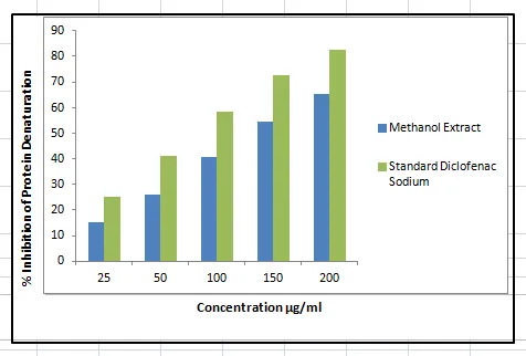
Figure 1: Percentage Inhibition of Anti-Inflammatory Activity
The IC50 values were determined by linear regression analysis. The methanolic flower extract of Senna surattensis exhibited an IC50 of 143.80 µg/mL, whereas the standard drug Diclofenac sodium showed an IC50 of 88.78 µg/mL. The lower IC50 value of Diclofenac sodium indicates its higher anti-inflammatory potency. Although the extract showed comparatively lower activity, its dose-dependent increase in percentage inhibition 15.20% to 65.44% demonstrates significant anti-inflammatory potential (fig. 2).
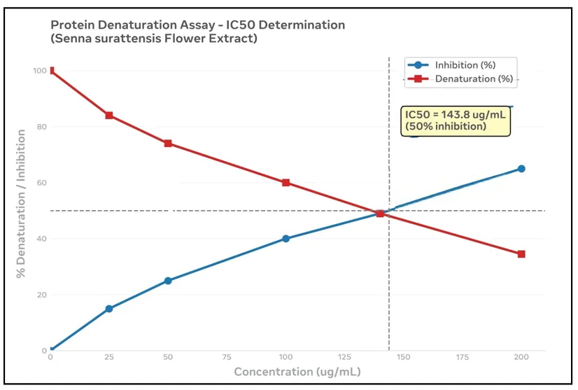
Figure 2: Determination of IC50 Value of Methanolic Flower Extract of Senna Surattensis by Protein Denaturation Inhibition Assay.
3.2 Anti-Microbial Activity
3.2.1 Antibacterial Test
The methanolic extract of Senna surattensis flowers was screened for antibacterial potential using agar well diffusion against four clinical isolates including Gram-positive Staphylococcus aureus and Bacillus subtilis, and Gram-negative Escherichia coli and Pseudomonas aeruginosa. Activity was assessed in terms of MIC and zone of inhibition at 100, 200 and 300 µg/mL, using Ciprofloxacin as reference standard. The extract displayed dose-dependent, broad-spectrum inhibition against all tested pathogens.
(i) Minimum Inhibitory Concentration (MIC):
The antibacterial activity of Senna suratensis was evaluated by determining the minimum inhibitory concentration (MIC) against selected Gram-positive and Gram-negative bacterial strains. MIC values varied between 61 ± 0.27 and 170 ± 0.01 µg/mL. B. subtilis was found to be most sensitive with the lowest MIC 61 ± 0.27 µg/mL, followed by P. aeruginosa 108 ± 0.78 µg/mL and S. aureus 150 ± 0.03 µg/mL. E. coli showed highest resistance 170 ± 0.01 µg/mL. A lower MIC value indicates higher antibacterial efficacy. In the present study, B. subtilis was found to be most susceptible with an MIC of 61 µg/mL, indicating that S. surattensis extract is most potent against B. subtilis (Table 2). Hence, the susceptibility pattern was: B. subtilis > P. aeruginosa > S. aureus > E. coli.
Zone of inhibition (ZOI)
The extract displayed a concentration-dependent increase in antibacterial activity across 100-300 µg/mL against all tested pathogens. At the highest concentration (300 µg/mL), the zone of inhibition was maximum for Staphylococcus aureus at 6.2 mm, followed by Bacillus subtilis at 5.6 mm, Escherichia coli at 5.4 mm and Pseudomonas aeruginosa at 5.2 mm, indicating the highest susceptibility of S. aureus among the tested strains (fig.3,4,5)
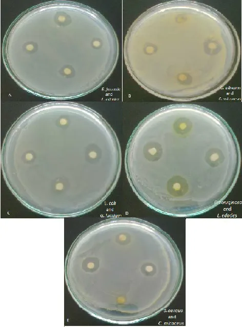
Figure 3: Concentration Dependent Antibacterial Activities of Senna surattensis Methanolic Flower Extract
E. coli
Table 2: MIC Value of Extract Against Different Bacterial Strains
| Bacterial strain | MIC (µg/mL) | Zone of Inhibition (mm) | |||
|---|---|---|---|---|---|
| 100 µg/mL | 200 µg/mL | 300 µg/mL | Ciprofloxacin | ||
| Staphylococcus aureus | 150 ± 0.03 | 2.8 | 3.2 | 6.2 | 3.1 |
| Escherichia coli | 170 ± 0.01 | 3.9 | 4.2 | 5.4 | 2.8 |
| Pseudomonas aeruginosa | 108 ± 0.78 | 3.5 | 4.3 | 5.2 | 3.1 |
| Bacillus subtilis | 61 ± 0.27 | 3.1 | 4.2 | 5.6 | 2.7 |
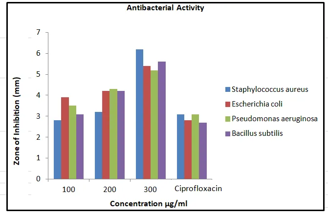
Figure 4: Anti-bacterial Activities of Senna surattensis Methanolic Flower Extract at Different Concentration
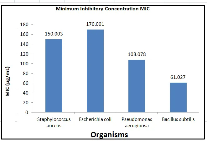
Figure 5: Minimum Inhibitory Concentration of Flower Extract of Senna Surattensis Against the Test Bacteria
3.2.2 Antifungal activity
The methanolic extract of Senna surattensis exhibited antifungal activity against Trichoderma asperellum and Candida parapsilosis. The observed antifungal activity suggests that the extract may contain bioactive compounds capable of inhibiting fungal growth (Fig) .
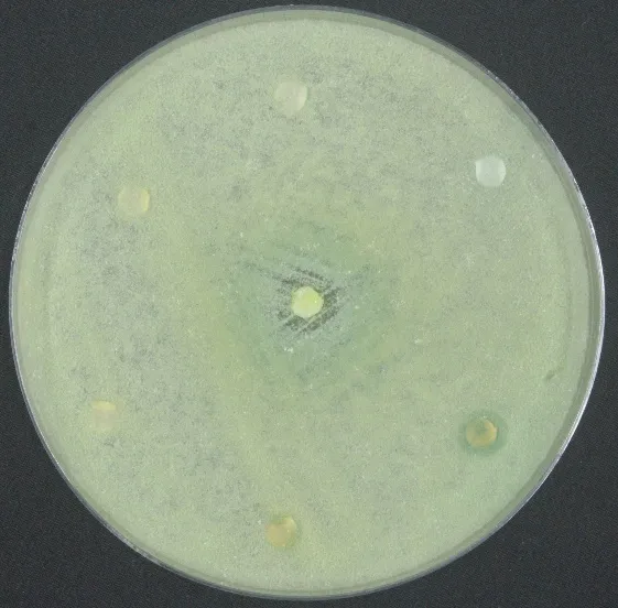
Figure 6: Antifungal Activity of Senna surattensis Against the Test Fungal
T.asperellum
DISCUSSION
Inflammation is a multifaceted physiological response triggered by tissue injury, microbial infection or immunological stimuli, associated with protein denaturation, altered vascular permeability and mediator release. Denaturation of endogenous proteins has been implicated as a major contributor to inflammatory and arthritic pathologies, leading to generation of auto-antigens and exacerbation of tissue damage (Bargah, et al.2025)[12]. Therefore, compounds capable of inhibiting protein denaturation and stabilizing proteins are considered promising candidates for the development of novel anti-inflammatory therapeutics.
The higher potency of diclofenac, as evidenced by its lower IC50 value, is anticipated since it is a purified synthetic non-steroidal anti-inflammatory drug (NSAID) with a defined mechanism involving direct cyclooxygenase (COX) inhibition (Vincent Ngouana et al.2025)[13]. In contrast, the substantial activity demonstrated by the crude flower extract is significant, considering it is a complex mixture of diverse secondary metabolites rather than a single isolated entity (Salha Mahdjar, et al. 2020)[14] . The anti-inflammatory effect of S. surattensis can be attributed to its abundance of bioactive phytochemicals such as flavonoids, phenolic compounds, anthraquinones and tannins, which are characteristic constituents of Senna species. Flavonoids including quercetin, kaempferol and rutin are documented to exert anti-inflammatory actions through inhibition of prostaglandin biosynthesis, lysosomal membrane stabilization, and prevention of protein denaturation via hydrophobic interactions and hydrogen bonding with albumin (Krishnaveni, et al 2021)[15]. Therefore, the presence of these polyphenolic compounds in the methanolic flower extract likely contributes to albumin stabilization and protection against heat-induced denaturation.
The present study investigates the antimicrobial potential of the methanolic flower extract of Senna surattensis, with particular attention to its antibacterial activity against Staphylococcus aureus, Escherichia coli, Pseudomonas aeruginosa and Bacillus subtilis, and its antifungal activity against Trichoderma asperellum and Candida parapsilosis. Medicinal plants are widely investigated as potential sources of antimicrobial compounds because they contain diverse secondary metabolites that may inhibit microbial growth through different mechanisms (Cowan, 1999; Zouine et al., 2024)[6,16]. However, antimicrobial activity depends on several factors, including plant species, plant part, extraction solvent, extract concentration and the susceptibility of the tested microorganism (Zouine et al., 2024; Bargah et al.2014)[16-17].
The use of methanol as an extraction solvent can help recover a range of polar and moderately polar phytochemicals, including phenolic compounds and flavonoids. Some plant-derived compounds have been associated with antimicrobial effects through mechanisms such as disruption of microbial membranes and interference with cellular enzymes and proteins (Cowan, 1999; Doughari et al.2008)[6,18]. Nevertheless, the presence of specific phytochemicals in S. surattensis should be confirmed through phytochemical screening or analytical techniques rather than inferred solely from antimicrobial activity.
The antibacterial findings can be considered in the context of previous medicinal-plant research. Bargah (2020)[19] reviewed the antimicrobial properties of plant-derived compounds and described several mechanisms through which they may inhibit microorganisms. Research on Moringa oleifera has also investigated its antibacterial potential and highlighted variation in reporteSd outcomes among studies (Pareek et al., 2023; van den Berg & Kuipers, 2022) [20-21]. Similarly, research on Piper betle has documented antibacterial and antifungal potential, supporting further investigation of medicinal plants as sources of biologically active compounds (Nayaka et al., 2021; Bargah et al.2024)[22-23]. These studies provide context for investigating S. surattensis, but direct comparisons require similar extraction procedures, test concentrations, microbial strains and assay conditions.
The observed antifungal activity of the methanolic extract against T. asperellum and C. parapsilosis may indicate the presence of compounds that affect fungal growth. Plant-derived compounds have been investigated for antimicrobial properties against a range of microorganisms, although their effects vary depending on the fungal species and extract composition (Vincent Ngouana et al.2025; Nayaka et al., 2021)[13,22]. Because T. asperellum and C. parapsilosis are different organisms, activity against one should not be assumed to predict the same response in the other. Quantitative measurements, including inhibition-zone diameters and minimum inhibitory concentrations (MICs), are needed to evaluate and compare their susceptibility.
The findings should also be interpreted in view of methodological limitations. Disc-diffusion results can be influenced by the ability of extract constituents to diffuse through agar, so inhibition-zone size alone does not provide a definitive measure of antimicrobial potency. Standardized dilution methods are therefore useful for determining MIC values and comparing antimicrobial activity under controlled conditions (Zouine et al., 2024)[16]. Published studies should be compared cautiously because differences in extraction solvents, concentrations, microbial strains and experimental procedures can produce different results (van den Berg & Kuipers, 2022, Vaou et al..2021)[21,24].
The observed antimicrobial activity supports further investigation of S. surattensis as a potential source of bioactive compounds. Future work should establish quantitative MIC values, confirm activity using standardized methods, characterize the extract's phytochemical composition and evaluate its safety. These studies would help determine whether the extract has reproducible antimicrobial effects and whether particular constituents contribute to the observed activity.
CONCLUSION
The methanolic flower extract of Senna surattensis demonstrated dose-dependent anti-inflammatory activity by inhibiting protein denaturation with an IC50 of 143.8 µg/mL, compared to diclofenac sodium 88.78 µg/mL. The extract prevented protein denaturation in a dose-dependent manner, though less potent than diclofenac. This supports its traditional use in inflammatory disorders and indicates presence of bioactive anti-inflammatory metabolites requiring further characterization. It also exhibited broad-spectrum antibacterial potential against S. aureus, E. coli, P. aeruginosa and B. subtilis with MIC values of 61-170 µg/mL, showing highest efficacy against B. subtilis. The observed zones of inhibition 5.2-6.2 mm at 300 µg/mL, support its antimicrobial nature. The exhibition of antimicrobial activity by the plant against some pathogenic bacteria and fungi is an indication that the plant can be used for sourcing novel antimicrobial substances for drug development for the treatment of dysentery, typhoid fever, wound and unitary tract infections and mycotic diseases. The activity of this plant against a wider range of bacteria and fungi should be investigated. Overall, the extract represents a promising natural source for anti-inflammatory and antimicrobial agents, warranting further phytochemical isolation and toxicity validation for pharmaceutical development.
6. CONFLICTS OF INTEREST
The authors declare that they have no conflict of interest in the publication.
REFERENCES

Heeralal Singh, Arun Kumar Kashyap, Rohit Kumar Bargah, Evaluation of Anti-inflammatory, Anti-microbial Activity of Senna Suratensis Flower Extract, Int. J. of Pharm. Sci., 2026, Vol 4, Issue 10, 4882-4892. https://doi.org/10.5281/zenodo.23829883
 10.5281/zenodo.23829883