
We use cookies to ensure our website works properly and to personalise your experience. Cookies policy
Sage Institute of Technology Pharmacy, Sanjeev Agrawal Global Education University, Bhopal.
Neurodegenerative disorders such as Alzheimer’s disease are strongly associated with oxidative stress and cholinergic dysfunction, leading to progressive cognitive decline. The present study evaluated the antioxidant and anti-cholinesterase potential of methanolic fruit extract of Coccinia grandis (MECG) in Wistar rats. Phytochemical screening confirmed the presence of alkaloids, flavonoids, phenolics, tannins, and terpenoids. The extract exhibited significant antioxidant activity with a total phenolic content of 89.31 mg GAE/g and flavonoid content of 70.13 mg QE/g. In vitro assays demonstrated dose-dependent DPPH radical scavenging (IC?? = 503.52 µg/mL) and acetylcholinesterase inhibition (IC?? = 430.85 µg/mL). Acute toxicity testing revealed safety up to 2000 mg/kg. In vivo evaluation using a D-galactose-induced oxidative stress model showed that MECG improved catalase (CAT) and superoxide dismutase (SOD) activities in a dose-dependent manner, with 200 mg/kg extract approaching the efficacy of vitamin E. These findings suggest that Coccinia grandis fruit extract possesses neuroprotective potential through combined antioxidant and cholinergic modulation, supporting its possible role in managing oxidative stress-related neurodegenerative disorders such as Alzheimer’s disease.
Neurodegenerative disorders such as Alzheimer’s disease (AD) are characterized by progressive cognitive decline, memory impairment, and neuronal dysfunction. Among the various pathogenic mechanisms implicated, oxidative stress and cholinergic dysfunction play pivotal roles in disease progression. Excessive generation of reactive oxygen species (ROS) leads to lipid peroxidation, protein oxidation, and DNA damage, ultimately compromising neuronal survival [1,2]. Simultaneously, reduced activity of acetylcholine, a key neurotransmitter involved in learning and memory, further exacerbates cognitive deficits [3]. Hence, therapeutic strategies that combine antioxidant defense with cholinergic modulation are of considerable interest in the management of neurodegenerative conditions.
Medicinal plants have long been recognized as valuable sources of bioactive compounds with neuroprotective potential. Coccinia grandis (family: Cucurbitaceae), commonly known as ivy gourd, is widely distributed in tropical regions and traditionally used in Indian medicine for its antidiabetic, anti-inflammatory, and hepatoprotective properties [4,5]. Phytochemical investigations reveal that its fruits and leaves are rich in flavonoids, phenolic compounds, and alkaloids, which are known to exert antioxidant and neuroprotective effects [6,7]. Flavonoids and phenolic compounds, in particular, have been shown to modulate oxidative stress, prevent neurotoxic protein aggregation, and support mitochondrial function, thereby offering neuroprotection [8,9].
Despite its extensive ethnomedicinal use, limited scientific evidence exists regarding the role of Coccinia grandis in modulating cholinergic function and oxidative stress in the central nervous system. The present study was designed to evaluate the cholinergic and antioxidant potential of fruit extract of Coccinia grandis in Wistar rats. By assessing its ability to influence acetylcholinesterase activity and oxidative stress markers, this investigation aims to provide mechanistic insights into the neuroprotective efficacy of Coccinia grandis. The findings may contribute to the development of plant‑based therapeutic interventions for neurodegenerative disorders, particularly those involving cholinergic deficits and oxidative damage.
MATERIAL AND METHODS
Extraction of fruit
The fruits of Coccinia grandis were purchased from road side local vendors of Bhopal and have been identified and authenticated by botanist at RB Science, Bhopal. The fruits were washed with tap water, shade dried and powdered. The powdered plant material (65 g) was defatted with hexane at room temperature for 24 h. The marc was dried and packed in the extractor of the soxhlet apparatus and extracted with methanol by hot continuous extraction process for approximately 7.5 h. The extract was filtered and the solvent was evaporated on water bath. The extract obtained were collected and placed in desiccator to get rid of the excess moisture content [10-12]. The dried extract was stored in desiccator for phytochemical screening and pharmacological evaluation. The methanolic extract was tested for the presence of various phytochemicals using the reported methods [13,14].
Total Phenolic Content
The total phenolic content in the leaf extract was determined quantitatively using Folin-Ciocalteu reagent method, using gallic acid as the reference standard. For total phenolic content determination, 200 μL of sample was mixed with 1.4 mL purified water and 100 μL of Folin-Ciocalteu reagent. After incubating at room temperature for 15 min, 300 μL of 20% Na2CO3 aqueous solution was added and the mixture was allowed to incubate at room temperature for 2 h. The absorbance of the solution was measured at 760 nm with a UV-Vis spectrophotometer [15]. Results were expressed as milligrams of gallic acid equivalent (GAE) per 100 g of the dry sample.
Quantitative estimation of total flavonoids
Determination of total flavonoids content was based on aluminium chloride method. 50 mg quercetin was dissolved in 50 ml methanol, and various aliquots of 25- 150μg/ml were prepared in methanol. 0.1 g of dried extract was extracted with 10 ml methanol, filtered, and make up the volume up to 100 ml. One ml (1mg/ml) of this extract was for the estimation of flavonoid by incubation with sodium nitrate solution. 1 ml of 2% AlCl3 methanolic solution was added to 1 ml of extract or standard and allowed to stand for 60 min at room temperature; absorbance was measured at 415 nm [16].
In vitro antioxidant activity of extract by DPPH assay
The antioxidant action of the extract was determined using 2,2-Diphenyl-1-picrylhydrazyl (DPPH) radical scavenging assay. The free radical scavenging activity of the synthesized molecules was measured in terms of hydrogen donating or radical scavenging ability using the stable radical DPPH. The test samples (100 𝜇L, 100-500 µg/mL) were prepared in DMSO and were mixed with 1.0 mL of DPPH solution and filled up with methanol to a final volume of 4 mL. Absorbance of the resulting solution was measured at 517 nm in a visible spectrophotometer [17,18].
Evaluation of Anti-cholinesterase Activity
Preparation of test solutions
The extract suspended in required volume of dimethylsulfoxide (DMSO) and diluted using phosphate buffer solution (PBS) (pH = 7.8) for the final range of concentrations (100-500 µg/mL). Ellman’s reagent was used for AChE catalyzed hydrolysis. In fresh 96-well microplates, each well was filled with 40 μL of 50 mM Tris-HCl buffer pH 8.0, 20 μL of test sample solution, 100 μL of Ellman’s reagent (prepared by dissolving 18.5 mg reagent in 10 mL of buffer), and 20 μL of AChE solution (prepared by dissolving 0.26 U/mL enzyme in 15 mL buffer). The contents were mixed and incubated at 25°C for 15 min. The absorbance was measured at 412 nm. Now, 20 μL of 15 mM acetyl thiocholine (ACTh) (prepared by dissolving in water) was added as a substrate to the wells and the plate was incubated for 20 min at 37°C. Blank determination was done without test samples to obtain 100% AChE activity [19,20]. The absorbance was measured at 412 nm and the percentage AChE inhibition was calculated using formula:
%AChE inhibition = (Ablank-Asample)/Ablank *100
Acute Toxicity
A total of three animals were used which received a single oral dose (2000mg/kg). Animals were observed individually at least once during the first 30 min after dosing, periodically during the first 24 h and daily thereafter for a period of 14 days [21].
In vivo antioxidant action
Experiment Design
The animals (Swiss albino rat) were divided into 5 groups of each 6 animals each.
|
Group |
Type |
Treatment |
|
1 |
Normal Control |
Normal Diet and Water |
|
2 |
Toxic Control |
25 mg/Kg D-galactose for 21 days |
|
3 |
Standard |
25 mg/kg of vitamin E orally for 21 days + 25 mg/kg of D-galactose i.p for 21 days |
|
4 |
MECG (100 mg/Kg) |
Low dose of extract for 21 days + 25 mg/kg of D-galactose i.p for 21 day |
|
5 |
MECG (200 mg/Kg) |
High Dose for 21 days + 25 mg/kg of D-galactose i.p for 21 day |
Catalase assay
The animal of each group were sacrificed on 22nd day. The livers were dissected out immediately, washed with ice-cold saline and homogenized in phosphate buffer solution (PH 7.4) to prepare 10% homogenates. The homogenate were centrifuged at 1700 rpm for 10 min at 4°C using a refrigerated centrifuged [22].
The homogenate were assayed for antioxidant capacity by catalase measurement method. Catalase activity can be determined in homogenate using Aebi’s method. Fifty microliter of the lysate is added to a cuvette containing 2 mL of phosphate buffer (pH 7.0) and 1 mL of 30 mM H2O2. Catalase activity is measured at 240 nm for 1 min using spectrophotometer. The molar extinction coefficient of H2O2, 43.6 M cm-1 was used to deter- mine the catalase activity. One unit of activity is equal to 1 mmol of H2O2 degraded per minute and is expressed as units per milligram of protein.
Superoxide Dismutase (SOD) assay
To 50 µL of the tissue homogenate, 75 mM of Tris–HCl buffer (pH 8.2), 30 mM EDTA and 2 mM of pyrogallol are added. An increase in absorbance is recorded at 420 nm for 3 min by spectrophotometer. One unit of enzyme activity is 50% inhibition of the rate of autooxidation of pyrogallol as determined by change in absorbance/min at 420 nm. The activity of SOD is expressed as units/mg protein [22].
Statistical Analysis of data
The results will be expressed as mean ± SEM of six independent experiments. Statistical significance between the groups would be evaluated by one-way analysis of variance (ANOVA) followed by Dunett’s test. A significance value of p<0.05 was considered as statistically significant.
RESULTS AND DISCUSSION
The extraction yield of the fruit powder using petroleum ether and methanol was found to be 1.08 and 17.95% respectively. The preliminary phytochemical screening suggests the presence of alkaloids, saponin, tannins, phenolics, terpenoids, sterols, and flavonoids in the fruit of the plant.
The total phenolic content of methanolic extract of Coccinia grandis was found to be 89.31 GAE mg/g. The total flavonoid content of methanolic extract of Coccinia grandis was found to be 70.13 QE mg/g. The in vitro anti-oxidant action was studied by DPPH radical inhibition assay and the IC50 of DPPH radical inhibition by the extract of Coccinia grandis was found to be 503.52 µg/mL (Figure 1).
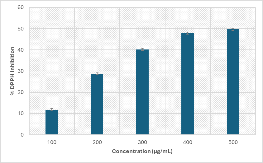
Figure 1. %DPPH radical inhibition by extract
The in vitro anti-cholinesterase activity was studied by Ellman’s method and the extract was able to inhibit AChE activity dose dependently from 16.63 to 58.66%. The IC50 was calculated from inhibition percentage and was found to be 430.85 µg/mL (Figure 2).
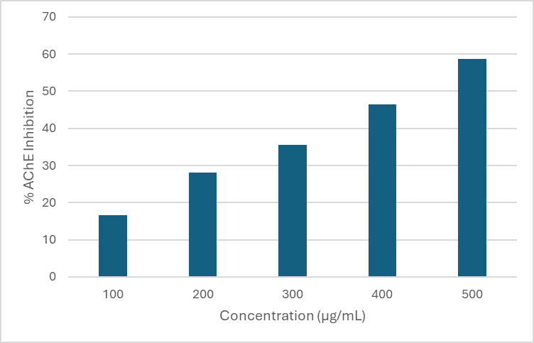
Figure 2. Percent AChE Inhibition
Acute Toxicity
The acute toxicity test was performed by using extract at concentration of 2000 mg/kg to the test animal, administered orally. No animal died and hence the dose of upto 2000 mg/Kg was considered to be safe. As none of the animals died, the LD50 was considered to be more than 2000 mg/Kg and any dose less than 2000 mg/Kg would be considered for evaluation of wound healing action.
In vivo antioxidant action
It was noticed that the SOD and CAT activities were considerably reduced in the toxic control group in comparison to the normal control group. The ability to scavenge oxygen-free radicals in vivo was also correspondingly reduced. It indicates that intraperitoneal injection of D-galactose solution could successfully create a mouse model of oxidative damage. The activities of antioxidant enzymes in mice were improved after the use of the standard drug as well as the extract (Figure 3 and 4).
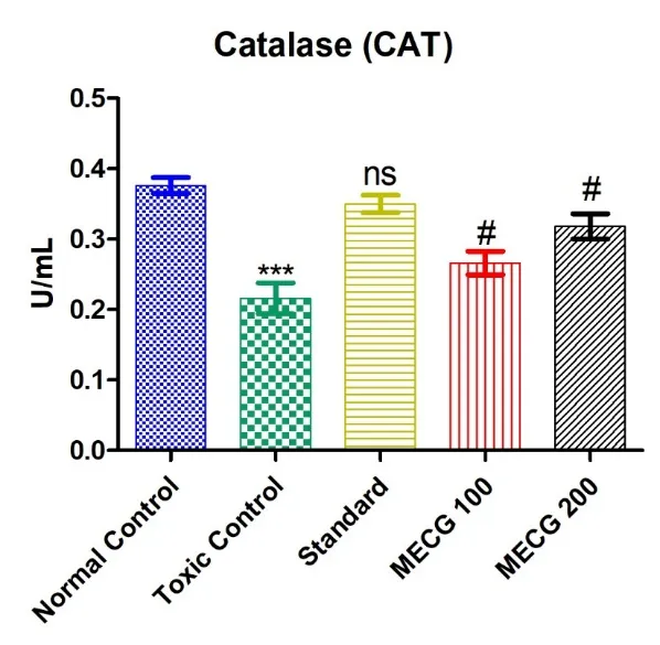
Figure 3. Effect on catalase activity. ns – not significant (p<0.001), significant difference ***p<0.05, #p<0.01 in comparison to control group
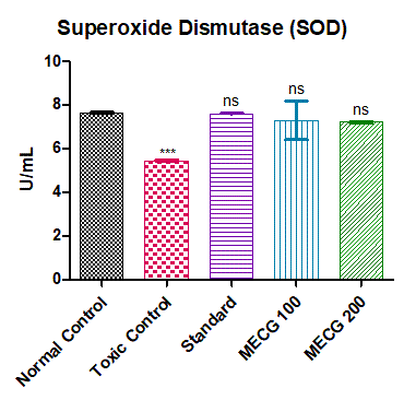
Figure 4. Effect on Superoxide dismutase activity. ns – not significant (p<0.001), significant difference ***p<0.05 in comparison to control group
The Toxic control (Group II) shows a marked reduction in both CAT and SOD compared to control, confirming oxidative stress. Group III (standard) is not significantly different (very close to control) suggesting restoring of the enzyme levels nearly to control, showing protective effect. MECG 100 (Group IV) provides partial protection but remains significantly lower than control whereas MECG 200 (Group V) shows better restoration than MECG 100, approaching control values. This shows dose-dependent protective effects of MECG, with 200 mg/kg approaching the efficacy of the standard treatment. Catalase (CAT) is an enzymatic antioxidant widely distributed in all animal tissues. It decomposes hydrogen peroxide and protects the tissue from highly reactive hydroxyl radical. Catalase activity varies greatly from tissue to tissue, the highest activity is found in liver and kidney, whereas the lowest activity is seen in the connective tissue. Inhibition of this enzyme may enhance sensitivity to free radical-induced cellular damage. Therefore, reduction in the activity of CAT may leads to deleterious effects as a result of superoxide and hydrogen peroxide assimilation. Superoxide Dismutase (SOD) activity is a cornerstone of the body's antioxidant defense, acting as a primary enzyme to neutralize harmful superoxide radicals by converting them into less reactive hydrogen peroxide, which is then further broken down by catalase and glutathione peroxidase into water and oxygen, protecting cells from oxidative stress, inflammation, and damage in various diseases.
CONCLUSION
The objective of the present study was to assess the anti-oxidant and anti-acetylcholine esterase potential of methanolic extract of the fruit of Coccinia grandis using the DPPH radical scavenging assay method and Ellman’s method respectively. Additionally, in vivo anti-oxidant activity was evaluated using D-galactose induced oxidation and measurement of CAT and SOD activity in liver homogenate of mice. The methanolic extract of the plant was found possess anti-oxidant and anticholinesterase action. Further investigations need to be carried out for determining the active principle in extract responsible of its actions and assessment of the extract in treatment of Alzheimer’s disease.
REFERENCES

Sachin Prajapati, Abhishek Shrivastav, Jitendra Banweer, Evaluation Of Cholinergic and Antioxidant Potential of Fruit Extract of Coccinia Grandis in Wistar Rats, Int. J. of Pharm. Sci., 2026, Vol 4, Issue 6, 1531-1538, https://doi.org/10.5281/zenodo.20567557
 10.5281/zenodo.20567557