

We use cookies to ensure our website works properly and to personalise your experience. Cookies policy
Rajgad Dnyanpeeth's College of Pharmacy, Bhor, Pune, Maharashtra 412206
Exosomes are nanosized extracellular vesicles that have emerged as biologically derived nanocarriers for targeted drug delivery. Their lipid-bilayer architecture, endogenous membrane proteins, low intrinsic immunogenicity, ability to protect labile cargo, and capacity to interact with recipient cells provide a distinct platform for transporting small molecules, proteins, messenger RNA, microRNA, short interfering RNA, and genome-editing components. This review summarizes recent advances from 2020 to 2026, with particular emphasis on 2023–2026, in exosome biogenesis, isolation, characterization, cargo-loading strategies, surface engineering, targeting mechanisms, therapeutic applications, and clinical translation. Endogenous loading through donor-cell engineering and exogenous techniques such as incubation, electroporation, sonication, extrusion, freeze–thaw cycling, and microfluidic or acoustofluidic processing are compared. Surface-functionalization approaches—including ligand display, antibody or aptamer conjugation, peptide insertion, genetic fusion, click chemistry, and hybridization with synthetic nanomaterials—are discussed in relation to tumor, brain, liver, bone, inflammatory and infectious disease targeting. Recent evidence shows that engineered exosomes can improve cellular uptake, tissue penetration, controlled intracellular release and therapeutic index relative to free drugs, although performance remains highly dependent on donor-cell source, isolation process, loading method, cargo physicochemical properties and disease model. The major barriers to clinical translation are heterogeneity, incomplete product definition, limited loading reproducibility, low yield, scalability, storage stability, biodistribution, potency testing, safety evaluation and evolving regulatory classification. Standardized manufacturing under good manufacturing practice, orthogonal characterization, quantitative potency assays and well-designed clinical studies are therefore essential. Exosomes are best viewed as promising but not yet universally validated precision nanomedicines; their future success will depend on reproducible engineering and clinically meaningful quality-by-design strategies.
The therapeutic performance of a drug is determined not only by molecular potency but also by its solubility, stability, biodistribution, cellular uptake, release kinetics and exposure at the intended site of action. Conventional dosage forms and many synthetic nanocarriers can improve one or more of these properties, yet non-specific distribution, premature clearance, immune recognition, dose-limiting toxicity and poor penetration into difficult tissues remain important limitations. These problems are particularly relevant for nucleic acids, proteins, peptides and hydrophobic anticancer agents, whose activity may be reduced by enzymatic degradation, inadequate intracellular delivery or accumulation in healthy organs.
Exosomes are a subset of small extracellular vesicles released after fusion of multivesicular bodies with the plasma membrane. They are surrounded by a lipid bilayer and carry proteins, lipids, metabolites and nucleic acids. Their biological role in intercellular communication gives them an inherent capacity for membrane interaction and cell entry. In drug-delivery research, this natural interface has been exploited to protect therapeutic cargo, improve circulation or tissue access, and facilitate uptake by selected recipient cells [1–5].
The field has progressed from using unmodified vesicles to engineering producer cells, modifying isolated vesicles, constructing exosome–liposome hybrids and applying microfluidic manufacturing. Between 2023 and 2026, the literature has increasingly focused on precision surface modification, large-molecule and gene-editing cargo, scalable bioprocessing, single-vesicle characterization and clinical translation [6–12]. However, the term exosome is still used inconsistently in the literature, and many studies report mixed extracellular-vesicle populations. Accordingly, this review uses exosome when the source study identifies an endosomal small-EV product, while recognizing the broader extracellular-vesicle terminology recommended by current consensus guidance [13].
The objective of this review is to provide an M.Pharm-level pharmaceutics synthesis of exosomes as advanced nanocarriers for targeted drug delivery, emphasizing formulation-relevant principles, recent evidence, translational constraints and future development priorities.
2. SCOPE AND LITERATURE-REVIEW APPROACH
This narrative review was prepared using recent peer-reviewed literature published from January 2020 through September 2026, with emphasis on 2023–2026 publications. Sources included PubMed/PMC-indexed reviews, systematic reviews, original preclinical studies, clinical-trial analyses and methodological consensus papers. Search concepts included exosome, extracellular vesicle, targeted drug delivery, engineered exosome, cargo loading, surface modification, bioprocessing, clinical translation and nanocarrier. The present article is a structured narrative review rather than a PRISMA systematic review; therefore, quantitative meta-analysis was not attempted.
3. EXOSOMES: BIOLOGICAL BASIS AND PHARMACEUTICAL RELEVANCE
3.1 Biogenesis and composition
Exosome formation begins with endocytosis and inward budding of the endosomal membrane, followed by the formation of intraluminal vesicles within multivesicular bodies. These bodies either fuse with lysosomes for degradation or with the plasma membrane for release of intraluminal vesicles into the extracellular space. ESCRT-dependent and ESCRT-independent pathways, tetraspanins, ceramide metabolism and Rab/SNARE-mediated trafficking contribute to vesicle formation and secretion [3,14,15].
The exosomal membrane commonly contains tetraspanins such as CD9, CD63 and CD81, adhesion proteins, integrins, heat-shock proteins, phosphatidylserine and glycosylated surface molecules. The lumen can contain mRNA, microRNA, siRNA, long non-coding RNA, circular RNA, proteins and small metabolites. This composition is not fixed: it depends on donor-cell identity, culture conditions, hypoxia, inflammation, disease state, passage number, serum source and isolation method [4,16].
From a pharmaceutics perspective, the lipid bilayer protects hydrophilic cargo from extracellular nucleases and proteases, while the membrane can accommodate hydrophobic molecules. The nanoscale size and biological surface can promote tissue penetration and cellular uptake. Nevertheless, the same biological complexity that gives exosomes their functionality also creates batch variability and complicates quality control.
3.2 Advantages and Limitations Versus Synthetic Nanocarriers
Table 1. Pharmaceutical Advantages and Limitations of Exosomes Compared with Synthetic Nanocarriers.
|
Feature |
Potential Advantage of Exosomes |
Important Limitation |
|
Biocompatibility |
Biological membrane and endogenous proteins may reduce acute material-associated toxicity. |
Source- and disease-dependent cargo may cause unintended biological effects. |
|
Cargo protection |
Lipid bilayer protects RNA, proteins and some small molecules. |
Loading capacity and encapsulation efficiency are variable. |
|
Targeting |
Native tropism and engineered ligands can support cell-specific delivery. |
Natural targeting is often incomplete; liver, spleen and lung uptake remain common. |
|
Biological barriers |
Some preparations can cross or interact with the blood–brain barrier and mucus layers. |
Barrier crossing is context-dependent and difficult to quantify. |
|
Immunogenicity |
Often lower than viral vectors and some cationic systems. |
Repeated dosing, impurities or allogeneic components may activate immunity. |
|
Manufacturing |
Producer cells can generate complex biologic vesicles. |
Low yield, heterogeneity and difficult GMP standardization. |
|
Clinical translation |
Multiple EV clinical studies demonstrate feasibility of administration and monitoring. |
Few products have reached definitive regulatory approval as drug carriers. |
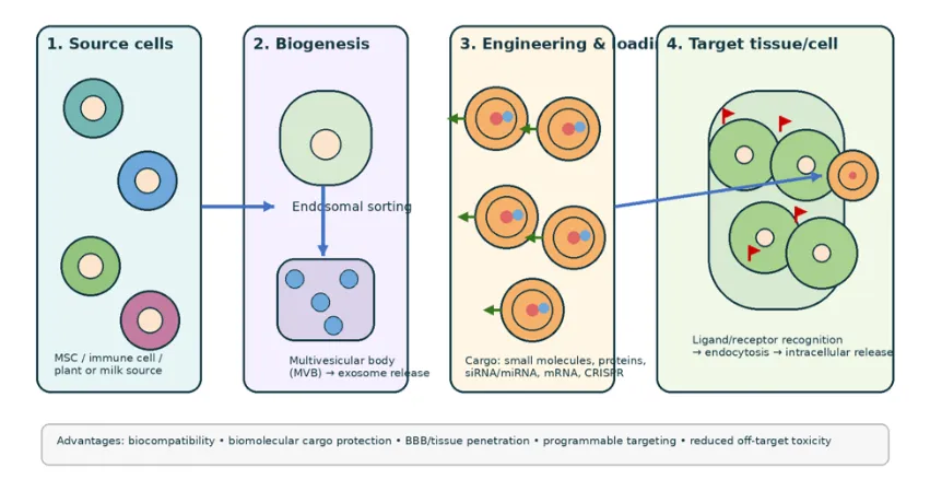
Figure 1. Schematic workflow of exosome generation, cargo loading, surface engineering and target-cell delivery. The figure is an original schematic prepared for this review.
4. ISOLATION, PURIFICATION AND CHARACTERIZATION
4.1 Isolation and purification
Exosome isolation is a critical formulation step because recovery, purity, membrane integrity and biological activity depend on the process. Differential ultracentrifugation remains widely used, but shear stress, aggregation, operator dependence and co-isolation of protein or lipoprotein contaminants limit reproducibility. Density-gradient ultracentrifugation can improve purity but is time-consuming. Ultrafiltration and tangential-flow filtration support larger volumes, while size-exclusion chromatography is relatively gentle and preserves vesicle structure but can dilute the product. Polymer precipitation is simple and scalable but frequently co-precipitates non-vesicular proteins. Immunoaffinity capture offers high specificity for selected markers but is costly and poorly suited to bulk manufacturing. Microfluidic and acoustofluidic systems are emerging for rapid, low-volume and potentially automated processing [17–22].
Table 2. Common Exosome Isolation Approaches and Their Pharmaceutical Implications.
|
METHOD |
STRENGTHS |
LIMITATIONS |
FORMULATION RELEVANCE |
|
Differential ultracentrifugation |
Common, no specialized consumables |
Low throughput; aggregation; possible damage |
Useful for discovery; less suitable as a stand-alone GMP process |
|
Density-gradient centrifugation |
Improved purity and separation by density |
Long processing time; limited scale |
Good for mechanistic studies and reference batches |
|
Ultrafiltration / TFF |
Scalable concentration and buffer exchange |
Membrane fouling; size-similar contaminants |
Promising for process intensification |
|
Size-exclusion chromatography |
Gentle; good recovery of vesicle integrity |
Sample dilution; limited loading capacity |
Useful polishing step |
|
Polymer precipitation |
Simple and low equipment burden |
Protein/lipoprotein contamination |
Generally requires orthogonal polishing |
|
Immunoaffinity |
High marker specificity |
Expensive; capture bias; not bulk scalable |
Useful for analytical subpopulations |
|
Microfluidics / acoustofluidics |
Rapid, low-volume, potentially automated |
Device cost and scale-up validation |
Emerging platform for integrated isolation and loading |
4.2 Characterization and Release Testing
A single analytical method cannot define an exosome product. A fit-for-purpose panel should include particle concentration and size distribution by nanoparticle-tracking analysis or an equivalent technique; morphology by transmission electron microscopy or cryo-electron microscopy; protein identity by immunoblotting, flow cytometry or high-sensitivity immunoassay; cargo profiling by qPCR, sequencing, proteomics or mass spectrometry; and impurity testing for host-cell proteins, DNA, albumin, lipoproteins, endotoxin, mycoplasma and residual process reagents. Recent consensus guidance emphasizes reporting the source, collection conditions, isolation method, storage, particle-to-protein ratio, markers and functional potency rather than relying on the word exosome alone [13,23].
Identity: positive EV-associated markers such as CD9, CD63, CD81 and source-specific markers; absence or reduction of non-EV contaminants.
Purity: particle/protein ratio, removal of serum proteins and lipoproteins, endotoxin and microbial testing.
Physical quality: particle size distribution, concentration, morphology, zeta potential where relevant and aggregation state.
Cargo quality: drug content, encapsulation efficiency, free-drug fraction, RNA/protein integrity and potency.
Functional potency: target-cell uptake, intracellular release, gene-silencing or protein activity, and disease-relevant bioassays.
Stability: particle recovery, size, cargo retention and potency after intended storage, freeze–thaw and administration conditions.
5. CARGO-LOADING STRATEGIES
Cargo loading can be classified as pre-loading (endogenous loading during vesicle biogenesis) or post-loading (direct incorporation into isolated vesicles). The optimal approach depends on molecular size, hydrophobicity, charge, membrane permeability, sensitivity to shear or electrical stress, and the desired release profile [24–27].
Table 3. Exosome cargo-loading strategies relevant to targeted drug delivery.
|
Strategy |
Typical Cargo |
Advantages |
Limitations |
|
Passive incubation / co-incubation |
Hydrophobic small molecules, some drugs |
Simple; mild; preserves membrane structure |
Often low loading for hydrophilic or macromolecular cargo |
|
Donor-cell incubation or transfection |
miRNA, siRNA, mRNA, proteins, small molecules |
Uses natural biogenesis; good cargo bioactivity |
Variable sorting; donor-cell stress and heterogeneous product |
|
Electroporation |
RNA, oligonucleotides, some proteins |
Rapid; can improve loading of hydrophilic cargo |
Aggregation, RNA precipitation and membrane damage possible |
|
Sonication |
Small molecules, siRNA and proteins |
High loading potential; relatively fast |
May alter membrane proteins and vesicle integrity |
|
Extrusion |
Small molecules, proteins, nucleic acids |
Can generate uniform vesicles and facilitate loading |
Mechanical stress; possible loss of native properties |
|
Freeze–thaw cycling |
Small molecules and some proteins |
Low equipment burden |
Low or variable efficiency; damage to labile cargo |
|
Microfluidics / acoustofluidics |
Complex and combination cargo |
Controlled mixing, short processing time, improved reproducibility |
Device design and scale-up remain developmental |
|
Hybridization / membrane fusion |
Drug-loaded liposomes, nanoparticles, polymers |
Combines exosome biology with synthetic loading capacity |
More complex characterization and regulatory pathway |
5.1 Small-Molecule Drugs
Hydrophobic drugs such as doxorubicin, paclitaxel, docetaxel, curcumin, berberine, quercetin, luteolin, rapamycin and atovaquone have been incorporated into cell-, milk- or plant-derived vesicles. Recent studies report improved solubility, protection from premature metabolism, enhanced uptake and reduced exposure of healthy cells. Examples include docetaxel-loaded exosomes for non-small-cell lung cancer, curcumin-loaded plant or MSC-derived vesicles for inflammatory diseases, rapamycin-loaded vesicles for glioblastoma or uveitis, and drug-loaded vesicles for antimicrobial or antifungal therapy [28–37].
5.2 Nucleic Acids, Proteins And Genome-Editing Cargo
Exosomes are especially attractive for RNA delivery because the lipid bilayer can protect nucleic acids from nucleases and support cytosolic uptake. Engineered vesicles carrying siRNA against KRASG12D, BACE1, CTGF or other disease targets, as well as miRNA, mRNA and antisense oligonucleotides, have demonstrated gene-silencing or protein-replacement effects in preclinical models. Surface display of RVG, GPC3, ApoB100-derived peptides, integrin-binding ligands and other targeting elements has been used to direct RNA or protein cargo to neurons, hepatocytes, tumors or immune cells [38–45]. Delivery of CRISPR–Cas systems is an emerging area, but it requires careful control of cargo size, editing specificity, off-target activity and long-term safety.
6. TARGETING AND SURFACE-ENGINEERING STRATEGIES
Targeting may be passive, endogenous or actively engineered. Passive targeting results from circulation, vascular permeability, tissue retention and uptake by the mononuclear phagocyte system. Endogenous targeting is determined by donor-cell origin, membrane proteins, integrins, glycans and chemokine receptors. Active targeting introduces a ligand that recognizes a disease-associated receptor. Recent reviews distinguish pre-isolation engineering—genetic, metabolic or donor-cell membrane engineering—from post-isolation modification—chemical conjugation, lipid insertion, click chemistry, physical fusion or hybridization [8,46–50].
Table 4. Representative surface-engineering strategies for targeted delivery.
|
Engineering Approach |
Representative Targeting Element |
Potential Application |
Key Concern |
|
Genetic fusion to exosome membrane proteins |
Lamp2b–RVG; Lamp2b–anti-GPC3 scFv; CD63 fusions |
Brain, hepatocellular carcinoma and gene therapy |
Expression level, orientation and immunogenicity |
|
Peptide or aptamer display |
RGD, RVG, AS1411, bone-targeting peptides |
Tumor, brain, bone and inflammatory sites |
Ligand density and receptor heterogeneity |
|
Antibody or nanobody display |
EGFR, HER2 or tumor-associated antigens |
Precision oncology and theranostics |
Cost, stability and Fc-related immune effects |
|
Chemokine-receptor engineering |
CXCR4 and related receptors |
Inflammatory tissue and tumor microenvironment |
Potentially altered biodistribution |
|
Metabolic labeling / click chemistry |
Azide–DBCO or other bioorthogonal pairs |
Tracking and modular targeting |
Reaction control and residual reagents |
|
Lipid insertion / post-isolation conjugation |
Hydrophobic peptide, PEG-lipid or ligand conjugate |
Rapid surface functionalization |
Membrane perturbation and ligand shedding |
|
Hybrid exosome–liposome or nanoparticle system |
Drug-loaded liposome, SPION or polymer component |
Higher loading, magnetic guidance or combination therapy |
Product complexity and regulatory classification |
6.1 Cellular Uptake and Intracellular Trafficking
Following circulation and tissue distribution, exosomes interact with target cells through receptor–ligand binding, electrostatic interactions, phagocytosis, macropinocytosis, clathrin-mediated endocytosis, caveolae-mediated uptake or membrane fusion. The final therapeutic outcome depends not only on uptake but also on endosomal escape and cytosolic release. Therefore, high fluorescence uptake does not necessarily prove functional cargo delivery; potency should be measured using a biological readout such as gene silencing, protein activity, apoptosis or disease-specific repair [51–54].
7. THERAPEUTIC APPLICATIONS
7.1 Oncology
Cancer is the most extensively studied application because exosomes can carry chemotherapeutics, gene-silencing agents, immunomodulators and photosensitizers. Preclinical studies have reported enhanced delivery of doxorubicin, paclitaxel, docetaxel, gemcitabine, cisplatin, bleomycin, berberine, curcumin and combination payloads. Examples include SDF1–CXCR4-mediated osteosarcoma targeting, RGD or antibody targeting of glioblastoma and colorectal cancer, macrophage-derived vesicles for breast cancer, and TRAIL-engineered exosomes carrying cisplatin for cervical cancer. Combination strategies, such as simultaneous delivery of a cytotoxic drug and miRNA or a photosensitizer and immunomodulator, are increasingly prominent [31,34,55–61].
7.2 Central Nervous System Delivery
The blood–brain barrier makes CNS delivery a major target for exosome engineering. RVG-functionalized vesicles, Angiopep-2-modified exosomes, neural-stem-cell-derived vesicles and exosome–drug combinations have been investigated for glioblastoma, Alzheimer’s disease, Parkinson’s disease, stroke and CNS infections. Reported cargos include siRNA, miRNA, rifampicin, donepezil, quercetin, rapamycin, cetuximab and doxorubicin. Although several studies demonstrate improved brain accumulation or therapeutic response in rodents, translation requires rigorous assessment of human BBB transport, dose scaling, off-target neural effects and repeated-administration safety [39,40,62–65].
7.3 Inflammatory, Autoimmune and Regenerative Disorders
MSC-, macrophage-, platelet-, plant- and milk-derived vesicles have been investigated for spinal cord injury, rheumatoid arthritis, osteoarthritis, periodontitis, alopecia areata, uveitis, diabetes, wound healing, corneal injury and inflammatory bowel disease. The therapeutic effect may arise from the delivered drug, endogenous vesicle cargo, immunomodulatory membrane components or a combination. Hydrogel–exosome systems and microneedle arrays are being developed to increase local retention and prolong release, which is particularly useful for skin, bone and wound applications [32,36,66–71].
7.4 Infectious Diseases
Macrophage-derived exosomes loaded with atovaquone, lysostaphin and vancomycin, iPSC-derived exosomes carrying amphotericin B, and plant or bacterial vesicles containing antibiotics illustrate the potential for intracellular antimicrobial delivery. These systems may exploit natural uptake by macrophages or infected cells and can increase drug concentration at intracellular pathogen reservoirs. However, source-cell safety, endotoxin control, antimicrobial resistance, immunological effects and scale-up require careful evaluation.
8. Clinical Translation and Regulatory Considerations
Clinical translation has accelerated, but the field remains early. A 2024 systematic review of EV clinical trials found substantial variability in source, isolation, characterization and reporting, with only a minority of studies using defined EV subpopulations. A 2026 analysis of ClinicalTrials.gov identified 355 human-derived EV trials initiated through the end of 2025, including 148 interventional studies; MSC-derived EVs were common in interventional work, while oncology, inflammatory disorders, respiratory disease and regenerative medicine were recurrent areas [10,11]. These data demonstrate clinical interest but do not equate to regulatory approval of exosome drug-delivery products.
Table 5. Key translational risks and quality-by-design responses.
|
Translation Domain |
Current Issue |
Recommended Pharmaceutical Response |
|
Source material |
Donor-cell and culture-condition variability |
Define a qualified master cell bank, passage limits and culture medium. |
|
Isolation and purification |
Different methods yield different particle populations |
Use a validated, scalable and orthogonal purification train. |
|
Product identity |
“Exosome” may include heterogeneous small EVs |
Report MISEV-aligned source, markers, size, purity and function. |
|
Cargo loading |
Variable encapsulation and free-drug contamination |
Quantify total, encapsulated and free cargo with validated assays. |
|
Potency |
No universal release assay |
Use mechanism-linked cell-based potency assays. |
|
Sterility and safety |
Endotoxin, mycoplasma, residual DNA and immunogenicity concerns |
Establish release specifications and repeat-dose toxicology. |
|
Stability |
Loss of membrane integrity or activity during storage |
Develop cryoprotectant, lyophilization or controlled-temperature strategies. |
|
Regulatory classification |
Biologic, advanced therapy, drug or combination-product status may vary |
Engage regulators early and prepare a complete CMC package. |
9. MAJOR CHALLENGES AND FUTURE DIRECTIONS
Heterogeneity: donor-cell source, disease state and culture conditions change membrane composition and cargo.
Scalability: ultracentrifugation and affinity capture are difficult to translate directly to high-volume GMP production.
Loading reproducibility: encapsulation efficiency and cargo localization differ between methods and laboratories.
Targeting specificity: ligand-receptor heterogeneity, protein corona formation and reticuloendothelial uptake can reduce precision.
Endosomal escape: internalization does not guarantee cytosolic delivery or functional release.
Storage stability: freezing, thawing, agitation, concentration and container interactions can alter size and potency.
Analytical standardization: single-particle measurements, orthogonal methods and potency assays are required.
Safety and immunology: allogeneic source components, oncogenic cargo, residual DNA, thrombogenicity and repeat-dose effects need evaluation.
Regulatory science: product definition, comparability after process changes and clinical endpoints remain incompletely harmonized.
9.1 Promising development directions
Quality-by-design manufacturing using stirred-tank or perfusion bioreactors, tangential-flow filtration and in-line analytics.
Precision producer-cell engineering to control cargo sorting and membrane ligand display.
Microfluidic and acoustofluidic systems that integrate isolation, loading and surface modification.
Hybrid exosome–liposome or exosome–polymer systems to increase drug loading while retaining biological uptake.
Lyophilized or spray-dried formulations for improved storage and distribution, provided membrane functionality is preserved.
Single-vesicle multi-omics and machine-learning models to link composition with potency and biodistribution.
Clinically relevant large-animal studies and dose-ranging trials with validated pharmacokinetic and pharmacodynamic biomarkers.
10. CONCLUSION
Exosomes have evolved from biological messengers into highly adaptable nanocarriers capable of transporting small molecules, proteins and nucleic acids to selected tissues and cells. Their major pharmaceutical advantages are biocompatibility, endogenous membrane functionality, cargo protection, potential barrier penetration and the possibility of genetic or chemical surface engineering. Recent work from 2023–2026 has expanded the field toward ligand-displayed vesicles, gene-editing cargo, hybrid nanocarriers, continuous manufacturing and clinical-trial analysis. Nevertheless, exosome delivery is not yet a universally standardized technology. Heterogeneity, limited yield, inconsistent loading, incomplete potency assays, biodistribution uncertainty, storage instability and regulatory ambiguity continue to restrict clinical translation. For M.Pharm pharmaceutics, the most important development principle is that exosomes should be treated as complex biological drug products requiring a defined source, controlled process, validated characterization, mechanism-based potency and rigorous safety evaluation. With these controls, exosome-based targeted delivery may become a practical component of precision nanomedicine rather than remaining only a promising preclinical concept.
REFERENCES

Tanuja Jadhav, Kakasaheb Kore, Dr. Rajkumar Shete, Exosomes as Advanced Nanocarriers for Targeted Drug Delivery: Recent Progress, Engineering Strategies and Translational Challenges, Int. J. of Pharm. Sci., 2026, Vol 4, Issue 9, 3598-3608. https://doi.org/10.5281/zenodo.23019163
 10.5281/zenodo.23019163