

We use cookies to ensure our website works properly and to personalise your experience. Cookies policy
Shantha College of Pharmacy, Peresandra, Chikkabalapur, Karnataka, India 562104
Background: Cancer remains a leading cause of mortality worldwide, necessitating the discovery of novel therapeutic agents. Neolinderatin, a naturally occurring phytoconstituent, has attracted attention due to its potential pharmacological activities. Objective: This study aimed to clarify the underlying molecular mechanisms and therapeutic targets of Neolinderatin against cancer using network pharmacology, molecular docking, and in silico ADMET analyses. Methods: Potential targets of Neolinderatin were retrieved from public databases and intersected with cancer-related genes to identify common targets. Protein-protein interaction (PPI) the analysis was conducted using the STRING database and Cytoscape. Gene Ontology (GO) and KEGG pathway enrichment analyses were conducted using Enrichr. Hub genes were identified through CytoHubba. Molecular docking was performed using AutoDock Vina and Discovery Studio Visualizer against selected hub proteins. Drug-likeness and pharmacokinetic properties were evaluated using SwissADME and pkCSM. Results: A total of 110 overlapping targets between Neolinderatin and cancer-associated genes were identified. PPI network analysis revealed CASP3, PTGS2, MMP9, MAPK14, ERBB2, CDK2, CDK4, CASP9, HSP90AA1, and MMP2 as key hub genes. Enrichment analysis demonstrated significant involvement in pathways associated with apoptosis, cancer signaling, IL-17 signaling, p53 signaling, and interleukin-mediated pathways. Molecular docking demonstrated favorable binding affinities of Neolinderatin with PTGS2 (5IKR, ?8.3 kcal/mol), MMP9 (1GKC, -8.0 kcal/mol), MAPK14 (1A9U, -7.7 kcal/mol), and CASP3 (1PAU, -6.2 kcal/mol). SwissADME and pkCSM analyses indicated acceptable pharmacokinetic and toxicity profiles. Conclusion: The findings suggest that Neolinderatin exerts anticancer effects through multi target interactions involving apoptosis, inflammation, and cell proliferation pathways. These results provide a theoretical basis for further experimental validation of Neolinderatin as a potential anticancer agent.
Cancer remains one of the most significant global health challenges and is a leading cause of morbidity and mortality worldwide1,2. Despite substantial advances in chemotherapy, radiotherapy, targeted therapy, and immunotherapy, the effectiveness of current treatment strategies is often limited by drug resistance, severe adverse effects, and tumor recurrence25,26. Consequently, the search for safer and more effective therapeutic agents continues to be a major focus of cancer research.
Natural products have long served as valuable sources of anticancer drugs and lead compounds due to their structural diversity and broad spectrum of biological activities3. Numerous plant-derived compounds have demonstrated promising anticancer potential by modulating multiple molecular targets involved in tumor initiation, progression, and metastasis3. Neolinderatin, a naturally occurring phytochemical isolated from medicinal plants, has attracted considerable attention because of its reported pharmacological activities, including antioxidant and anti-inflammatory effects. However, the molecular mechanisms underlying its potential anticancer activity remain poorly understood.
Network pharmacology has emerged as a powerful systems-based approach for elucidating the complex interactions among bioactive compounds, target proteins, genes, and disease pathways4,5. Unlike the traditional one-drug–one-target paradigm, network pharmacology emphasizes multitarget and multipathway regulation, making it particularly suitable for investigating natural compounds with diverse biological effects. By integrating bioinformatics databases and computational analyses, network pharmacology enables the identification of key therapeutic targets and signaling pathways involved in disease treatment.
Molecular docking is widely employed in computer-aided drug discovery to predict the binding interactions and affinities between small molecules and target proteins16,29,30. In addition, in silico absorption, distribution, metabolism, excretion, and toxicity (ADMET) analyses provide valuable insights into the pharmacokinetic and safety profiles of candidate compounds18,19. The integration of network pharmacology, molecular docking, and ADMET evaluation offers a comprehensive strategy for exploring the therapeutic potential of bioactive molecules and accelerating the drug discovery process.
Therefore, the present study aimed to systematically investigate the anticancer mechanisms of Neolinderatin using an integrated computational approach involving network pharmacology, protein–protein interaction (PPI) network construction, Gene Ontology (GO) and Kyoto Encyclopedia of Genes and Genomes (KEGG) enrichment analyses, molecular docking, and in silico ADMET evaluation16,18,19. The findings of this study may provide valuable insights into the molecular basis of Neolinderatin's anticancer activity and offer a theoretical foundation for its future development as a potential anticancer agent.
2.1 Identification of Neolinderatin Targets
The chemical structure of Neolinderatin was obtained from the PubChem database. Potential targets of Neolinderatin were predicted using Swiss Target Prediction and related target prediction databases6.
2.2 Collection of Cancer-Associated Targets
Cancer-related genes were collected from Gene Cards, OMIM, and DisGeNET databases using the keyword "Cancer"7,8.
2.3 Identification of Common Targets
The overlapping targets between Neolinderatin-associated genes and cancer-associated genes were identified using Venny/GeneVenn.
2.4 Protein–Protein Interaction Network Construction
The common targets were imported into STRING database to generate a PPI network9. The network was visualized and analysed using Cytoscape software10.
2.5 Hub Gene Analysis
CytoHubba plugin was used to identify the top hub genes according to degree centrality11.
2.6 GO and Pathway Enrichment Analysis
Enricher was employed to perform Gene Ontology (GO) and pathway enrichment analyses12. GO annotation was performed according to the Gene Ontology database14,15. While pathway enrichment analysis was conducted based on the KEGG database13.
2.7 Molecular Docking Studies
Protein structures were obtained from the Protein Data Bank.
Docking studies were performed using AutoDock Vina16, and interaction analyses were conducted using Discovery Studio Visualizer17.
2.8 ADMET Analysis
Swiss ADME and pkCSM servers were used to evaluate physicochemical properties, pharmacokinetics, and toxicity profiles of Neolinderatin18,19.
3.1 Common Target Identification
A total of 110 common targets were identified between Neolinderatin and cancer-associated genes. These overlapping targets suggest the multitarget therapeutic potential of Neolinderatin.
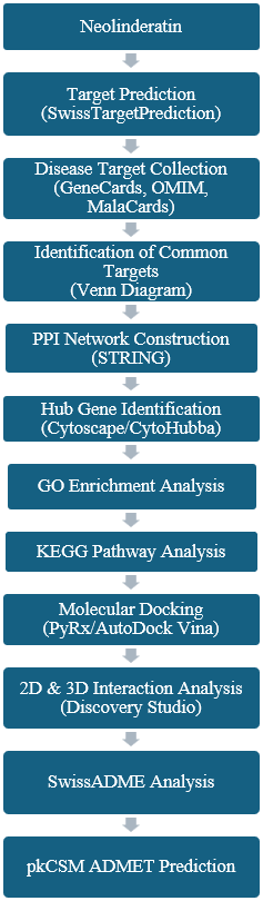
Figure 1. Study workflow of the network pharmacology and molecular docking approach.
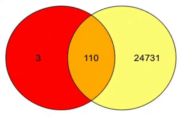
Figure 2. Venn diagram showing 110 overlapping targets between Neolinderatin targets and cancer-related genes.
3.2 PPI Network and Hub Gene Analysis
STRING analysis generated a highly connected PPI network consisting of 110 nodes and 522 edges. The network exhibited significant enrichment (PPI enrichment p-value < 1.0 × 10⁻¹⁶).
CytoHubba analysis identified CASP3, PTGS2, MMP9, MAPK14, ERBB2, CDK2, CDK4, CASP9, HSP90AA1, and MMP2 as key hub genes. These proteins are critically involved in apoptosis, inflammation, tumor invasion, angiogenesis, and cell cycle regulation.
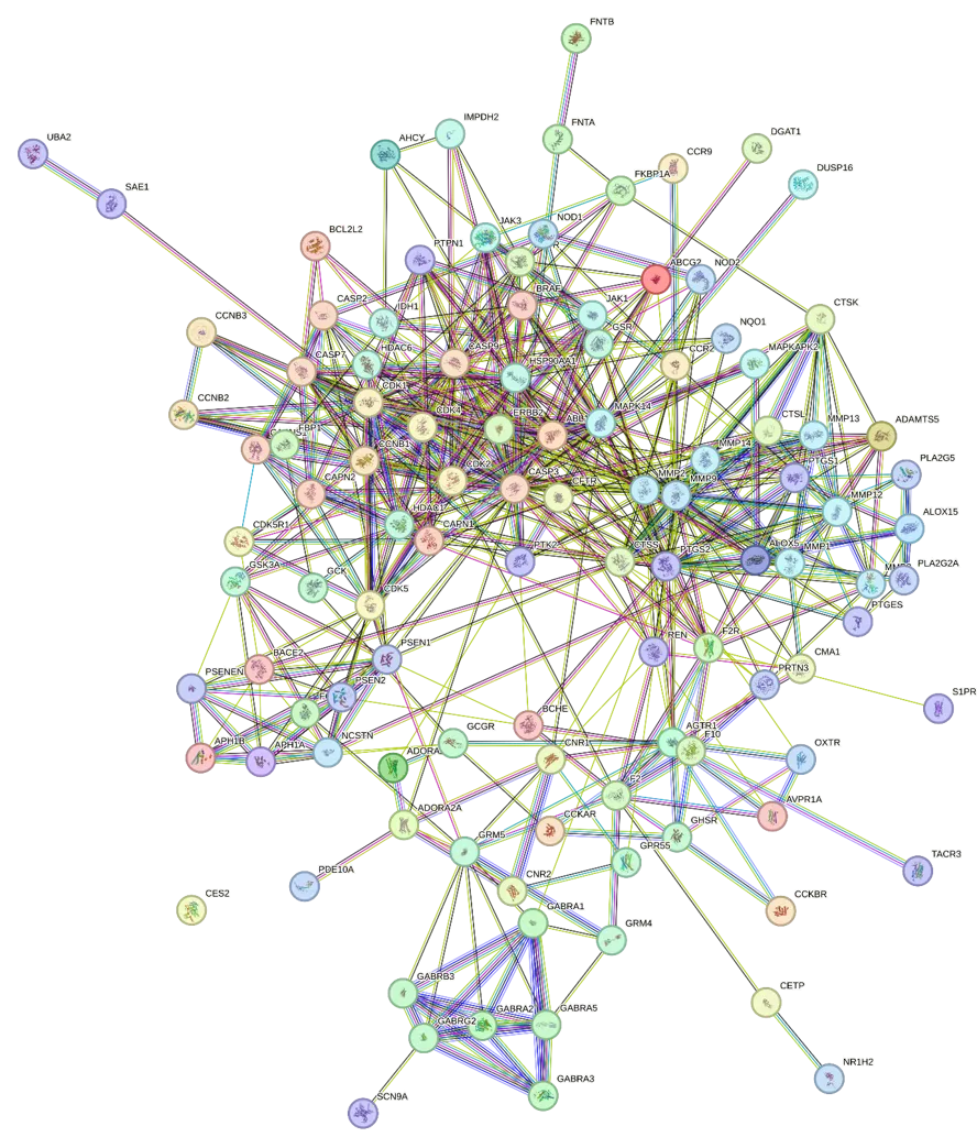
Figure 3. STRING PPI network of 110 common targets.
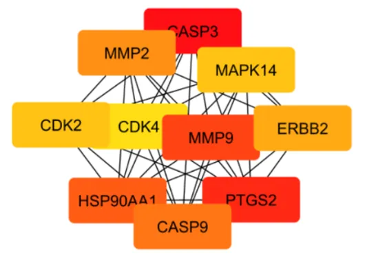
Figure 4. Top 10 hub genes identified by CytoHubba.
Table 1. Top hub genes and their degree scores.
|
Rank |
Gene |
Degree |
|
1 |
CASP3 |
92 |
|
2 |
PTGS2 |
74 |
|
3 |
MMP9 |
70 |
|
4 |
HSP90AA1 |
66 |
|
5 |
CASP9 |
56 |
|
6 |
MMP2 |
52 |
|
7 |
ERBB2 |
50 |
|
8 |
CDK2 |
46 |
|
9 |
MAPK14 |
46 |
|
10 |
CDK4 |
42 |
3.3 GO Enrichment Analysis
GO biological process analysis indicated significant enrichment in:
GO molecular function analysis revealed enrichment in:
These results indicate the involvement of Neolinderatin in apoptosis and cell signaling regulation.
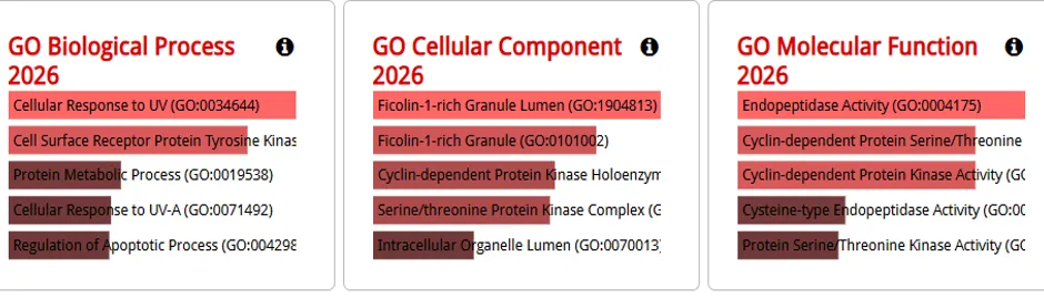
Figure 5. Gene Ontology (GO) enrichment analysis of the identified hub genes, showing the top enriched Biological Process (BP), Cellular Component (CC), and Molecular Function (MF) categories.
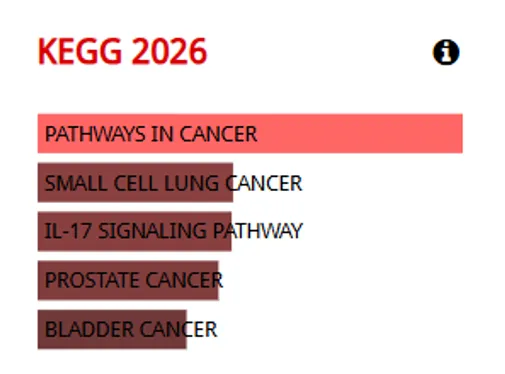
Figure 6. KEGG pathway enrichment analysis of the identified hub genes, highlighting the top significantly enriched signaling and cancer-related pathways.
3.4 Pathway Enrichment Analysis
Pathway analysis revealed enrichment in:
These pathways are closely associated with tumor growth, inflammation, and metastasis.
3.5 Molecular Docking Analysis
Neolinderatin demonstrated strong binding interactions with the selected target proteins.
Table 2. Molecular docking scores of Neolinderatin with selected hub proteins.
|
Target |
PDB ID |
Binding Affinity (kcal/mol) |
|
PTGS2 |
5IKR |
-8.3 |
|
MMP9 |
1GKC |
-8.0 |
|
MAPK14 |
1A9U |
-7.7 |
|
CASP3 |
1PAU |
-6.2 |
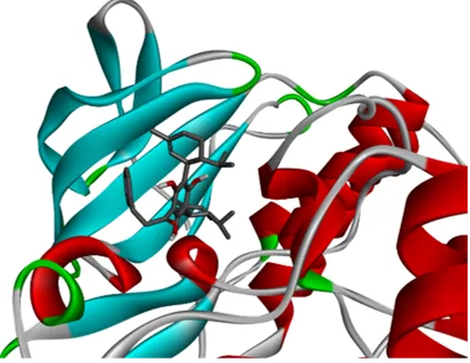
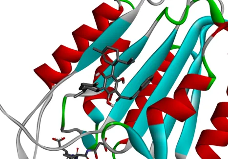
The ligand formed hydrogen bonds, hydrophobic interactions, and van der Waals interactions with key amino acid residues, suggesting stable protein-ligand complexes.
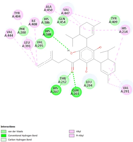
Figure 7. 2D interaction diagram of Neolinderatin with PTGS2 (5IKR).
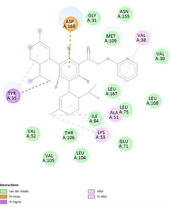
Figure 8. 2D interaction diagram of Neolinderatin with MAPK14 (1A9U).
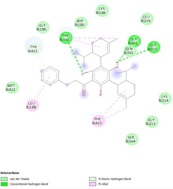
Figure 9. 2D interaction diagram of Neolinderatin with MMP9 (1GKC).
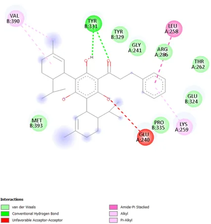
Figure 10. 2D interaction diagram of Neolinderatin with CASP3 (1PAU).
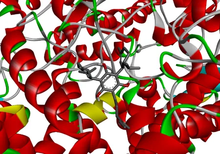
(A) PTGS |
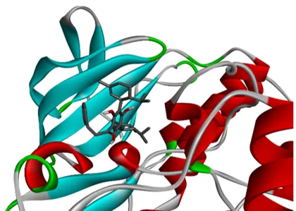
(B) MAPK14 |
(C) HSP90AA |
(D) CASP3 |
Figure 11. Representative 3D receptor–ligand binding poses of Neolinderatin with (A) PTGS2, (B) MAPK14, (C) HSP90AA1, and (D) CASP3.
3.6 ADMET Analysis
SwissADME analysis indicated:
pkCSM analysis revealed:
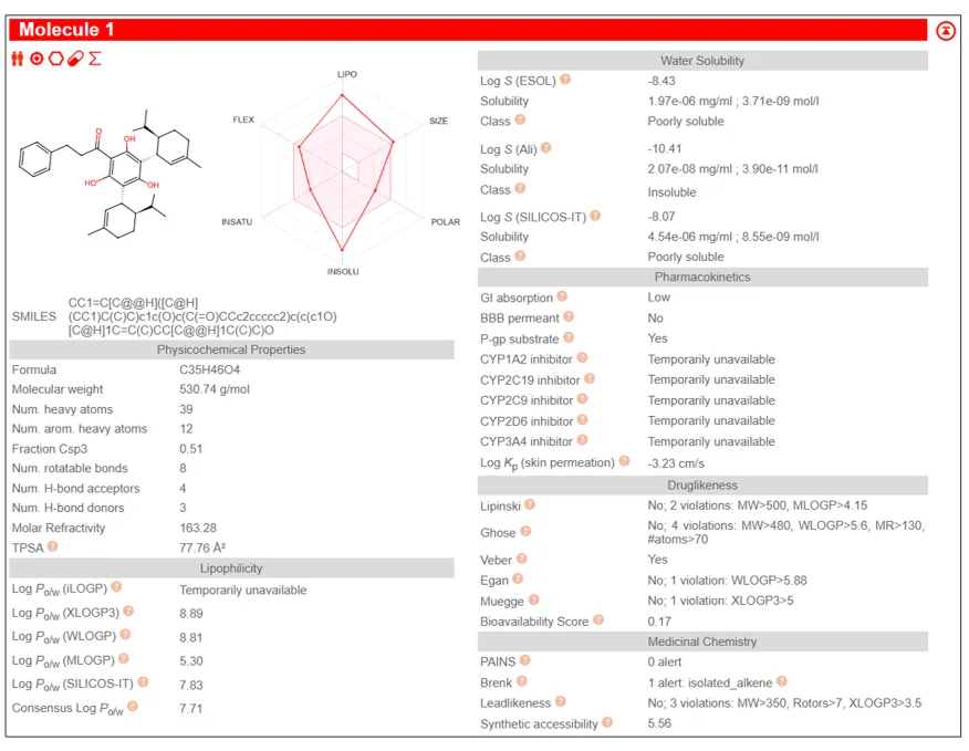
Figure 12. Swiss ADME bioavailability radar of Neolinderatin.
Table 3. Swiss ADME physicochemical and drug-likeness properties.
|
Property |
Value |
|
Molecular Formula |
C35H46O4 |
|
Molecular Weight (g/mol) |
530.74 |
|
Heavy Atoms |
39 |
|
Fraction Csp3 |
0.51 |
|
Rotatable Bonds |
8 |
|
H-Bond Acceptors |
4 |
|
H-Bond Donors |
3 |
|
Topological Polar Surface Area (TPSA, Ų) |
77.76 |
|
Consensus Log P |
7.71 |
|
Water Solubility Class |
Poorly Soluble |
|
GI Absorption |
Low |
|
BBB Permeant |
No |
|
P-gp Substrate |
Yes |
|
Lipinski Rule |
Violated (2 violations) |
|
Ghose Filter |
Violated (4 violations) |
|
Veber Rule |
Passed |
|
Egan Rule |
Violated (1 violation) |
|
Muegge Rule |
Violated (1 violation) |
|
Bioavailability Score |
0.17 |
|
PAINS Alert |
0 |
|
Brenk Alert |
1 |
|
Lead-likeness |
No (3 violations) |
|
Synthetic Accessibility |
5.56 |
Table 4. pkCSM-predicted ADMET properties of Neolinderatin.
|
Category |
Parameter |
Predicted Value |
|
Absorption |
Water Solubility (log mol/L) |
-4.138 |
|
|
Caco-2 Permeability (log Papp in 10⁻⁶ cm/s) |
0.545 |
|
|
Human Intestinal Absorption (%) |
91.474 |
|
|
Skin Permeability (log Kp) |
-2.736 |
|
|
P-glycoprotein Substrate |
Yes |
|
|
P-glycoprotein I Inhibitor |
Yes |
|
|
P-glycoprotein II Inhibitor |
Yes |
|
Distribution |
Volume of Distribution (VDss, log L/kg) |
-0.411 |
|
|
Blood–Brain Barrier Permeability (log BB) |
-0.767 |
|
|
CNS Permeability (log PS) |
-1.293 |
|
Metabolism |
CYP2D6 Substrate |
No |
|
|
CYP3A4 Substrate |
Yes |
|
|
CYP1A2 Inhibitor |
No |
|
|
CYP2C19 Inhibitor |
Yes |
|
|
CYP2C9 Inhibitor |
No |
|
|
CYP2D6 Inhibitor |
No |
|
|
CYP3A4 Inhibitor |
No |
|
Excretion |
Total Clearance (log ml/min/kg) |
0.353 |
|
Toxicity |
AMES Toxicity |
No |
|
|
hERG I Inhibitor |
No |
|
|
hERG II Inhibitor |
Yes |
|
|
Hepatotoxicity |
No |
|
|
Skin Sensitisation |
No |
|
|
Maximum Tolerated Dose (Human, log mg/kg/day) |
0.062 |
|
|
Oral Rat Acute Toxicity (LD₅₀,mol/kg) |
1.886 |
|
|
Oral Rat Chronic Toxicity (LOAEL) |
1.245 |
These findings indicate an acceptable safety profile despite limitations in solubility and bioavailability.
This study systematically explored the anticancer potential and underlying molecular mechanisms of Neolinderatin through an integrated network pharmacology, molecular docking, and in silico ADMET approach. A total of 110 common targets associated with Neolinderatin and cancer were identified, and protein–protein interaction network analysis revealed CASP3, PTGS2, MMP9, MAPK14, ERBB2, CDK2, CDK4, CASP9, HSP90AA1, and MMP2 as key hub genes. GO and KEGG enrichment analyses demonstrated that these targets are primarily involved in apoptosis, p53 signaling, IL-17 signaling, inflammatory responses, and cancer-related pathways.
Molecular docking analysis showed strong binding affinities of Neolinderatin toward major hub proteins, particularly PTGS2, MMP9, MAPK14, and CASP3, supporting its potential therapeutic relevance in cancer treatment. Furthermore, Swiss ADME and pkCSM predictions indicated favorable drug-likeness, pharmacokinetic behavior, and acceptable toxicity profiles.
Collectively, these findings suggest that Neolinderatin exerts anticancer effects through multitarget and multipathway modulation and may serve as a promising lead compound for anticancer drug development. Nevertheless, further in vitro and in vivo studies are required to validate the predicted targets, biological activities, and therapeutic efficacy of Neolinderatin
ACKNOWLEDGMENTS
The authors acknowledge the use of Swiss Target Prediction, STRING, Cytoscape, Enrichr, Auto Dock Vina, Discovery Studio Visualizer, SwissADME, and pkCSM databases and software tools.
CONFLICT OF INTEREST
The authors declare no conflict of interest.
REFERENCES





Brunda M A, Mukthiyar Ahamed, Bindu M A, B. Subhashini, Dr. Gopinath, Exploring the Anticancer and Mechanisms and Therapeutic Targets of Neolinderatin through Network Pharmacology and Molecular Docking, Int. J. of Pharm. Sci., 2026, Vol 4, Issue 8, 4490-4501. https://doi.org/10.5281/zenodo.22122651
 10.5281/zenodo.22122651