

We use cookies to ensure our website works properly and to personalise your experience. Cookies policy
1,2,3,4,5 Faculty of Pharmacy, Marwadi University, Rajkot, India
1,3,4 School of Pharmacy, RK University, Bhavnagar Highway, Rajkot, Gujarat, India, 360020
Diabetes mellitus is a major metabolic disorder in which inhibition of carbohydrate-digesting enzymes represents one approach for controlling postprandial blood glucose levels. The present study aimed to formulate and evaluate herbal nutraceutical gummies containing Vernonia amygdalina, Trigonella foenum-graecum, and cinnamon extracts and to investigate their preliminary antidiabetic potential through in vitro enzyme inhibition and molecular docking studies. The plant materials were subjected to physicochemical evaluation and preliminary phytochemical screening, followed by hydroalcoholic extraction using 70% methanol. The ?-amylase inhibitory activity of the extracts was evaluated at concentrations of 10–200 µg/mL using a CNPG3-based colorimetric assay, with Acarbose as the standard. Three gummy formulations containing different concentrations of gelatin were prepared and evaluated for physicochemical characteristics. The optimized formulation was further assessed for total flavonoid content using quercetin as the reference standard. Molecular docking was performed to investigate the predicted interactions of selected V. amygdalina phytoconstituents with human pancreatic ?-amylase and lysosomal acid ?-glucosidase. All three extracts demonstrated ?-amylase inhibitory activity, with fenugreek showing the highest inhibition (43.15%), followed by bitter leaf (41.05%) and cinnamon (38.95%) at 200 µg/mL, while Acarbose showed 66.11% inhibition. None of the plant extracts reached 50% inhibition within the tested concentration range. F2, containing 10% gelatin, was selected as the optimized gummy formulation based on its physicochemical characteristics. The optimized gummy showed a total flavonoid content of 0.07705 mg QE/g. Molecular docking indicated favorable predicted interactions of several V. amygdalina phytoconstituents with the selected enzyme targets, while caffeic acid, apigenin and kaempferol showed favorable overall in silico profiles. The findings provide preliminary evidence supporting the potential of the selected plant extracts as sources of bioactive constituents for nutraceutical development, although further studies are required to establish their biological efficacy and safety.
Diabetes mellitus, particularly type 2 diabetes mellitus, is a major metabolic disorder characterized by persistent elevation of blood glucose and is associated with long-term complications affecting multiple organs. The increasing prevalence of diabetes has created a need for complementary approaches that can support conventional management. Nutraceuticals derived from plant sources have attracted considerable interest because they contain bioactive compounds that may influence carbohydrate metabolism and other pathways associated with glycemic control [1,2].
Several medicinal plants have been investigated for their potential role in glucose regulation. Vernonia amygdalina Delile (Bitter Leaf) has been reported to possess hypoglycemic and antidiabetic properties, which have been associated with its diverse phytochemical constituents, including flavonoids, alkaloids, phenolic compounds and saponins [3,4]. Trigonella foenum-graecum L. (Fenugreek) contains soluble dietary fiber and several bioactive constituents and has been investigated for its effects on glycemic control. Clinical and meta-analytic evidence has also suggested beneficial effects of fenugreek consumption on blood glucose parameters [5]. Cinnamon (Cinnamomum spp.) contains several bioactive compounds, including cinnamaldehyde, and has likewise been investigated for its potential effects on glucose metabolism and glycemic control [6].
One approach for investigating the potential of plant extracts in glycemic management is inhibition of carbohydrate-digesting enzymes. α-Amylase is an important digestive enzyme responsible for the hydrolysis of dietary starch into smaller carbohydrates. Inhibition of this enzyme can reduce the rate of carbohydrate digestion and may therefore help moderate postprandial increases in blood glucose. Plant-derived extracts and phytochemicals have demonstrated varying degrees of α-amylase inhibitory activity, providing a useful preliminary in vitro approach for evaluating their potential in glucose management[7].
Although these plants have individually been investigated for their biological activities, there is a need to explore their incorporation into a convenient and palatable nutraceutical dosage form. Herbal preparations may be limited by taste, handling and dosing convenience. Gummy formulations provide an alternative oral delivery system that can improve palatability and consumer acceptability while allowing incorporation of plant-derived ingredients [8,9]. However, formulation characteristics such as gelatin concentration, pH, swelling behavior, syneresis and disintegration need to be evaluated to identify a suitable formulation.
Phytochemical characterization can provide additional information about the constituents present in plant extracts, while quantitative determination of total flavonoid content can provide a measure of one important group of bioactive compounds. In addition, molecular docking can be used as a computational approach to investigate the predicted interaction of selected phytoconstituents with biological targets involved in carbohydrate digestion. Such computational findings can complement experimental enzyme inhibition results, although docking predictions alone cannot establish biological activity.
Therefore, the present study was undertaken to formulate and evaluate herbal nutraceutical gummies containing V. amygdalina, T. foenum-graecum, and Cinnamon extracts. The study aimed to provide preliminary experimental and computational evidence for the potential of the selected formulation and its phytochemical constituents.
The specific objectives were to:
2. MATERIALS AND METHODS
2.1. Plant material and identification
Fresh Bitter Leaf was procured from MedicinaLive, Tamil Nadu, India; Fenugreek seeds were obtained from a local market (Gauridad Market, Rajkot); and Cinnamon powder was obtained from the Faculty of Pharmacy, Marwadi University. Identification and authentication were performed at the Faculty of Pharmacy laboratory using established pharmacognostic and physicochemical procedures [10,11].
2.2. Physicochemical evaluation of plant materials
The selected plant materials were evaluated for moisture content, total ash value, water-soluble extractive value, alcohol-soluble extractive value, foreign matter, and microscopic characteristics [10,11].
2.2.1. Moisture content
Moisture content was determined by the loss-on-drying method. Approximately 5 g of the powdered plant material was accurately weighed in a previously dried Petri dish and dried in an oven at 105 °C until a constant weight was obtained. The percentage loss in weight was calculated as the moisture content of the sample [10,11].
2.2.2. Total ash value
Approximately 3 g of each powdered plant material was accurately weighed into a previously ignited and tarred crucible. The sample was gently heated to remove organic matter and subsequently incinerated in a muffle furnace at 500–600 °C until a constant weight of ash was obtained. The crucible was cooled in a desiccator and weighed, and the total ash value was calculated as a percentage of the original sample weight [10,11].
2.2.3 Water-soluble extractive value
Approximately 2 g of the air-dried, coarsely powdered plant material was macerated with 100 mL of distilled water in a stoppered flask for 24 h, with frequent shaking during the first 6 h. The mixture was filtered, and 25 mL of the filtrate was evaporated to dryness in a previously weighed shallow dish. The residue was dried at 105 °C to constant weight, cooled in a desiccator, and weighed to determine the water-soluble extractive value [10,11].
2.2.4 Alcohol-soluble extractive value
Approximately 2 g of the air-dried, coarsely powdered material was macerated with 100 mL of 90% alcohol for 24 h, with frequent shaking during the first 6 h. The mixture was filtered and an aliquot of the filtrate was evaporated to dryness and dried at 105 °C to constant weight. The alcohol-soluble extractive value was calculated with reference to the air-dried plant material [10,11].
2.2.5 Foreign matter
The plant materials were visually examined for extraneous matter, including soil, insects, and other foreign plant materials [10,11].
2.2.6 Microscopic examination
Microscopic examination was performed to characterize the diagnostic anatomical features of the plant material. The observed characteristics included epidermal cells, stomata, glandular and non-glandular trichomes, palisade and spongy mesophyll, vascular bundles, crystals, and starch grains [10,11].
2.3. Phytochemical screening
Qualitative phytochemical screening was performed on the plant extracts to detect major classes of secondary metabolites. Alkaloids were tested using Dragendorff's reagent, flavonoids by the Shinoda test, tannins using ferric chloride, saponins by the foam test, terpenoids by the Salkowski test, steroids by the Liebermann–Burchard test, and phenolic compounds using the Folin–Ciocalteu reaction. Mucilage was assessed using the swelling-index method, while cinnamaldehyde was examined using Fehling's test. The development of characteristic colour changes, precipitates, or persistent foam was used as an indication of the respective constituents [10,11].
2.4. Preparation of hydroalcoholic extracts
Hydroalcoholic extraction was performed by maceration. Approximately 30 g of each powdered plant material; Bitter Leaf, Fenugreek, and Cinnamon, was separately extracted with 150 mL of methanol (70:30, v/v). The mixtures were maintained in stoppered containers at room temperature for 72 h with occasional shaking. Following extraction, the mixtures were filtered and the filtrates were concentrated to dryness to obtain the respective crude extracts, which were used for further analysis and formulation [10].
2.5. In vitro α-amylase inhibition assay
α-Amylase inhibitory activity of the three extracts (10–200 µg/mL) was assessed using a CNPG3 (2-chloro-4-nitrophenyl-α-maltotrioside)-based colorimetric assay, with Acarbose as the positive control. Extract or standard solution (0.5 mL) was incubated with α-amylase (250 µL, 1 µg/mL in 40 mM phosphate buffer, pH 6.9) at 37°C for 10 minutes. CNPG3 substrate (1 mL) was then added, and the mixture was incubated for a further 10 minutes at 37°C. The reaction was terminated with 1% acetic acid (250 µL), and the absorbance was measured at 405 nm [7,12,13].
The percentage inhibition of α-amylase activity was calculated relative to the control using the following equation:
IC50=C1 +50-%I1%I2 -%I1 ×(C2-C1

Where:


The inhibitory activity was evaluated at concentrations ranging from 10 to 200 µg/mL. Since 50% inhibition was not achieved by the plant extracts at the highest experimentally tested concentration of 200 µg/mL, an experimentally determined IC₅₀ value could not be obtained within the tested concentration range.
2.6. Formulation of herbal nutraceutical gummies
Three gummy formulations (F1, F2, and F3) were prepared by varying the concentration of gelatin while maintaining the quantities of the three herbal extracts. Each formulation contained 2.5 g each of Fenugreek, Cinnamon, and Bitter Leaf extracts. Gelatin concentrations of 5%, 10%, and 15% were used for F1, F2, and F3, respectively. Citric acid, sodium benzoate, and stevia were incorporated as acidulant, preservative, and sweetening agent, respectively. Purified water was used as the vehicle. The total batch quantity for each formulation was 50 g.
The ingredients were mixed in a beaker containing warm water to obtain a homogeneous gummy mixture. The prepared mass was subsequently transferred into suitable moulds and allowed to set before evaluation. The composition of the three formulations is presented in Table 1.
Table 1. Composition of the herbal nutraceutical gummy formulations (F1–F3)
|
Ingredient |
F1 (5% Gelatin) |
F2 (10% Gelatin) |
F3 (15% Gelatin) |
|
Fenugreek extract (g) |
2.5 |
2.5 |
2.5 |
|
Cinnamon extract (g) |
2.5 |
2.5 |
2.5 |
|
Bitter Leaf extract (g) |
2.5 |
2.5 |
2.5 |
|
Gelatin (g) |
2.5 |
5 |
7.5 |
|
Citric acid (g) |
0.75 |
0.75 |
0.75 |
|
Sodium benzoate (g) |
0.075 |
0.075 |
0.075 |
|
Stevia (g) |
0.05 |
0.08 |
0.1 |
|
Purified water (mL) |
40 |
35.8 |
30 |
|
Total quantity (g) |
50 |
50 |
50 |
2.7. Evaluation of herbal gummies
The formulated gummies were evaluated for physical appearance 17,18.
2.7.1 Physical appearance
The prepared gummies were visually examined for colour, transparency, homogeneity, and uniformity. The texture was assessed manually for grittiness and stickiness.
2.7.2 pH
The pH of the gummy formulation was determined using a digital pH meter. A portion of the gummy was dispersed in distilled water, and the pH was measured after immersion of the electrode into the resulting dispersion.
2.7.3 Swelling ratio
The swelling behaviour of the gummies was determined by recording their initial weight (W₀), immersing the gummies in 100 mL of purified water for 10 s at room temperature, removing excess surface water using filter paper, and recording the final weight (Wₛ). The swelling ratio was calculated from the change in weight before and after immersion.
2.7.4 Syneresis
Syneresis was evaluated by monitoring changes in the gummy matrix during storage at room temperature for 48 h. The gummies were examined for water release, shrinkage, and changes in consistency. The amount of syneresis was determined from the weight difference associated with water released from the gel matrix.
2.7.5 Disintegration time
Disintegration behaviour was evaluated by placing a gummy in 50 mL of purified water maintained at 37 °C with agitation using a magnetic stirrer. The time required for complete dispersion of the gummy was recorded as the disintegration time.
2.8. Determination of total flavonoid content
Total flavonoid content of the formulated gummy was determined using a UV-Visible spectrophotometric method with quercetin as the reference standard. A quercetin stock solution was prepared by dissolving 10 mg of quercetin in 100 mL of methanol. Serial dilutions were prepared to obtain concentrations of 6.25, 12.5, 25, 50, 80, and 100 µg/mL for construction of the calibration curve.
For sample preparation, 100 mg of the dried methanolic gummy extract was dissolved in 5 mL of methanol and transferred to a 10 mL volumetric flask, followed by dilution to volume with methanol. For the assay, 0.5 mL of standard or sample solution was mixed with 1.5 mL methanol, 0.1 mL of 10% aluminium chloride, 0.1 mL of 1 M potassium acetate, and 2.8 mL distilled water. The mixtures were allowed to stand for 30 min at room temperature and filtered. Absorbance was measured at 415 nm against an appropriate blank. Total flavonoid content was calculated from the quercetin calibration curve and expressed as quercetin equivalents.
The calibration equation obtained in the study was y = 0.1932x + 0.0216, with R² = 0.9979 [14,15].
2.9. Molecular docking and in silico profiling
Molecular docking was performed to investigate the potential interactions of selected phytoconstituents of Vernonia amygdalina with carbohydrate-digesting enzymes. The three-dimensional crystal structures of human pancreatic α-amylase (PDB ID: 1HNY) and human lysosomal acid α-glucosidase (PDB ID: 5NN8) were obtained from the Protein Data Bank [16–18]. Protein structures were prepared using Discovery Studio and AutoDock Tools (ADT 1.5.7). Water molecules and non-essential heteroatoms were removed, hydrogen atoms were added, and Kollman charges were assigned before conversion to PDBQT format [19].
Sixteen phytoconstituents reported from V. amygdalina were selected based on previously published GC-MS studies [20]. Their two-dimensional structures were obtained from PubChem, prepared using ChemDraw, converted to PDB format, geometrically optimized using Discovery Studio, and subsequently converted to PDBQT format.
Docking simulations were performed using AutoDock 4.2 with the Lamarckian Genetic Algorithm. Grid parameter and docking parameter files were prepared to define the docking space, and multiple docking runs were performed for each ligand. The resulting poses were ranked according to predicted binding free energy (ΔG, kcal/mol), and the most favorable poses were further analyzed using Discovery Studio Visualizer for ligand–protein interactions.
Ligand efficiency (LE) was calculated as a measure of binding affinity relative to ligand size. It was determined from the docking-derived binding energy and the number of heavy atoms in each ligand. Higher LE values were considered indicative of more efficient binding relative to molecular size.
The pharmacokinetic and drug-likeness characteristics of the selected phytoconstituents were evaluated using SwissADME [21]. Parameters including molecular weight, lipophilicity, water solubility, gastrointestinal absorption, topological polar surface area, cytochrome P450 inhibition and compliance with Lipinski's Rule of Five were assessed. Compounds showing favorable docking and ligand-efficiency profiles were further evaluated using ProTox-III for prediction of acute oral toxicity and related toxicological endpoints [22].
3. RESULTS
3.1. Physicochemical evaluation of the raw materials
The physicochemical evaluation of the herbal material showed a moisture content of 8.21% and a total ash value of 12.13%. The water-soluble and alcohol-soluble extractive values were 28.00% and 19.60%, respectively. These values were comparable with the reference values reported in the thesis, supporting the quality and suitability of the selected plant materials for further processing (Table 2).
Table 2. Physicochemical evaluation of the herbal plant materials
|
Test |
Inference |
Std. Reference (31) |
|
Moisture content (LOD) |
8.21% |
8.3% |
|
Total Ash value |
12.13% |
12.5% |
|
Water soluble extractive |
28.00% |
Not less than 20.50% |
|
Alcohol soluble extractive |
19.60% |
Not less than 17.01% |
3.2. Preliminary phytochemical screening
Qualitative phytochemical screening indicated the presence of alkaloids, flavonoids, tannins, saponins, terpenoids, steroids, phenolic compounds and mucilage in the selected herbal materials. Cinnamaldehyde was detected in Cinnamon but was not detected in Fenugreek or Bitter Leaf.
3.3. In vitro α-amylase inhibition and IC₅₀
All three plant extracts demonstrated α-amylase inhibitory activity. The inhibitory effect increased with increasing extract concentration, with the highest inhibition observed at 200 µg/mL. At this concentration, fenugreek extract exhibited the highest inhibitory activity among the plant extracts, with 43.15% inhibition, followed by bitter leaf extract (41.05%) and cinnamon extract (38.95%). Acarbose, used as the positive control, showed 66.11% inhibition at 200 µg/mL.
Although the three extracts demonstrated inhibitory activity against α-amylase, none of the plant extracts reached 50% inhibition at the highest tested concentration of 200 µg/mL. Therefore, an experimentally determined IC₅₀ could not be established within the concentration range investigated. Acarbose showed greater α-amylase inhibition than all three plant extracts at 200 µg/mL (Table 3, Figure 1).
Table 3. α-Amylase inhibitory activity of herbal extracts and Acarbose
|
Phytoconstituents |
Concentration (ug/ml) |
Absorbance (nm) |
% Inhibition |
|
Acarbose (std) |
200 |
0.322 |
66.11 |
|
Bitter leaf |
10 |
0.91 |
4.21 |
|
|
25 |
0.89 |
6.32 |
|
|
50 |
0.8 |
15.79 |
|
|
100 |
0.72 |
24.21 |
|
|
200 |
0.56 |
41.05 |
|
Fenugreek |
10 |
0.944 |
0.63 |
|
|
25 |
0.85 |
10.53 |
|
|
50 |
0.7 |
26.32 |
|
|
100 |
0.62 |
34.74 |
|
|
200 |
0.54 |
43.15 |
|
Cinnamon |
10 |
0.9 |
5.26 |
|
|
25 |
0.82 |
13.68 |
|
|
50 |
0.73 |
23.15 |
|
|
100 |
0.66 |
30.52 |
|
|
200 |
0.58 |
38.95 |
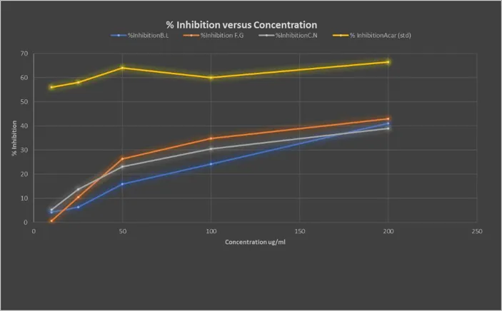
Figure 1. α-Amylase inhibitory activity of Bitter Leaf, Fenugreek, Cinnamon and Acarbose at different concentration
3.4. Gummy formulation and physicochemical evaluation
Three gummy formulations (F1, F2 and F3) were prepared using different gelatin concentrations and evaluated for appearance, pH, swelling ratio, syneresis and disintegration time. All formulations exhibited a brown colour and heart-shaped appearance (Figure 2). F1 was non-sticky, whereas F2 and F3 showed slightly sticky textures.
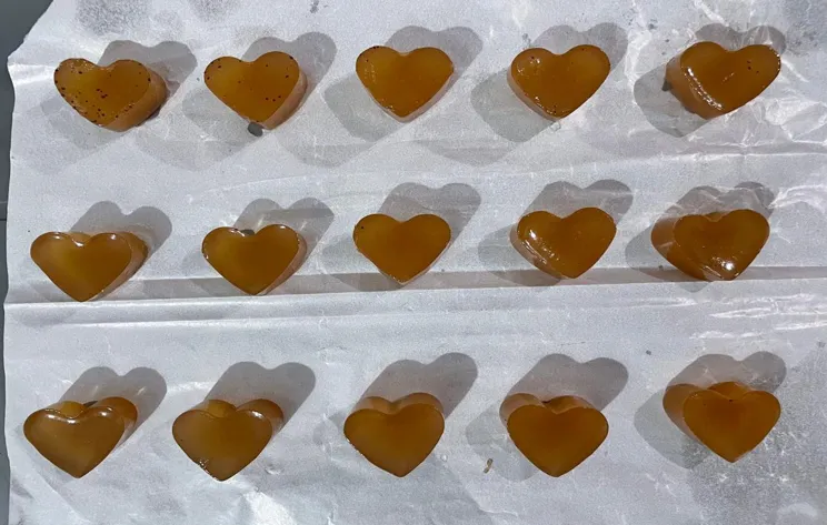
Figure 2. Formulated herbal nutraceutical gummies containing Bitter Leaf, Fenugreek and Cinnamon extracts.
F1 had a pH of 5.92 ± 0.356 and a swelling ratio of 1.0234 ± 0.2301. Syneresis was observed and the disintegration time was 7 ± 1.21 min. F2 had a pH of 5.5, the highest swelling ratio (1.1125), no observable syneresis and the shortest disintegration time (5 min). F3 had a pH of 4.8, a swelling ratio of 1.0052, slight syneresis and a disintegration time of 6 min.
Based on the overall physicochemical characteristics, F2 was selected as the optimized formulation because it showed the highest swelling ratio, absence of syneresis and the shortest disintegration time among the three formulations (Table 4).
Table 4. Physicochemical evaluation of the formulated herbal gummies (F1–F3)
|
Parameter evaluated |
F1 |
F2 |
F3 |
|
Physical appearance |
Brown; heart shape; non-sticky texture |
Brown; heart shape; slightly sticky texture |
Brown; heart shape; slightly sticky texture |
|
pH |
5.92 ± 0.356 |
5.5 (within 3–6 range) |
4.8 (within 3–6 range) |
|
Swelling ratio |
1.0234 ± 0.2301 |
1.1125 |
1.0052 |
|
Syneresis study |
Syneresis observed |
No syneresis observed |
Slight syneresis observed |
|
Disintegration time |
7 ± 1.21 minutes |
5 minutes |
6 minutes |
3.5. Total flavonoid content
The total flavonoid content of the formulated gummy was determined spectrophotometrically using quercetin as the reference standard. The calibration curve showed good linearity over the tested range, with the equation y = 0.1932x + 0.0216 and R² = 0.9979. The gummy extract showed an absorbance of 1.51 at 415 nm (Table 5).
Table 5. Total flavonoid content of the formulated herbal gummy
|
|
Concentration (ug/ml) |
Absorbance |
|
Quercetin (STD) |
0 |
0 |
|
|
0.65 |
0.12 |
|
|
1.25 |
0.27 |
|
|
2.5 |
0.54 |
|
|
8 |
1.52 |
|
|
10 |
1.96 |
|
Gummy (extract) |
8 |
1.51 |
The total flavonoid content was calculated as 0.07705 mg quercetin equivalent (QE) per gram of gummy.
3.6. Molecular docking and ADMET evaluation
The docking analysis demonstrated favorable predicted interactions between several V. amygdalina phytoconstituents and both target enzymes, α-amylase (1HNY) and acid α-glucosidase (5NN8) (Figure 3).
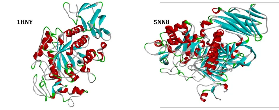
Figure 3. Molecular docking interactions of selected phytoconstituents with α-amylase (PDB ID: 1HNY) and lysosomal acid α-glucosidase (PDB ID: 5NN8).
Luteolin 7-O-β-glucoside showed the most favorable predicted binding energy against 5NN8 (−11.35 kcal/mol), while its predicted binding energy against 1HNY was −10.07 kcal/mol. Vernonioside B2 also showed strong predicted binding, with values of −10.78 and −10.75 kcal/mol against 5NN8 and 1HNY, respectively (Table 6).
Table 6. Binding energies and ligand efficiencies of selected phytoconstituents against 5NN8 and 1HNY
|
Phytoconstituent (ligand) |
5NN8 B.E. (kcal/mol) |
5NN8 L.E. |
1HNY B.E. (kcal/mol) |
1HNY L.E. |
|
Acarbose (std) |
-8.85 |
0.201 |
-4.43 |
0.101 |
|
3,5-Dicaffeoylquinic acid |
-9.72 |
0.207 |
-8.81 |
0.238 |
|
Apigenin |
-8.54 |
0.427 |
-7.51 |
0.376 |
|
Caffeic acid |
-6.81 |
0.524 |
-7.66 |
0.589 |
|
Chlorogenic acid |
-6.81 |
0.524 |
-7.64 |
0.587 |
|
Diosgenin |
-9.57 |
0.383 |
-10.5 |
0.306 |
|
Kaempferol |
-8.73 |
0.397 |
-7.55 |
0.477 |
|
Luteolin 7-O-β-glucoside |
-11.35 |
0.378 |
-10.07 |
0.252 |
|
Luteolin 7-O-β-D-glucuronide |
-10.35 |
0.323 |
-9.52 |
0.325 |
Several compounds demonstrated more favorable predicted binding energies than Acarbose. However, ligand efficiency analysis provided a different ranking. Caffeic acid and chlorogenic acid demonstrated particularly high ligand efficiencies, with values of 0.524 against 5NN8 and 0.589 and 0.587 against 1HNY, respectively.
Following docking and ligand-efficiency screening, seven compounds were shortlisted for further in-silico evaluation. SwissADME analysis identified caffeic acid, apigenin and kaempferol as the most favorable candidates, with zero predicted Lipinski violations and predicted bioavailability scores of ≥0.55 (Table 7).
Table 7. ADME and drug-likeness characteristics of selected Vernonia amygdalina phytoconstituents
|
Ligand name |
Mw (g/mol) |
Mlogp |
TPSA (Ų) |
H-donors |
H-acceptors |
LogS (solubility) |
Lipinski violations |
Bioavailability |
|
3,5-Dicafeoyl quinic acid |
516.45 |
-0.35 |
211.28 |
7 |
12 |
-3.65 |
3 |
0.11 |
|
Apigenin |
270.24 |
0.52 |
90.9 |
3 |
5 |
-3.94 |
0 |
0.55 |
|
Caffeic acid |
180.16 |
0.7 |
77.76 |
3 |
4 |
-1.89 |
0 |
0.56 |
|
Chlorogenic acid |
354.31 |
-1.05 |
164.75 |
6 |
9 |
-1.62 |
1 |
0.11 |
|
Diosgenin |
414.62 |
4.94 |
38.69 |
1 |
3 |
-5.98 |
1 |
0.55 |
|
Kaempferol |
302.24 |
0.12 |
120.36 |
4 |
7 |
-3.38 |
0 |
0.55 |
|
Luteolin 7-O-beta-glucoside |
432.38 |
-1.09 |
177.14 |
7 |
10 |
-2.79 |
1 |
0.55 |
|
Luteolin 7-O-beta-D-glucuronide |
462.36 |
-2.12 |
207.35 |
7 |
12 |
-3.41 |
2 |
0.11 |
|
Luteolin |
286.24 |
-0.03 |
111.13 |
4 |
6 |
-3.71 |
0 |
0.55 |
|
Quercetin |
302.24 |
-0.56 |
131.36 |
5 |
7 |
-3.16 |
0 |
0.55 |
|
Trigonelline |
137.14 |
0.33 |
44.01 |
0 |
2 |
-1.39 |
0 |
0.55 |
|
Vernodalin |
360.36 |
1.42 |
99.13 |
1 |
7 |
-1.94 |
0 |
0.55 |
|
Vernomygdin |
394.46 |
1.81 |
94.59 |
1 |
7 |
-3.69 |
0 |
0.55 |
|
Vernodalol |
388.45 |
2.08 |
89.9 |
1 |
6 |
-3.65 |
0 |
0.55 |
|
Vernolepin |
276.28 |
1.42 |
72.83 |
1 |
5 |
-2.35 |
0 |
0.55 |
|
Vernonioside B2 |
682.8 |
-0.33 |
207.99 |
8 |
13 |
-3.62 |
3 |
0.17 |
ProTox-III predicted an oral LD₅₀ of 3919 mg/kg for kaempferol (Class 5), while caffeic acid and apigenin were predicted to have LD₅₀ values of 1190 mg/kg (Class 4). Apigenin was additionally associated with predicted hepatotoxicity, neurotoxicity and immunotoxicity alerts. These are in silico predictions and do not establish clinical safety.
4. DISCUSSION
The present study evaluated the in vitro α-amylase inhibitory activity of fenugreek, bitter leaf and cinnamon extracts as part of their potential antidiabetic activity. All three extracts demonstrated inhibitory activity against α-amylase at the tested concentrations, although the degree of inhibition differed among the extracts. At 200 µg/mL, fenugreek showed the highest inhibition (43.15%), followed by bitter leaf (41.05%) and cinnamon (38.95%). Acarbose, used as the positive control, produced a higher inhibition of 66.11% at the same concentration. These findings indicate that the three plant extracts possess the ability to inhibit α-amylase in vitro, although their activity under the present experimental conditions was lower than that of the standard drug.
Fenugreek showed the highest α-amylase inhibition among the three extracts. This finding is consistent with previous reports demonstrating the ability of Trigonella foenum-graecum to inhibit carbohydrate-hydrolyzing enzymes. A study investigating fenugreek leaf extracts reported concentration-dependent inhibition of α-amylase and α-glucosidase, with the ethyl acetate extract showing greater α-amylase inhibition than the aqueous extract.¹⁴ The antidiabetic activity of fenugreek has also been associated with several mechanisms, including inhibition of α-amylase and maltase, enhancement of glucose utilization and protection of pancreatic β-cells.¹⁵ The comparatively greater activity observed for fenugreek in the present study may therefore be related to the presence of bioactive constituents capable of interacting with carbohydrate-digesting enzymes. However, the extent of inhibition can vary according to the plant part, extraction solvent, phytochemical composition and assay conditions, making direct comparison between studies difficult.
Bitter leaf (Vernonia amygdalina) also demonstrated appreciable α-amylase inhibition, producing 41.05% inhibition at 200 µg/mL. This finding is supported by previous in vitro investigations of V. amygdalina, in which phenolic extracts significantly inhibited α-amylase and α-glucosidase activities in a concentration-dependent manner.¹⁶ More recent work has also identified α-amylase inhibitory activity in V. amygdalina extracts and associated the activity with secondary metabolites including flavonoids, phenolic compounds, tannins, alkaloids and saponins.¹⁷ Therefore, the inhibitory activity observed in the present study provides further support for the potential contribution of bitter leaf to the modulation of carbohydrate digestion. Nevertheless, the inhibition obtained in the present experiment was lower than that reported for some solvent fractions of V. amygdalina. This difference may be attributed to differences in extraction procedures, extract composition, plant material and experimental conditions.
Cinnamon produced 38.95% inhibition at 200 µg/mL, which was slightly lower than the activities observed for fenugreek and bitter leaf in the present study. Nevertheless, this result supports previous evidence that cinnamon can inhibit enzymes involved in carbohydrate digestion. Cinnamon extracts have been reported to inhibit α-amylase and α-glucosidase, although the magnitude of inhibition varies considerably among Cinnamomum species.¹⁸ A comparative study of four commercially used cinnamon species demonstrated marked species-dependent differences in α-amylase inhibition, with Cinnamomum cassia showing greater activity than some of the other species examined.¹⁸ This provides a possible explanation for differences between the present finding and previously reported activities because the botanical species, plant origin and phytochemical composition of cinnamon can influence its enzyme-inhibitory properties.
The lower inhibition observed for all three extracts compared with Acarbose is expected because Acarbose is a clinically established carbohydrate-digesting enzyme inhibitor specifically used to delay carbohydrate digestion and reduce postprandial glucose elevation. The stronger inhibition produced by Acarbose in the present assay confirms the responsiveness of the assay system and provides a useful reference for comparison with the plant extracts. Importantly, the fact that the extracts showed lower activity than Acarbose does not exclude their potential antidiabetic value. Plant extracts contain multiple constituents that may act through several complementary mechanisms beyond direct α-amylase inhibition, including effects on α-glucosidase, glucose uptake, insulin secretion, oxidative stress and glucose metabolism. Fenugreek, for example, has been reported to influence several pathways involved in glucose homeostasis rather than acting solely through digestive enzyme inhibition.¹⁵
An important observation from the present study is that none of the three plant extracts achieved 50% inhibition at the highest experimentally tested concentration of 200 µg/mL. Consequently, an experimentally determined IC₅₀ could not be established within the concentration range investigated. This should be considered when interpreting the magnitude of the inhibitory activity. Although the extracts clearly demonstrated measurable α-amylase inhibition, the present results do not support assigning a numerical IC₅₀ value without additional concentration-response data extending beyond 200 µg/mL. This also represents a limitation of the present in vitro evaluation and indicates that future studies should include a broader concentration range if accurate IC₅₀ determination is required.
The differences observed among the extracts may reflect variations in their phytochemical composition. Phenolic compounds, flavonoids, tannins and other secondary metabolites have been associated with inhibition of carbohydrate-hydrolyzing enzymes. Such compounds may interact with enzyme proteins through hydrogen bonding, hydrophobic interactions and other non-covalent interactions, thereby reducing catalytic activity. However, the present assay evaluated crude plant extracts and therefore cannot establish which individual compounds were responsible for the observed inhibition. Phytochemical characterization and subsequent enzyme-inhibition studies using isolated or enriched fractions would be required to establish the compounds contributing to the activity.
Overall, the findings demonstrate that fenugreek, bitter leaf and cinnamon possess measurable in vitro α-amylase inhibitory activity, with fenugreek showing the greatest inhibition among the tested extracts. The results are broadly consistent with previous reports describing the ability of these plants to interfere with carbohydrate-digesting enzymes, while the differences in the magnitude of inhibition highlight the influence of plant material, phytochemical composition and experimental conditions. The findings provide experimental support for the potential use of these plant materials as sources of α-amylase-inhibitory constituents, although the activity was lower than that of Acarbose under the conditions used. Further studies involving phytochemical standardization, α-glucosidase inhibition, expanded concentration-response analysis and appropriate in vivo models would be necessary to determine whether the observed in vitro activity translates into meaningful antidiabetic effects.
5. CONCLUSION
The present study successfully investigated the development of a herbal nutraceutical gummy containing Vernonia amygdalina, Trigonella foenum-graecum, and cinnamon extracts. The selected plant materials showed the presence of several major phytochemical groups and demonstrated measurable in vitro α-amylase inhibitory activity. Among the three extracts, fenugreek showed the highest inhibition at 200 µg/mL (43.15%), followed by bitter leaf (41.05%) and cinnamon (38.95%), although all three showed lower inhibition than Acarbose (66.11%). Since 50% inhibition was not achieved within the tested concentration range, an experimental IC₅₀ could not be established.
Three gummy formulations were prepared by varying the gelatin concentration, and F2 containing 10% gelatin was selected as the optimized formulation based on its higher swelling ratio, absence of syneresis and shorter disintegration time. The optimized formulation also demonstrated a measurable total flavonoid content of 0.07705 mg QE/g. Molecular docking further indicated favorable predicted interactions between selected V. amygdalina phytoconstituents and α-amylase and lysosomal acid α-glucosidase, with caffeic acid, apigenin and kaempferol showing favorable overall in silico characteristics.
Overall, the study provides preliminary experimental and computational evidence for the potential of the selected plant extracts as ingredients for herbal nutraceutical development. However, the findings are based primarily on in vitro and in silico investigations and should not be interpreted as evidence of clinical antidiabetic efficacy. Further studies involving expanded concentration-response analysis, phytochemical standardization, additional enzyme assays, in vivo evaluation and safety assessment are required to establish the therapeutic potential of the developed formulation.
6. ACKNOWLEDGEMENTS
The authors thank the Faculty of Pharmacy, Marwadi University, Rajkot, for providing the laboratory facilities and resources necessary to carry out this research.
Author contributions: To be completed by the authors using the journal’s preferred contribution statement.
Funding: None to declare.
Conflict of interest: None to declare.
Ethics approval: Not applicable. This study did not involve human participants or animal subjects.
REFERENCES


Jochebed Dzyeenom Joel, Emmanuel M Shabani, Fathiah Oreoluwa Oladele, Gloria Ebisentei Ebimo-Moko, Hiral Kapuriya, Formulation and Standardization of Herbal Nutraceutical Gummies Containing Bitter Leaf, Fenugreek, and Cinnamon Extracts with In Vitro ?-Amylase Inhibitory Activity, Int. J. of Pharm. Sci., 2026, Vol 4, Issue 10, 718-733. https://doi.org/10.5281/zenodo.23170038
 10.5281/zenodo.23170038