
We use cookies to ensure our website works properly and to personalise your experience. Cookies policy
RJS College of Pharmacy, Kokamthan, Kopargaon.
The present study focuses on the formulation and evaluation of a Fluconazole-loaded liposomal gel for topical drug delivery in the treatment of fungal infections. Conventional topical antifungal formulations often show limited skin penetration and reduced drug retention, leading to decreased therapeutic effectiveness. To overcome these limitations, liposomes were used as novel drug carriers due to their ability to enhance drug permeation, improve stability, and provide controlled drug release. Fluconazole-loaded liposomes were prepared by the thin film hydration method using phospholipids, stearic acid, chloroform, and ethanol. The prepared liposomal suspension was incorporated into a Carbopol 934 gel base to obtain the liposomal gel formulation. The developed formulation was evaluated for physicochemical and pharmaceutical parameters including particle shape and size, pH, zeta potential, entrapment efficiency, spreadability, washability, drug content uniformity, irritancy, and antifungal activity. The prepared liposomal vesicles were spherical in shape with satisfactory stability and an entrapment efficiency of 86.27%. The liposomal gel exhibited good spreadability, smooth consistency, easy washability, skin-compatible pH, and non-irritant characteristics. Drug content uniformity was found to be 85.01%, indicating uniform distribution of Fluconazole within the gel formulation. Antifungal studies against Candida albicans demonstrated effective antifungal activity of the developed formulation. The study concludes that Fluconazole-loaded liposomal gel can serve as a promising topical drug delivery system with improved therapeutic efficacy, better skin penetration, prolonged drug retention, and reduced systemic side effects compared to conventional formulations.
These days, one of the most prevalent dermatological issues is fungal skin infections. Solid, semisolid, and liquid dose formulations are among the many therapeutic options available to doctors. Clear transparent gels are the most extensively used topical formulation in both cosmetics and medications. Liposomal formulations have been widely used in the last 10 years to improve the effectiveness of medication delivery via a variety of administration routes. In many cases, liposomal medication formulations in any dose form have proven to be significantly better than traditional dosage forms, particularly for topical and intravenous drug administration. Topical liposomal medication formulations are superior to conventional dosing forms primarily due to their ability to:
The development of controlled drug delivery systems (CDDS) has received more attention in recent years. Better plasma level profiles, higher patient compliance, reduced dosage and toxicity, targeted potential, and more effective active agent use are just a few of the many benefits of CDDS over traditional therapy. One of the best methods for delivering drugs to the intended organ while reducing their spread to non-target tissues is liposomes. There is a lot of promise for topical delivery with novel drug delivery systems (NDDS).
Since the past ten years, lipidic and non-lipidic vesicular systems, such as liposomes, niosomes, and ethosomes, have been employed to address the drawbacks of traditional topical formulations. Because these vesicular systems offer controlled medication release, they are found to be far more effective. This is because they create a depot in the epidermis that maintains the therapeutic medication concentration for an extended amount of time. The use of topically applied liposomal formulations for fungal infections utilizing fluconazole as the preferred medication is covered in later chapters of this study.
Fluconazole would be used in the topical antifungal formulation to treat deeper fungal infections such as candidiasis. Because liposomal topical gel has so many advantages over traditional dosing forms, it was developed. The route of penetration, the mechanism by which liposomes aid in medication deposition into the skin, and its possible uses are all covered. For both local and systemic treatment, topical medication distribution is a very practical and appealing method. Drugs applied topically have been found to be an effective treatment for localized dermatological conditions. One synthetic antifungal drug that is derived from triazoles is fluconazole. In addition to other severe systemic candidal infections, it is used to treat oropharyngeal, esophageal, or candidiasis. Additionally, it works well against dermatophytoses and superficial fungal infections.
Conventional formulations may not be as effective as topical liposome formulations. Fluconazole molecules will be assisted in penetrating the skin layers by liposomal carriers, which are well-known for their potential in topical medication administration and their drug release efficiency. It is anticipated that these vesicles will offer lipid-enriched hydration conditions to keep the medication molecules inside the layers of skin. In order to achieve this goal, fluconazole-loaded liposomal systems will be created, and their topical efficacy will be contrasted with that of non-liposomal fluconazole-containing systems.
LIPOSOMES AS A NEW MEDICATION DELIVERY SYSTEM
For a successful therapy, an ideal release mechanism distributes the medication to the body at a predetermined pace within a certain amount of time. Medications used to treat different fungal diseases can be extremely harmful to healthy tissues. Reducing the amount of these medications' distribution to healthy cells and tissues could lessen their toxicity. One of the best models of cell membranes is thought to be liposomes. Effective drug delivery, which is commonly employed in cutaneous applications, is explained. Alec D. Bangham, a British haematologist, originally described liposomes at the Babraham Institute in Cambridge in 1961 (published in 1964)
Liposomes are tiny spherical vesicles with an aqueous core that are composed of one or more lipid bilayers. The pharmaceutical and cosmetic industries have long utilized liposomes as carriers for a variety of compounds. Food and underdeveloped sectors employ liposome encapsulation as a grow delivery technique that can contain unstable substances and protect their functioning. Both hydrophobic and hydrophilic substances can be caught by liposomes, which can then release the trapped substances at specific locations while preventing the disintegration of the entrapped mixtures. Because of its biocompatibility, biodegradability, low toxicity, and ability to capture both hydrophilic and lipophilic medicines and facilitate site-specific drug delivery to tumor tissues, liposomes have become more popular as a drug delivery device. Liposomes have been the subject of numerous investigations aimed at reducing medication toxicity or focusing on particular cells Increasing the drug's therapeutic index while reducing its negative effects is one of the primary goals of any drug-using behavior.
The majority of conservative chemotherapeutics' clinical use is limited by their inability to deliver therapeutic drug concentrations to the target soft tissue or by their toxic side effects on healthy organs and tissues. Many strategies have been developed to overcome this challenge by delivering the medication "selectively" to the intended location; the best solution would be to only target the cells, tissues, and organs that are impacted by the illness. Liposomes are important because of their makeup, which makes them biocompatible and biodegradable.
There are numerous ways to administer the medications contained in liposomes, including intravenous, oral, nasal, intramuscular, pulmonary, topical, and ophthalmic.
Numerous vesicles, such as creams, ointments, capsules, solutions, sprays, etc., can be used to deliver them. Numerous illnesses, including endocrine, arthritis, cancer, bacterial, fungal, ophthalmic, vaccinations, fibrinolysis, asthma, diabetes, immune system disorders, painkillers, and topical anesthetic, can be treated with liposomes. Liposomes are categorized according to their composition, manufacture method, and use in biology, biochemistry, cosmetics, and medicine. Both manufactured and natural phospholipids have the ability to produce liposomes. Although they can be found in plant oils like olive oil, the primary sources of natural phospholipids are yolk eggs and soy beans.
Conventional liposomes, pH-sensitive liposomes, cationic liposomes, immunoliposomes, temperature- or heat-sensitive liposomes, magnetic liposomes, and sterically stabilized "stealth" liposomes are examples of liposomes that can be kept private. Further developments in liposome research have made it possible for liposomes to evade immune system identification, particularly by reticuloendothelial system (RES) cells. These liposomes are called "stealth liposomes" because the membrane's outside is studded with PEG (polyethylene glycol). The medication delivery device has a prolonged circulation life because to the PEG coating, which is inert in the body. However, current study aims to determine how much PEG coating actually prevents the liposome from attaching to the delivery site.
To facilitate binding via a particular expression on the targeted drug delivery location, most stealth liposomes feature a biological species connected as a ligand to the liposome in addition to a PEG coating. These targeting ligands could be particular antigens, vitamins, or monoclonal antibodies that form an immunoliposome. Drugs that would normally be administered systemically can be transported via targeted liposomes, which can target almost any type of cell in the body. When making liposomes, the following phospholipids are most frequently utilized: Phosphatidylinositol (PI), phosphatidylcholine (PC), phosphatidylserine (PS), and phosphatidylethanolamine (PE) Phosphatidylcholine and cholesterol are the typical components of conservative liposomes. Conversely, "stealth" liposomes are lipid bilayers that have been coated with polyethylene glycol. These liposomes offer the entrapped molecules more protection and improved stabilization.
LIPOSOME CLASSIFICATION USING STRUCTURE PARAMETERS
They enable the medication delivery system to circulate for a longer period of time. Continued drug release can accomplish the significance of a protracted duration. Liposomes have little intrinsic toxicity and are made of recognized phospholipids that are weakly immunogenic and physiologically inactive. Additionally, medications with varying lipophilicities can be encapsulated in liposomes: highly hydrophilic drugs are located entirely in the aqueous compartment, whereas extremely lipophilic drugs are almost fully confined in the lipid bilayer.
LIPOSOME STRUCTURE AND COMPOSITION
Liposomes appear to have the greatest potential among the range of innovative drug delivery systems because they can hold both water-soluble and lipid-soluble compounds, protecting the drug encapsulated in the liposome from metabolic degradation and serving as a delivery tool by releasing active ingredients gradually and under control. Phospholipid, a component of the liposome lipid bilayer, is often derived from egg yolk or soy bean oil. It is composed of two hydrocarbon tails that represent the lipophilic portion and a hydrophilic head section that is covalently connected. The hydrophilic head groups in a bilayer structure aggregate by moving toward the aqueous environment. while retaining the hydrocarbon chains that are lipophilic within.
When the polar head groups and hydrophobic contacts of the lipid chain are solvated, the formation of such a shape gives the vesicle the lowest potential energy state. The primary component of many liposomal formulations is natural phosphatidylcholine that is derived from egg yolk, soy bean oil, or its semisynthetic counterparts. A glycerol moiety is joined to two acyl chains, which can be saturated or unsaturated, in the chemical structure of naturally occurring phosphatidylcholine. The hydrophobic (lipophilic) section of the particle may consist of ten to twenty-four carbon atoms. The hydrophilic "head" is made up of the phosphate and choline moieties.
Depending on their length and level of saturation, the fatty acid chains can reside in either the more fluid liquid crystalline phase or the gel phase, where the lipids are stiff, impermeable, and readily agglomerate during storage. The transition temperature is the temperature at which the gel phase changes into the liquid crystalline phase. Most liposomal formulations often contain trace amounts of cholesterol to improve the fluidity of the liposomal gel phase and the retention of hydrophilic particles and to stabilize the bilayer membrane in a way that resembles biological membranes
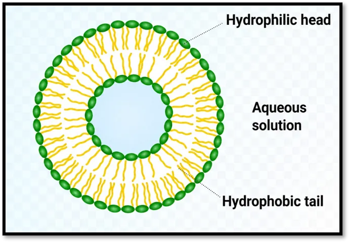
Fig no 01: Structure of Liposomes.
METHODS OF PREPARATION OF LIPOSOMES:
1. Thin Film Hydration Method
2. Sonication Method
3. French Pressure Method
4. Ether Infusion Method
5. Ethanol Injection Method
6. Reverse Phase Evaporation Method
1. Thin Film Hydration Method: The most used approach for MLV research. The process entails drying a lipid solution until a thin film form at the bottom of a round-bottom flask. The film is then hydrated by adding water buffer and briefly vortexing the dispersion. The temperature used for the hydration process is higher than the gel-liquid crystalline transition. temperature Tc of the lipid or higher than the Tc of the lipid mixture's maximal melting component. Depending on their solubilities, the chemicals to be encapsulated are added to either an organic solvent containing lipids or an aqueous buffer.This approach makes MLV easy to organize, and these liposomes can be used to encapsulate a wide range of compounds. limited internal volume, limited encapsulation efficiency, and a heterogeneous size distribution are the process's drawbacks
2. Sonication Method: Under an inert atmosphere, MLVs are solicited using either a probe sonicator or a bath type sonicator. This method's primary disadvantages are extremely low interior volume/encapsulation efficiency, potential degradation of phospholipids and compounds to be encapsulated, exclusion of big molecules, metal pollution from the probe tip, and the presence of MLV in addition to SUV.
3. French Pressure Method: MLV is extruded through a tiny aperture at 20,000 psi and 4°C. Compared to the sonication approach, the method has several advantages. The process is straightforward, quick, repeatable, and requires handling unstable materials gently. Compared to sonicated SUVs, the resultant liposomes are a little bigger.
4. Ethanol Injection Method: A large excess of buffer is quickly injected with a lipid ethanol solution. The MLVs are created right away. The method's disadvantages include the population's heterogeneity (30-110 nm), the liposomes' extreme dilution, the difficulty of eliminating all ethanol due to its azeotrope formation with water, and the potential for different biologically active macromolecules to become inactive in the presence of even minute amounts of ethanol.
5. Ether Infusion Method: An aqueous solution of the item to be encapsulated is slowly injected with a solution of lipids dissolved in diethyl ether or an ether/methanol combination at 55–65°C or under decreased pressure. Liposomes are created when ether is then removed under vacuum. The population's heterogeneity (70–190 nm) and the chemicals to be encapsulated's exposure to organic solvents or high temperatures are the method's primary disadvantages.
6. Reverse Phase Evaporation Method: First water in oil emulsion is formed by sonication of a two-phase system containing phospholipids in organic solvent and aqueous buffer. A viscous gel is created when the organic solvents are extracted under low pressure. When leftover solvent is eliminated through continuous rotating evaporation at lower pressure, liposomes are created. This technique can achieve high encapsulation competency of up to 65% in a low ionic strength media, such as 0.01 M NaCl. Small, big, and macromolecules have all been encapsulated using this technique. The exposure of the materials to be encapsulated to organic solvents and short sonication times is the method's primary drawback.
MATERIAL AND METHOD
MATERIAL –
Chemicals:
Flucanazole:
Vaginal thrush, oral thrush, esophageal candidiasis, and cryptococcal meningitis are among the fungal and yeast infections that can be treated and prevented using the prescription antifungal drug fluconazole.
It works by preventing the growth of fungus, and symptoms usually go away in 24 to 3 days.
Phospholipids:
The basic structural components of all biological cell membranes are phospholipids, a type of compound lipids.
They have two hydrophobic (water-repelling) "tails" and a hydrophilic (water-attracting) "head" because they are amphipathic molecules.
In aquatic settings, their special dual characteristic enables them to spontaneously self-assemble into a lipid bilayer, thereby regulating what enters and leaves a cell.
Chloroform:
Chloroform (systematic IUPAC name: trichloromethane) is a chemical compound that is transparent, colorless, and extremely volatile. It smells sweet and ether-like.
It was independently discovered in 1831 and became well-known for being the first inhaled medicinal anesthetic.
Its clinical use has been completely discontinued in contemporary medicine due to its extreme toxicity profile and tendency to cause organ damage.
It is now mostly used as a chemical solvent and heavy-duty industrial precursor.
Methylparaben:
A common preservative in food, cosmetic, and pharmaceutical items is methylparaben.
Methylparaben is a preservative used in emulgel compositions to stop bacteria and fungi from growing.
Triethanolamine:
pH adjuster Triethanolamine is used, especially in medicines and cosmetics, to change the pH of compositions.
Triethanolamine, an emulsifier, enhances the solubility of components and stabilizes emulsions.
Essential oils and perfumes are among the many compounds for which triethanolamine is employed as a solvent.
Ethanol:
The main purpose of ethanol in liposomes is to serve as a solvent for active substances, which aids in the development of a stable and uniform topical formulation. It can also serve as a co-surfactant and a penetration enhancer, and at a certain concentration, it can protect the network structure of the liposomes, increasing their freeze-thaw stability.
Carbapol934:
A synthetic high molecular weight polymer of acrylic acid, carbopol 934 is frequently employed as a gelling agent in cosmetic and medicinal applications.
Gelling Agent: Carbopol 934 is a transparent, stable gel that swells in water. neutralized (such as sodium hydroxide or triethanolamine).
For topical applications, it has a smooth, non-greasy feel. By avoiding phase separation, it aids in stabilizing suspensions or emulsions in gel compositions.
Glycol Propylene:
The main role of propylene glycol in emulgel is to improve the solubility and skin delivery of hydrophobic medications by acting as a co-surfactant and penetration enhancer.
Additionally, it absorbs water and aids in moisture retention by acting as a humectant.
It is frequently utilized in liposomalgel formulation to assist generate a stable, effective product by dissolving additional ingredients like parabens.
EXPERIMENTAL WORK
FORMULA
Formula for liposomes:
Table no: 01 Formula for liposomes: (For 20 gm)
|
Sr. no |
Ingredients |
Quantity |
|
1 |
Flucanazole |
0.2 gm |
|
2 |
Phospholipds |
0.2 gm |
|
3 |
Stearic Acid |
0.4 gm |
|
4 |
Chloroform |
08 ml |
|
5 |
Ethanol |
02ml |
|
6 |
Methyl Paraben |
0.2 gm |
|
7 |
Distilled Water |
q.s to 20 ml |
Formula for gel:
Table no: 02 Formula for gel: (For 20 gm)
|
Sr. no |
Ingredients |
Quantity |
|
1 |
Carbopol |
0.2 gm |
|
2 |
Methyl Paraben |
0.02 gm |
|
3 |
Triethanolamine |
pH adjusts to 6.4 to 7.4 |
|
4 |
Distilled Water |
q. s to 20 gm |
How to Make Liposomal Gel:
Forming the liposomes carrying the active substance and then integrating them into a gel base for topical administration are two separate procedures in the manufacture of a liposomalgel.
METHODS:
Step 1: Liposome Preparation As indicated in Table No. 01, the various liposome formulations were made by dissolving flucanazole, soy lecithin, and cholesterol in ethanol and chloroform. This mixture was placed in a 250 ml round-bottom flask. To create a thin lipid coating on the flask wall, evaporate the organic solvent. A milky white suspension was produced by progressively hydrating the dry lipid film with 10 milliliters of phosphate buffer pH 6.8 and rotating the mixture for two hours. To extract the uncapsulated medication, the formulation is centrifuged for 30 minutes at 3000 rpm.
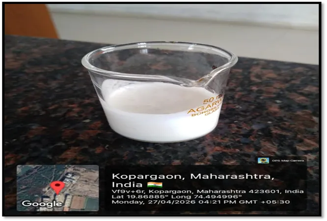
Fig no: 02 Liposomal Suspension
Step 2: Carbopol gel preparation the determined quantity of carbopol was added to the warm water while being continuously stirred at a moderate speed using a magnetic stirrer. Triethanolamine was used to change the pH of the Carbopol gel.
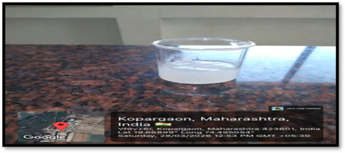
Fig no: 03 Carbopol gel
Step 3: Liopsomal gel formation to create the liposomal gel, the produced liposomal suspension and gel base were separately combined in the proper ratio while being continuously stirred. A container was filled with the prepared Liopsomal gel
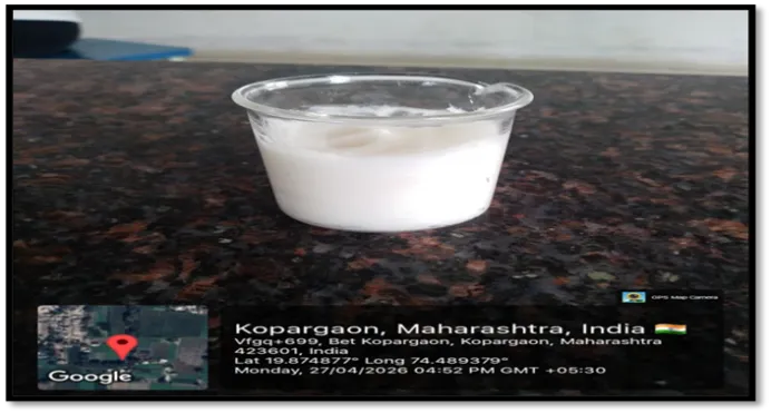
Fig no: 04 Liosomal gel
EVALUATION PARAMETER:
1.Evaluation of Liposome:
1.Colour
2. Particle shape and size analysis
3.pH of Liposomes
4. Zeta potential
5. Entrapment efficiency
2.Evaluation for Gel:
1.Colour
2.pH of gel
3.Grittiness
4.Spreadability
3.Evalution for Liposomal gel
1.Physical Examination
2. pH of Liposomal gel
3. Spreadability
4. Washability
5. Drug content
6. Irritancy Test
7. Anti fungal Activity
1. Evaluation of Liposome:
1. Color: Visual inspection can also be used to evaluate it.
2. Particle size and shape analysis: An optical microscope is used to examine the shape of the particles.
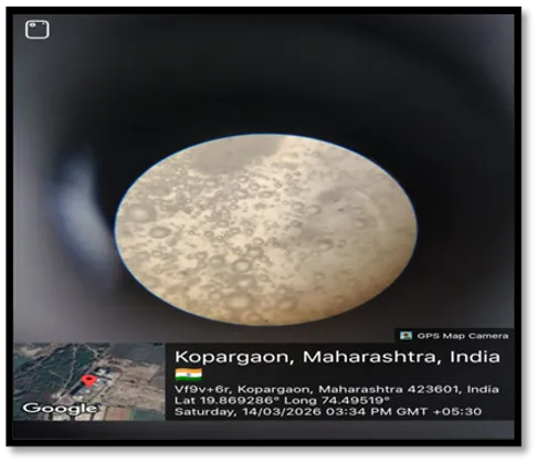
Fig no. 05: Particle shape
3. Liposome pH: A digital pH meter was used to measure the formulations' pH. The formulation's pH was measured in triplicate, and average results were computed.
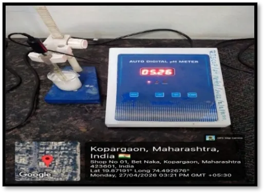
Fig no. 06: pH of Liposomal suspension
4. Zeta potential: The partical size reveals the uniformity of the size distribution and displays the average diameter of the liposomal vesicles. The vesicles' surface charge is represented by their zeta potential, which aids in predicting their stability.
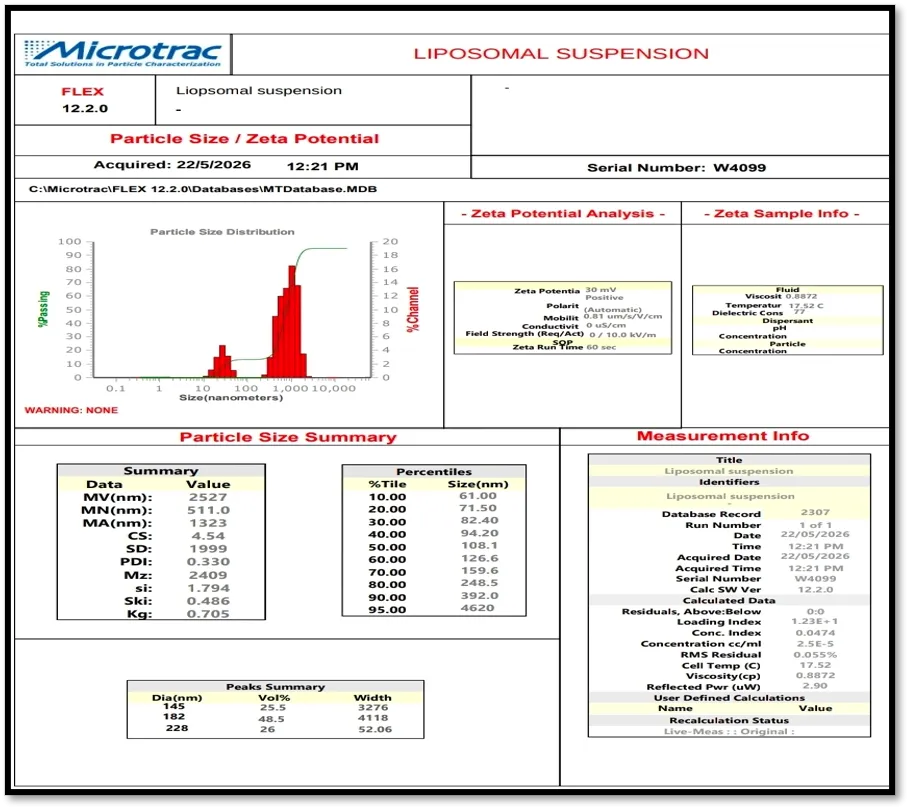
Fig no. 07: Particle size & zeta potential
5. Entrapment efficiency: The following formula can be used to calculate EE, which is the ratio of drug molecules encapsulated in liposomal particles to the total amount of medication used.
Entrapped drug divided by total drug times 100 is the entrapment efficiency.
Total medication = First medication added Free drug is the unentrapped drug found in the supernatant.
Entrapped drug is equal to total drug minus free drug. Following the preparation of liposomal solution, unentrapped drug is separated using centrifugation, gel filtering, and dialysis.
Centrifuge the sample for 60 minutes at 4°C at a high speed of 15,000 rpm. In order to separate the entrapped drug from the free (unentrapped) drug in the supernatant, carefully pipette or decant the supernatant without upsetting the pellet.
Evaluation for Gel:
1.Color: Visual inspection is another way to evaluate it.
2. Grit: Visual inspection and light finger rubbing were used to test the liposomal gel. A smooth texture and homogeneous dispersion of the medication and excipients were indicated by the formulation's lack of gritty particles. The formulation's fineness is confirmed by the lack of grittiness, which enables topical administration without irritating the skin.
3. pH of gel: A digital pH meter was used to measure the formulations' pH. The formulation's pH was measured in triplicate, and average results were computed.

Fig no. 08 pH of gel
4. Spreadability
Weigh out one gram of gel and put it in the middle of a pre-marked circle or the initial area of a glass plate. Put a second glass plate on top of the gel and apply weight. For five minutes, a 500-gram weight is applied to the upper plate in order to compress the gel into a homogenous film. Calculate the diameter. Once the weight has been applied for the predetermined amount of time, take it off and measure the spread gel circle's circumference. Determine spreadability: A formula can be used to determine spreadability.
S=Df-Di / Di
Were,
S= spredability
Df =Diameter after spreading
Di=Initial diameter
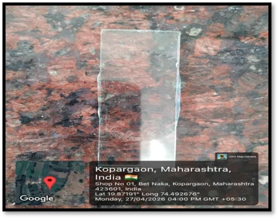
Fig no: 09 Spreadability of gel
1.Physical Assessment
Color: Visual inspection can also be used to evaluate it.
Grit: Visual inspection and light finger rubbing were used to test the liposomal gel. A smooth texture and homogeneous dispersion of the medication and excipients were indicated by the formulation's lack of gritty particles. The formulation's fineness is confirmed by the lack of grittiness, which enables topical administration without irritating the skin.
2. Liposomal gel pH: A digital pH meter was used to measure the formulations' pH. The formulation's pH was measured in triplicate, and average results were computed.
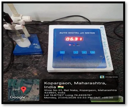
Fig no .10: pH of Liposomal gel
3. The ability to spread: Weigh out 1g of liposomal gel and put it in the middle of a pre-marked circle or the first spot on a glass plate. Put a second glass plate on top of the gel and apply weight. For five minutes, a 500-gram weight is applied on the upper plate in order to compress the liposomal gel into a homogenous layer. Calculate the diameter. Once the weight has been applied for the predetermined amount of time, take it off and measure the spread gel circle's circumference. Determine spreadability: A formula can be used to determine spreadability.
S=Df-Di / Di
Were,
S= spredability
Df =Diameter after spreading
Di=Initial diameter
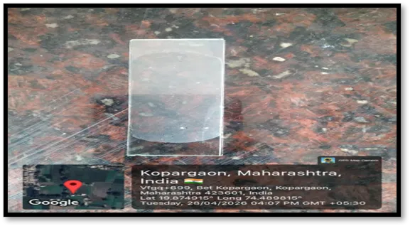
Fig no. 11: Spreadability of liposomal gel
4. Washability: A gel washability test evaluates a formulation's ease of removal from the skin with water. This is often done by putting a predetermined quantity of gel to the skin, rinsing it under running water, and tracking the removal. Because gels shouldn't leave any oily residue, it guarantees patient compliance.
5. Drug content: 20 ml of phosphate buffer (pH 5.5) and 1 g of liposomal gel formulation were added to a 100 ml volumetric flask, and the mixture was swirled for 30 minutes. One milliliter of the aforementioned solution was further diluted to fifty milliliters using phosphate buffer (pH 5.5) after the volume was increased to one hundred milliliters of ethanol. Whatman paper was used to filter the final solution. Using a UV spectrophotometer set to 260 nm and phosphate buffer pH 5.5 as a blank, the absorbance was measured. The drug content was calculated using the fluconazole standard calibration curve.
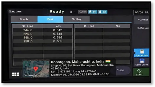
Fig. No. 12 Drug Content by UV Spectroscopy
6. Irritation Test: A shaved section of skin was treated with the liposomal gel. Any indications of redness, irritation, or itching were noted at the test location.
7. Anti-fungal Test: Fungal cultures were used to prepare the microorganism inoculum. Clean, sterile Petri dishes were filled with 15 milliliters of Saubroad agar (Hi media) medium, which was then left to cool and harden. Using a spreading rod, 100 µl of the fungal strain's broth was pipetted out and evenly distributed over the medium until it dried completely. A sterile cork borer was used to drill wells with a diameter of 6 mm. Compound solutions (100µl/ml) were made in DMSO, and the wells were filled with 100µl of the prepared test solutions and standard. The petri dishes were incubated for twenty-four hours at 37°C. DMSO was used as the negative control, and miconazole (1 mg/ml) was created as the positive control. The diameters of the zone of inhibitions (ZI) were measured to assess antifungal activity; all measurements were made in triplicate.
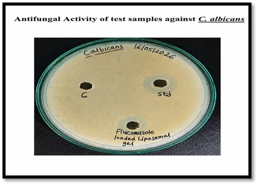
Fig no. 13: Atifungal Activity of test samples against C. albicans
RESULT AND DISCUSSION
Liposomal Suspension :
1. Particle shape analysis:
The Liposome prepared using flucanzole drug was studied under microscope to observe the formation of Liposomal vesicles. The Liposomal vesicles were found to be uniform in size and shape. The shape of the Liposomes was observed spherical.
2. pH :
The standard range of the pH of the Liposomal solution was 5.0- 7.4 and the pH of the Liposomal solution was found to be 5.22 which is ideal pH.
Table No. 03 pH of Liposomal suspension
|
Sr. No. |
Sample |
pH |
Mean |
|
1 |
Liposomal Suspension |
5.26 |
5.22 |
|
2 |
5.21 |
||
|
3 |
5.19 |
3. Zeta potential & Particle size:
The prepared Flucanazole -loaded Liposomes showed an average particle size of 182 nm, indicating larger vesicle formation. The zeta potential was found to be 30 mV, suggesting acceptable stability of the Liposomal suspension.
4. Entrapment efficiency :
Entrapped drug /Total drug× 100
=10 -1.373 / 10 × 100
=86.27 %
The entrapment efficiency of the fluconazole loaded Liposomal vesicles was found to be 86.27 %. For topical or transdermal delivery, an EE of 70-90% is desirable.
Gel :
1. pH : The standard range of the pH of the gel was 5.5 - 7.5 and the pH of the gel solution was found to be 5.83 which is ideal pH.
Table No. 04 pH of gel
|
Sr. No. |
Sample |
pH |
Mean |
|
1 |
Gel |
5.84 |
5.83 |
|
2 |
5.81 |
||
|
3 |
5.86 |
2. Spreadability:
S=Df-Di / Di
= 6-1 / 1
= 5 gm.cm/sec
Liposomal gel:
1. Physical Examination: The Liposomal gel was observed for colour, odour and appearance, which is shown in below table
Table no: 05 Observation of physical Examination
|
Sr. No. |
Properties |
Observation |
|
1 |
Color |
Milky White |
|
2 |
Odor |
Characteristic |
|
3 |
Clarity |
opaque |
|
4 |
Grittiness |
None |
|
7 |
Consistency |
Smooth |
2. pH : The standard range of the pH of the Liposomal gel was 5.1- 7.4 and the pH of the Liposomal gel solution was found to be 6.18 which is ideal pH.
Table No. 06 pH of Liposomal gel
|
Sr. No. |
Solution |
pH |
Mean |
|
1 |
Liposomal Gel |
6.31 |
6.18 |
|
2 |
6.10 |
||
|
3 |
6.15 |
3. Spreadability::
S=Df-Di / Di
=6.8-1/1
=5.8 gm.cm/sec
4. Washability:
The Liposomal gel was Easily Washable
5. Irritancy Test:
No any irritation or rashes seen on skin.
6. Antifungal Activity : The antifungal profile of Fluconazole Loaded Liposomal Gel was evaluated by measuring the zone of inhibition against fungal strains C albicans Via well diffusion method. The compounds Fluconazole Loaded Liposomal Gel exhibited good activity as compared to the standard Miconazole
Table no: 07: Antifungal Activity of test samples against C. albicans
|
Sr. No |
Samples |
Zone in diameter (mm) |
|
1 |
Control |
00 |
|
2 |
Standard (Miconazole) |
14 |
|
3 |
Fluconazole Loaded Liposomal Gel |
09 |
7. Drug Content:
Drug Content (%) = Sample absorbance / Standard absorbance × 100
Average absorbance
=0.512+0.518+0.526+0.535 / 4
=2.091 / 4
=0.52275 ≈ 0.522
If standard absorbance = 0.614, then
Drug Content (%) =0.522 / 0.614 × 100
=85.01%
The drug content was studied for the formulation prepared by Thin film hydration method. The drug content was found to be 85.01 %
Table no: 07: Evaluation Parameters and Standard Ranges
|
Sr. No |
Evaluation |
Standard Ranges |
Observation |
|
A |
Evaluation of Liposome |
|
|
|
A |
Colour |
Milky white, slightly translucent, or opalescent |
Milky White |
|
B |
Particle shape and size analysis |
spherical |
spherical |
|
C |
pH of Liposomes |
5.0- 7.4 |
5.22 |
|
D |
Zeta potential |
±30 and ±60 mv |
+30 mv |
|
E |
Entrapment efficiency |
70-90% |
86.27 % |
|
B |
Evaluation for Gel |
|
|
|
A |
Colour |
Completely colorless and crystal-clear |
crystal-clear |
|
B |
pH of gel |
5.5 - 7.5 |
5.83 |
|
C |
Spreadability |
5 to7 cm |
5 gm.cm/sec |
|
C |
Evalution for Liposomal gel |
|
|
|
A |
Colour |
milky white to off-white and opaque |
Milky White |
|
B |
pH of Liposomal gel |
5.1- 7.4 |
6.18 |
|
C |
Spreadability |
5 to7 cm |
5.8 gm.cm/sec |
|
D |
Washability |
Easily Washable |
Easily Washable |
|
E |
Drug content |
80 to 100 % |
85.01% |
|
F |
Irritancy Test |
Non-irritant |
No any irritation |
|
G |
Anti fungal Activity |
5 to 35 |
09 |
SUMMARY:
The present study focused on the formulation and evaluation of a Fluconazole-loaded liposomal gel for topical drug delivery. Liposomes were prepared by the thin film hydration method using phospholipids, stearic acid, chloroform, and ethanol, followed by incorporation into a Carbopol 934 gel base. The prepared formulation was evaluated for various parameters including particle shape, pH, zeta potential, spreadability, washability, drug content uniformity, irritancy, and antifungal activity.
The liposomal suspension showed spherical vesicles with acceptable stability and suitable pH. The prepared liposomal gel was milky white, smooth, homogeneous, non-gritty, and showed good spreadability and washability. The pH of the gel was found to be within the skin-compatible range, indicating suitability for topical application. Irritation studies confirmed that the formulation was non-irritant to the skin. The antifungal study demonstrated effective activity against Candida albicans.
CONCLUSION:
The study successfully formulated and evaluated a Fluconazole-loaded liposomal gel for topical drug delivery. The liposomal gel prepared by the thin film hydration method showed satisfactory physicochemical properties including suitable pH, smooth texture, good spreadability, good washability, and non-irritant nature. The prepared liposomes exhibited acceptable stability and effective drug entrapment. The antifungal study demonstrated that the formulation showed good activity against Candida albicans, confirming its effectiveness for topical antifungal therapy. The incorporation of Fluconazole into liposomes improve therapeutic efficacy and reduce systemic side effects.
Therefore, the developed Fluconazole-loaded liposomal gel can be considered a promising, safe, and effective topical drug delivery system for the treatment of fungal infections.
REFERENCES

Snehal Gondkar, Kanchan Gursal, Formulation and Evaluation of Fluconazole Loaded Liposomal Gel for Topical Drug Delivery, Int. J. of Pharm. Sci., 2026, Vol 4, Issue 6, 1780-1795. https://doi.org/10.5281/zenodo.20577956
 10.5281/zenodo.20577956