

We use cookies to ensure our website works properly and to personalise your experience. Cookies policy
Deparment Of Pharmaceutics, Sankaralingam Bhuvaneswari College Of Pharmacy (Affilated To The TN. Dr M.G.R. Medical University - Chennai). India
Herbal medicines have gained significant global recognition due to their therapeutic efficacy and comparatively minimal side effects. The present study focuses on the formulation and evaluation of an herbal microencapsulated product containing Emblica officinalis (Amla) and Ocimum gratissimum L (Tulsi), two well-known medicinal plants extensively used in traditional systems of medicine. Amla is a rich source of vitamin C, polyphenols, and potent antioxidants, whereas Tulsi exhibits adaptogenic, antimicrobial, anti-inflammatory, and immunomodulatory properties. The extracts were prepared using Soxhlet extraction and decoction methods, followed by preliminary phytochemical screening to confirm the presence of bioactive constituents. Microencapsulation was performed using sodium alginate, starch, and acacia as coating materials through the ionotropic gelation technique to enhance stability and controlled release. The prepared microcapsules were evaluated for organoleptic characteristics, ash values, loss on drying, encapsulation efficiency, drug content, FT-IR compatibility, antimicrobial activity, probiotic viability, and stability. The formulation demonstrated an encapsulation efficiency of 78% and drug content of 88.75%, confirming effective entrapment of phytoconstituents. Stability studies indicated no significant changes over 30 days. Overall, the developed formulation shows promising potential for gut-health and therapeutic applications.
HERBAL MEDICINE:
Herbal medicines, derived from whole plants and specific phytoconstituents, are increasingly
recognized for their therapeutic value, with around 50% of modern drugs originating from natural sources.1 Despite challenges in identifying active compounds, assessing their pharmacological activity, toxicity, and adhering to regulatory standards, herbal medicine remains popular globally. The World Health Organization highlights the need for quality control and efficacy evaluation.
TULSI:
Tulsi (Ocimum gratissimum), also known as holy basil, is a revered herb used in Ayurveda for over 3000 years, known for its healing properties against ailments like bronchitis and fever. It is recognized for its environmental benefits, being referred to as the "Queen of Herbs."2
AMLA:
Amla (Emblica officinalis), or Indian gooseberry, is significant in Ayurveda, used for treating conditions such as cold and liver disorders. It exhibits properties such as being a diuretic and antioxidant, with compounds like gallic acid providing anti-inflammatory and cardioprotective effects.3
GUT-BRAIN AXIS:
The gut-brain axis is a critical communication system between gut microbiota and the central nervous system, influencing digestion and mental well-being. Probiotics and prebiotics help maintain this balance, crucial for overall health, and are linked to various mental health conditions.4
MICROENCAPSULATION:
Microencapsulation involves enclosing materials within polymeric coatings to form microcapsules, allowing for controlled release and protection of core substances. This technique is essential for targeted drug delivery in pharmaceuticals.5
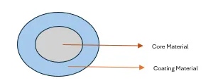
Figure-1: Microcapsule.
THE COMPONENTS AND TECHNIQUES OF MICROENCAPSULATION:
It defines core materials, which can be solid or liquid, and may consist of active ingredients, stabilizers, and other substances, depending on their intended use. The coating materials form the outer layer of microcapsules and must possess certain characteristics: they should be chemically compatible with the core, strong, flexible, impermeable, and stable to create a cohesive film. Examples of coating materials include various gums, carbohydrates, cellulose, lipids and proteins.6
SEVERAL TECHNIQUES FOR MICROENCAPSULATION:
1. Spray Drying:
A method where a mixture of core and wall materials is atomized into hot air, evaporating the solvent to create stable powders with controlled particle sizes, though it may not suit heat-sensitive materials.7
2. Spray Cooling:
Similar to spray drying but utilizes cold air, solidifying droplets, effective for heat-sensitive compounds and suitable for large-scale productions.8
3. Coacervation:
Forms a polymer-rich layer around the core by manipulating temperature, pH, or ionic strength, effective for hydrophilic molecules, commonly using sodium alginate and calcium chloride for microcapsule.9
4. Fluidized Bed Coating:
Core particles are suspended in air while coating material is sprayed, with efficiency influenced by air temperature and spray parameters.10
5. Extrusion:
Involves passing core and wall materials through nozzles to form droplets, producing large and denser microcapsules.11
6. Emulsification:
Disperses core material in an organic solvent with the wall polymer, allowing polymer shell formation after solvent evaporation, often used for enzymes and microorganisms.12
7. Cyclodextrin Inclusion:
Uses cyclodextrins to form inclusion complexes with hydrophobic compounds, enhancing their solubility and stability.13
8. Ionotropic Gelation Method:
Relies on polyelectrolytes crosslinking with counter ions to form hydrogel beads, ensuring controlled drug release and suitable for temperature-sensitive substances. This technique is applied to encapsulate proteins, enzymes, and biologically active compounds, offering sustained release and improved stability.14
FORMULATION DESIGN COMPONENTS:
Key Components:
Types:
Selection criteria:
METHODS OF MICROENCAPSULATION:
Process Parameters:
Core-to-coating ratio, particle size control, drying techniques, pH, and temperature optimization.18
Characterization of Microcapsules:
Includes morphology analysis, encapsulation efficiency, drug release studies, and stability testing.19
EXTRACTION METHODS IN PHARMACEUTICALS:
Involves separating medicinal compounds from plant/animal tissues using selective solvents.
Methods: Maceration, Percolation, Decoction, Soxhlet extraction, Hydro distillation with different solvents for various compounds.20
APPLICATIONS OF MICROENCAPSULATION:
BENEFITS OF MICROENCAPSULATION:
PLANT PROFILE:
PLANT PROFILE: (AMLA)23
Kingdom : Plantae
Subkingdom : Tracheobionta
Super division : Spermatophyta
Division : Magnoliophyta
Class : Magnoliopsida
Subclass : Rosidae
Order : Myrtales
Family : Phyllanthaceae
Genus : Phyllanthus
Species : Emblica officinalis (Gaertner)
Description:
Amla (Phyllanthus emblica L.) is a small to medium-sized deciduous tree, reaching a height of 8-18 meters. The bark is light brown or grayish with exfoliating thin scales. Leaves are simple, small, closely set, linear-oblong and light green, giving a feathery appearance. The flowers are greenish-yellow, unisexual and inconspicuous, appearing in clusters. The fruit is nearly spherical, light greenish-yellow, smooth and hard with six vertical furrows. It has a sour, astringent taste due to high vitamin C and tannin content. The seed is hard and enclosed within the fibrous pericarp.
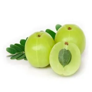
Figure-2: Phyllanthus emblica (Amla)
Medicinal Properties:
Bottom of Form
PLANT PROFILE: (TULASI)24
Kingdom : Plantae
Subkingdom : Tracheobionta
Super division : Spermatophyta
Division : Magnoliophyta
Class : Magnoliopsida
Subclass : Asteridae
Order : Lamiales Family: Lamiaceae Genus: Ocimum L.
Species : Ocimum sanctum L. (Tulasi / Holy Basil)
Description:
Tulasi is a small, erect, much-branched, fragrant, perennial herb growing up to 30-60 cm in height. The stems are hairy and quadrangular. Leaves are simple, opposite, ovate, 2-4 cm long with toothed margins, hairy on both sides and green or purple depending on the variety. Flowers are small, purplish or reddish, borne in close whorls on elongated racemes. The plant has a characteristic aromatic odour due to essential oils, mainly eugenol. Seeds are small, yellowish-brown and come mucilaginous when soaked in water.
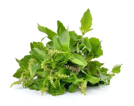
Figure-3: Ocimum sanctum (Tulasi)
Medicinal properties:
Tulasi is widely known for its therapeutic uses:
METHODOLOGY:
1.Extraction by Soxhlet Method:
This technique uses 70% ethanol to extract phenolics, flavonoids, tannins, and glycosides. The process involves heating ethanol in a round-bottom flask while the plant sample is placed in a cellulose thimble within a Soxhlet extractor. The solvent evaporates, condenses, and circulates through the sample, allowing for thorough extraction over 6-8 hours until the solvent is nearly colorless. The extract is then concentrated for yield calculation, benefiting from the solvent's dual ability to dissolve polar and non-polar compounds.25
2.Extraction by Decoction Method:
Specifically for Amla fruit (Emblica officinalis), this method involves boiling the fruit pieces in water to extract vitamin C, polyphenols, and tannins. The decoction is consumed warm or stored for later use, and is recognized for its potential health benefits, including improved digestion and immunity, with scientific evidence supporting its traditional applications.26
3.Physical Evaluation:
The extraction's physical properties, such as total ash, acid-insoluble ash, and loss on drying, are measured. The total ash is determined by incinerating the sample, while acid-insoluble ash is obtained after treatment with hydrochloric acid, and water-insoluble ash is calculated by boiling total ash with water. The loss on drying assesses the moisture content under specified conditions.27
Additionally, various qualitative tests for detecting fundamental phytochemicals such as alkaloids, flavonoids, phenols, terpenoids, tannins, saponins, and starch are described. These tests typically involve specific reagents that yield characteristic precipitates or color changes, confirming the presence of these compounds in the plant extracts.28
FORMULATION OF EMBLICA OFFICINALIS GAERTNER AND OCIMUM GRATISSIMUM L HERBAL MICROENCAPSULATION: PREPARATION OF MICROENCAPSULE:
The microencapsulation was prepared by using a combination of herbal extracts and polymeric excipients. Precisely, 0.5 g of Tulasi powder and 0.5 g of Amla powder were taken as the core materials due to their therapeutic potential. To form the encapsulating matrix, 2 g of sodium alginate was used as the primary coating polymer, while 1.5 g of starch and 1.5 g of acacia were incorporated as stabilizers and co-polymers to enhance encapsulation efficiency and control the release of active constituents. The mixture was homogenized thoroughly to ensure uniform dispersion of the drug within the polymeric solution, followed by the appropriate encapsulation process (such as ionic gelation or emulsification), leading to the formation of stable microcapsules containing Tulasi and Amla extracts. This preparation provides a protective barrier around the active ingredients, improving their stability, solubility and potential bioavailability.29
FORMULATION TABLE FOR AMLA AND TULASI HERBAL MICROENCAPSULTION:
Table no 1: Ingredients and Formulation ratio.
|
S/NO
|
INGREDIENTS |
FORMULATION |
|
1 |
Tulasi extract(g) |
0.5g |
|
2 |
Amla extract(g) |
0.5g |
|
3 |
Sodium alginate(g) |
2.5g |
|
4 |
Starch(g) |
1.5g |
|
5 |
Acacia(g) |
1.5g |
|
6 |
Calcium chloride (ml, 6%w/v) |
Q. S |
|
7 |
Purified water(ml) |
100ml |
EVALUVATION OF MICROENCAPSULATION:
PHYSICAL APPEARANCE:
The Preparation of microencapsulation containing amla and Tulasi are inspected visually for their colour, odour, size and morphology.30
MEASUREMENT OF PH:
The pH was determined by using a digital PH meter. pH measurement is the determination of the hydrogen ion concentration of the microencapsulated formulation. Disperse a specific number of microcapsules in distilled water (commonly 1% w/v suspension) and calculated the average values.31
MICROENCAPSULATION EFFICIENCY (ENCAPSULATION EFFICIENCY):
Encapsulation Efficiency (%)=Actual drug contentTheoretical drug content×10

MOISTURE CONTEENT:
Amount of residual moisture present in microcapsules. Determined by Karl Fischer titration or oven drying method at 105°C until constant weight. (Ideal moisture content: below 5%.)33
STABILITY STUDIES:
Determines the ability of microcapsules to maintain their physical and chemical properties over time. Store samples as per ICH conditions (25°C/60% RH and 40°C/75% RH).34
ANTI-VIABILITY STUDY:
Antimicrobial study is carried out to evaluate the ability of microencapsulated formulations to inhibit or kill microorganisms, confirming the retained antimicrobial activity of the encapsulated drug or herbal extract.
Method:
8.RESULT AND DISCUSSION:
The formulated microencapsulated herbal product containing Amla (Emblica officinalis) and Tulsi (Ocimum sanctum) was successfully prepared using sodium alginate and starch as coating materials. The prepared microcapsules were evaluated for various parameters such as percentage yield, particle size, drug content, in vitro drug release, antimicrobial activity, viscosity, and stability studies. The percentage yield was found to be satisfactory, indicating efficient encapsulation of the herbal extract. The particle size analysis showed that the microcapsules were uniform and spherical in shape. The drug content and encapsulation efficiency confirmed that the herbal actives were successfully entrapped within the polymeric matrix.
Figure-4: Microencapsule of Amla and Tulsi.
8. RESULT AND DISCUSSIONS:
8.1. PHYSICAL EVALUATION:
8.1.1. Organoleptic Properties:
The organoleptic properties of the crude drug of Amla and Tulsi were presented in Table No 9:
Table No 2: The Organoleptic Properties of Amla and Tulsi Extract
|
Parameters |
Characteristics |
|
|
Amla |
Tulsi |
|
|
Colour |
Brownish-green |
Dark green |
|
Odour |
Characteristic |
Aromatic |
|
Taste |
Sour and astringent |
Pungent and slightly bitter |
Discussion:
The organoleptic evaluation of Amla and Tulsi extract revealed that Amla had a brownish-green colour with a sour and astringent taste, while Tulsi showed a dark green colour with an aromatic odour and slightly bitter taste.
8.1.2. Ash value, Acid insoluble ash, Water soluble ash and Loss on drying:
The ash value, acid insoluble ash, water soluble ash and loss on drying of crude extract of Amla and Tulsi were presents in Table No:10
Table No 3: Ash content of Amla and Tulsi Extract
|
S. No. |
Parameters |
observation |
|
|
Amla Extract (% w/w) |
Tulsi Extract (% w/w) |
||
|
1 |
Total Ash Value |
5.2 |
6.1 |
|
2 |
Acid Insoluble Ash |
0.6 |
0.7 |
|
3 |
Water Soluble Ash |
2.8 |
3.2 |
|
4 |
Loss on Drying |
7.0 |
6.5 |
Discussion:
Table No 4: Ash content of Combined Capsule Extract
|
S.No
|
Parameters |
Observations (% w/w) |
|
1 |
Total Ash Value |
5.6 |
|
2 |
Acid Insoluble Ash |
0.6 |
|
3 |
Water Soluble Ash |
3.0 |
|
4 |
Loss on Drying |
4.3 |
The ash value and moisture content help to determine the purity and stability of the extracts. Amla and Tulsi showed acceptable ash values indicating minimal inorganic impurities, and moderate loss on drying indicates proper storage stability.
Discussion:
The combined Amla–Tulsi capsule showed acceptable physicochemical parameters. The ash values indicate minimal inorganic and siliceous matter, while the moderate loss on drying suggests adequate moisture control, ensuring good stability of the formulation.
8.1.3. PHYTOCHEMICAL SCREENING STUDY
The phytochemical screening results of the combined crude extract of Amla (Emblica officinalis) and Tulsi (Ocimum sanctum) were presented in Table No. 12;
Table No. 5: Phytochemical Screening Results of Amla and Tulsi Combined Capsule Extract.
|
S. No. |
Phytoconstituents |
Tests |
Results |
|
1 |
Alkaloids |
Wagner’s test Dragendorff’s test Mayer’s test Hager’s test |
+++- |
|
2 |
Flavonoids |
Lead acetate test Shinoda test |
++ |
|
3 |
Steroids |
Liebermann–Burchard test |
+ |
|
4 |
Terpenoids |
Salkowski test |
+ |
|
5 |
Tannins |
Ferric chloride test |
+ |
|
6 |
Phenols |
Ferric chloride test |
+ |
|
7 |
Saponins |
Froth test |
+ |
Note: + = Present - = Absent
Discussion:
From the results of the phytochemical screening, the Amla and Tulsi combined capsule showed the presence of various bioactive compounds such as alkaloids, flavonoids, steroids, terpenoids, tannins, phenols, saponins, carbohydrates, and glycosides.
These phytoconstituents are known for their antioxidant, antimicrobial, and immunomodulatory activities, contributing to the capsule’s therapeutic potential for gut and general health improvement.
8.2. MICROENCAPSULATION EFFICIENCY (ENCAPSULATION EFFICIENCY):
The encapsulation efficiency (EE%) of the Amla–Tulsi combined formulation was found to be high, indicating that most of the active phytochemicals were successfully entrapped inside the microcapsules. The obtained EE% value shows that the formulation effectively retains both hydrophilic (Amla) and lipophilic (Tulsi) constituents within the encapsulation matrix.
Encapsulation Efficiency (%)=Actual drug contentTheoretical drug content×10

DISCUSSION:
Table No 6: % of Encapsulation Efficiency of Amla and Tulsi Extract.
|
S. No |
Formulation |
Percentage of Encapsulation Efficiency |
|
1 |
F1 |
78% |
CONCLUSION:
The Amla–Tulsi formulation shows high encapsulation efficiency, proving successful entrapment of phytochemicals and good formulation stability.
8.3. ESTIMATION OF DRUG CONTENT:
The drug content of Amla–Tulsi microencapsule formulation are shown in table-14:
Table No. 7: Drug content of Amla and Tulsi Extract.
|
S. No |
Formulation |
Drug Content |
|
1 |
F1 |
88.75% |
Discussion:
The drug content estimation of the Amla–Tulsi microencapsule formulation (F1) showed a drug content of 88.75%, indicating effective entrapment and uniform distribution of the active constituents within the microcapsules. The high drug content suggests minimal loss of herbal actives during the microencapsulation process. This may be attributed to the suitability of the selected polymers and the optimized preparation method, which helped in retaining the phytoconstituents of Amla and Tulsi. The result confirms the reproducibility and reliability of formulation method and indicates that the microencapsulated formulation is suitable for further evaluation and therapeutic application.
STANDARD CALIBRATION CURVE OF AMLA -TULSI FORMULATION
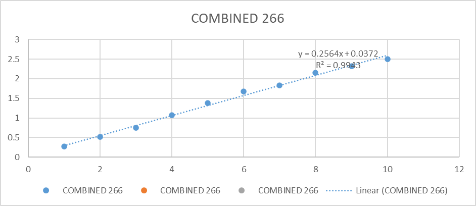
Figure - 5: Standard Calibration Curve of Amla -Tulsi Formulation
8.4. FT-IR Spectral Study:
FT-IR analysis was carried out to evaluate the compatibility and successful microencapsulation of Amla and Tulsi extracts in sodium alginate. The spectra of the microcapsules showed characteristic peaks of Amla and Tulsi, including O–H stretching (3200–3500 cm?¹), C–H stretching (2920–2850 cm?¹), and C=O/C=C vibrations (1700–1600 cm?¹), along with alginate carboxylate peaks around 1600 and 1410 cm?¹. Slight peak shifts and reduced intensities were observed due to hydrogen bonding and calcium–alginate crosslinking, while no new peaks appeared, indicating good drug–polymer compatibility and successful physical encapsulation without chemical interaction.
8.4.1. Amla FT-IR:
IR SPECTRUM OF AMLA
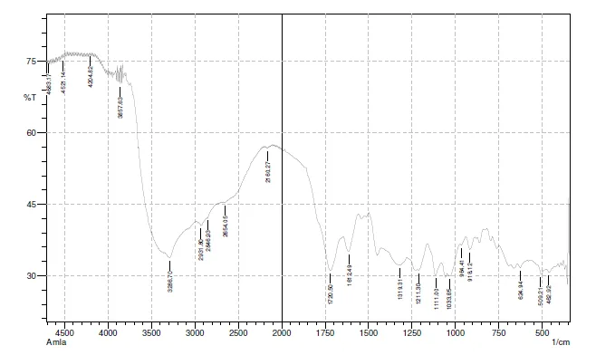
Figure -6: FT-IR spectral analysis of AMLA
FT-IR SPECTRAL ANALYSIS OF AMLA:
Table No 8: FT-IR analysis of amla
|
S.NO |
WAVENUMBER (CM-1) |
FUNCTIONAL GROUP |
|
1 |
3286 |
0-H (Stretching) |
|
2 |
1720 |
C=O (Stretching) |
|
3 |
1211 |
C-O (Stretching) |
|
4 |
2931 |
C-H (Stretching) |
|
5 |
1612 |
C=C (Aromatic) |
8.4.2. Tulsi FT-IR:
IR SPECTRUM OF TULSI
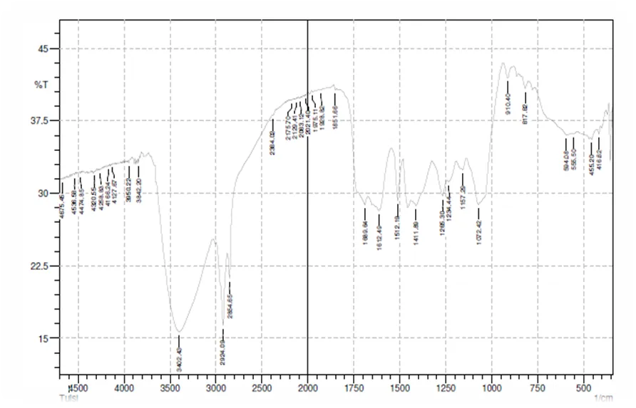
Figure -7: FT-IR spectral analysis of TULSI
FT-IR SPECTRAL ANALYSIS OF TULSI:
Table No 9: FT-IR analysis of Tulsi
|
S.NO |
WAVENUMBER (CM-1) |
FUNCTIONAL GROUP |
|
1 |
3402 |
0-H (Strong band) |
|
2 |
2924 |
C-H (Stretching) |
|
3 |
1612 |
C=C (Aromatic) |
|
4 |
1265 |
C-O (Stretching) |
|
5 |
1689 |
Carbonyl group |
IR SPECTRUM OF FORMULATION
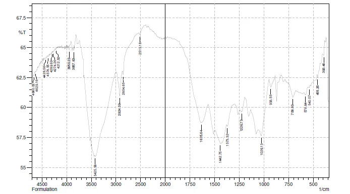
Figure -8: FT-IR spectral analysis of FORMULATION
FT-IR SPECTRAL ANALYSIS OF FORMULATION:
Table No 10: FT-IR analysis of Formulation
|
S.NO |
WAVENUMBER (CM-1) |
FUNCTIONAL GROUP |
|
1 |
3425 |
0-H (Stretching) Alcohol/Phenol |
|
2 |
2924 |
C-H (Stretching)Alkanes |
|
3 |
2854 |
C-H (Symmetric Stretch) |
|
4 |
1635 |
C-O (Stretch) |
|
5 |
1226 |
Carbonyl group |
DISCUSSION:
The FTIR spectra of Amla, Tulsi, and the combined herbal formulation were analyzed to identify functional groups and to evaluate compatibility among the components. All three spectra showed a broad absorption band in the region of 3400–3300 cm?¹, corresponding to O–H stretching vibrations, indicating the presence of phenolic compounds and alcohols responsible for antioxidant activity. The absorption peaks around 2924–2931 cm?¹ observed in Amla, Tulsi, and the formulation are attributed to C–H stretching vibrations of aliphatic hydrocarbons.
The formulation spectrum showed all major peaks present in both Amla and Tulsi without significant shifting or disappearance, indicating chemical compatibility and stability of the herbal formulation.
8.5. VIABILITY STUDY:
The viability study was performed to evaluate the survival of Lactobacillus in the presence of Amla–Tulsi microcapsules. The results showed that encapsulated formulations maintained higher viable counts during storage and under simulated gastrointestinal conditions compared to non-encapsulated cells. The alginate matrix provided a protective barrier against acidic pH and bile salts, while the bioactive components of Amla and Tulsi supported probiotic stability. These findings confirm that microencapsulation effectively enhances probiotic viability and functional performance.
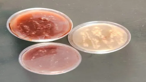
Figure-9: Viability study of Amla and Tulsi Extract.
RESULT:
The agar diffusion study showed good bacterial growth in the presence of Amla–Tulsi microencapsulated formulation. After incubation, no zone of inhibition was observed around the wells containing the microcapsules, indicating that the formulation did not exert any antimicrobial effect against the tested bacteria. Uniform and healthy bacterial growth around the wells confirmed that Amla and Tulsi microencapsulation is compatible with beneficial bacteria and supports their viability, making the formulation suitable for gut-health applications.
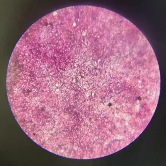
Figure-10: Microbial Growth of Amla and Tulsi Extract
Discussion:
The enhanced growth indicates that the combined Amla–Tulsi microcapsules provide a synergistic supportive effect for Lactobacillus. The presence of polyphenols and flavonoids from Amla along with flavonoids and essential oils from Tulsi, in microencapsulated form, did not inhibit bacterial growth. Instead, they created a favourable microenvironment, improving bacterial survival and proliferation compared to individual Amla or Tulsi microcapsules. This confirms that the Amla–Tulsi combination is the most compatible and effective for supporting Lactobacillus growth, highlighting its suitability for probiotic and gut-health formulations.
Top of Form8.6. Bottom of FormSTABILITY STUDY OF MICROENCAPSULATED AMLA AND TULSI CAPSULE:
The physical parameters such as colour, odour, pH, and moisture content of microencapsulated Amla and Tulsi capsules were observed for a period of 30 days under controlled conditions at 25°C ± 2°C / 60% ± 5% RH.
REFERENCES

Mrs.K.Sivaranjani*, Dr.M.Rajesh, S.Delsiya, D.Madhumitha, S.Logeshwari, P.Gopi Arun, Formulation And Evaluation Of Herbal Microcapsules Containing Amla (Emblica officinalis) And Tulsi (Ocimum gratissimum L) For Gastrointestinal Health , Int. J. of Pharm. Sci., 2026, Vol 4, Issue 9, 3580-3597. https://doi.org/10.5281/zenodo.23015099
 10.5281/zenodo.23015099