

We use cookies to ensure our website works properly and to personalise your experience. Cookies policy
Department of Pharmaceutics, Arunai College of Pharmacy, Tiruvannamalai-606 601.
The present study aimed to formulate and evaluate herbal transdermal patches containing the methanolic extract of Acalypha indica for sustained anti-inflammatory activity. The transdermal patches were prepared by the solvent casting method using hydrophilic polymers hydroxypropyl methylcellulose (HPMC) and polyvinylpyrrolidone (PVP), along with suitable plasticizers and penetration enhancers. The formulated patches were evaluated for physicochemical properties including thickness, weight variation, folding endurance, percentage elongation, drug content uniformity, moisture content, moisture uptake, flatness, and surface pH. The patches were smooth, flexible, and uniform, with satisfactory mechanical strength (folding endurance >500) and acceptable drug content (92%), indicating uniform distribution of the extract within the polymeric matrix.UV–Visible spectroscopic analysis revealed a prominent absorption maximum (?max) at 603 nm, confirming the presence of flavonoids and phenolic constituents responsible for anti-inflammatory activity. In vitro skin permeation studies using a Franz diffusion cell demonstrated sustained and controlled drug release, achieving 100% cumulative release within 240 minutes. Release kinetic modelling indicated that the formulation followed first-order kinetics with strong correlation to the Higuchi and Korsmeyer–Peppas models, suggesting diffusion-controlled and anomalous (non-Fickian) transport mechanisms.The in vitro anti-inflammatory activity evaluated by the protein denaturation assay exhibited concentration-dependent inhibition, with 69% inhibition at 1000 µg/mL compared to diclofenac sodium. Stability studies conducted as per ICH guidelines confirmed that the patches remained stable under accelerated conditions for 90 days without significant changes in physical properties or drug contentOverall, the formulated herbal transdermal patches of Acalypha indica demonstrated satisfactory physicochemical characteristics, sustained drug release, stability, and significant anti-inflammatory activity. The developed system represents a promising natural alternative for controlled transdermal anti-inflammatory therapy
Inflammation is a complex biological response of vascular tissues to harmful stimuli such as pathogens, damaged cells, irritants, or chemical mediators. Although inflammation is a protective mechanism, chronic inflammation is associated with several pathological conditions including arthritis, dermatitis, asthma, and cardiovascular diseases. Conventional anti-inflammatory drugs such as non-steroidal anti-inflammatory drugs (NSAIDs) are widely prescribed; however, their long-term use is often associated with adverse effects including gastrointestinal irritation, renal toxicity, and cardiovascular complications (1).
In recent years, herbal medicines have gained increasing attention due to their therapeutic efficacy, safety profile, affordability, and patient acceptability. Medicinal plants serve as a valuable source of bioactive compounds with anti-inflammatory, antioxidant, and analgesic properties. Among these, Acalypha indica (Family: Euphorbiaceae), commonly known as Indian Acalypha or Indian copperleaf, is a widely distributed medicinal herb in tropical regions of Asia and Africa. Traditionally, it has been used for the treatment of inflammatory conditions, skin disorders, bronchitis, rheumatism, and wounds. Phytochemical investigations have revealed the presence of flavonoids, alkaloids, tannins, saponins, and phenolic compounds, which contribute to its anti-inflammatory and antioxidant activities (2, 3).
Various experimental studies have demonstrated that extracts of Acalypha indica possess significant anti-inflammatory activity by inhibiting pro-inflammatory mediators and reducing oxidative stress. The flavonoid and phenolic constituents are believed to suppress cyclooxygenase (COX) and lipoxygenase pathways, thereby reducing prostaglandin synthesis and inflammatory responses (4).
Transdermal drug delivery systems (TDDS) have emerged as an effective alternative to oral and parenteral routes due to advantages such as avoidance of first-pass metabolism, sustained drug release, improved patient compliance, and reduced systemic side effects. Herbal transdermal patches provide controlled delivery of phytoconstituents directly through the skin, making them suitable for the management of localized inflammatory conditions. The formulation of herbal patches typically involves suitable polymers such as hydroxypropyl methylcellulose (HPMC), polyvinyl alcohol (PVA), or ethyl cellulose to ensure mechanical strength, flexibility, and controlled drug release (5).
Considering the therapeutic potential of Acalypha indica and the advantages of transdermal drug delivery, the present study aims to formulate and evaluate herbal transdermal patches containing Acalypha indica extract for anti-inflammatory activity. The prepared patches were evaluated for physicochemical parameters including thickness, weight variation, folding endurance, moisture content, drug content uniformity, in vitro drug release, and anti-inflammatory efficacy. This study intends to provide a novel herbal transdermal system that may offer safer and sustained management of inflammatory conditions.
METHODOLOGY
1. Collection and Authentication of Plant Material
Fresh whole plants of Acalypha indica (Family: Euphorbiaceae) were collected from Tiruvannamalai, Tamil Nadu, India, during the monsoon season (June – November). The collected plant materials were carefully examined to ensure free from disease, insect infestation, and physical damage. The material was washed thoroughly with running water followed by distilled water to remove adhering soil and extraneous matter, in accordance with WHO guidelines for good agricultural and collection practices (6).
Botanical authentication was carried out by a qualified taxonomist, Mr. J. Suresh Kumar, Department of Botany, Kalaignar Karunanidhi Government Arts College. Morphological characteristics including leaf morphology, stem structure, and inflorescence pattern were compared with standard taxonomic descriptions (7, 8). A voucher specimen was prepared and preserved in the departmental herbarium for future reference.
2. Preparation of Plant Material
The collected plant material was shade-dried at room temperature (7–10 days) with adequate ventilation to prevent degradation of thermolabile constituents. Shade drying preserves flavonoids and phenolic compounds from photodegradation (9). The dried material was coarsely powdered using a mechanical grinder and passed through a sieve no-40 to obtain uniform particle size. The powder was stored in airtight containers in a cool, dry place until further use (6).
3. Extraction Procedure
Approximately 150 g of air-dried powdered plant material was extracted using methanol in a Soxhlet apparatus for 24 hours at 60–70°C. Methanol was selected due to its efficiency in extracting polar phytoconstituents such as alkaloids, flavonoids, glycosides, and tannins (10). The extract was filtered and concentrated under reduced pressure using a rotary evaporator. The concentrated extract was stored in a desiccator until use (7).
Percentage yield was calculated using:
Percentage yield=Weight of extractWeight of crude drug×100

4. UV–Visible Spectroscopic Analysis
The methanolic extract was analysed using a UV–Visible spectrophotometer (Labindia UV 3200) in the wavelength range of 200–800 nm using methanol as blank. The absorption maxima (λmax) were recorded. UV–Visible spectroscopy is useful for detecting chromophoric compounds such as flavonoids and phenolics (11, 12).
5. Preliminary Phytochemical Screening
Qualitative phytochemical tests were performed to detect alkaloids, flavonoids, tannins, phenols, glycosides, saponins, steroids, terpenoids, carbohydrates, and amino acids using standard procedures (6, 7, 9). Observations were recorded based on colour change or precipitate formation.
6. Formulation of Herbal Transdermal Patch
Herbal transdermal patches were prepared by the solvent casting method as reported in earlier studies (13, 14). The required quantities of polymers hydroxypropyl methylcellulose (HPMC) and polyvinyl pyrrolidone (PVP) as shown in Table 1 were dissolved in distilled water with continuous stirring until a clear polymeric solution was obtained. Predetermined amounts of plasticizers such as polyethylene glycol (PEG) and glycerine were then incorporated to improve flexibility of the film. Penetration enhancers including propylene glycol and menthol/eucalyptus oil were added to enhance drug permeation through the skin. The measured quantity of the herbal extract was subsequently incorporated into the polymeric mixture and stirred thoroughly to obtain a uniform dispersion. The resulting homogeneous solution was poured carefully into a levelled glass petri dish and allowed to dry at room temperature. After complete drying, the prepared patches were peeled off, cut into uniform sizes of 2 × 2 cm, and stored in a desiccator until further evaluation.
Table 1: Composition of Anti-Inflammatory Herbal Transdermal Patch
|
S. No |
Ingredients |
Quantity |
Function |
|
1 |
Herbal extract (Acalypha indica) |
50 mg |
Anti-inflammatory agent |
|
2 |
HPMC |
200 mg |
Film-forming polymer |
|
3 |
PVP |
100 mg |
Secondary polymer |
|
4 |
PEG |
50 mg |
Plasticizer |
|
5 |
Glycerine |
50 mg |
Humectant/Plasticizer |
|
6 |
Menthol/Eucalyptus oil |
5 mg |
Penetration enhancer |
|
7 |
Propylene glycol |
20 mg |
Solvent/Penetration enhancer |
|
8 |
Distilled water |
q.s to 5 ml |
Solvent |
7. Evaluation of Herbal Transdermal Patches
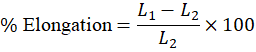
The prepared herbal transdermal patches containing Acalypha indica extract were systematically evaluated to ensure their physicochemical integrity, mechanical strength, uniformity, and suitability for transdermal drug delivery. Visual inspection was carried out to assess colour, transparency, smoothness, flexibility, and absence of surface imperfections such as air bubbles or cracks. The thickness of the patches was measured at three different positions using a calibrated digital micrometre to confirm uniform casting and reproducibility. Weight variation was determined by individually weighing ten patches and calculating the average weight to ensure uniform distribution of the drug and excipients within the polymeric matrix. Mechanical properties were evaluated by determining folding endurance, wherein a patch was repeatedly folded at the same point until breakage, and percentage elongation was calculated to assess elasticity and tensile strength.
% Elongation=L1-L2L2×100
Drug content uniformity was analysed by dissolving a known area of the patch in methanol, followed by spectrophotometric estimation at 281 nm to confirm consistent drug distribution throughout the formulation. Moisture content and moisture uptake studies were conducted to evaluate the stability of the patches under dry and humid conditions, respectively, while flatness testing was performed to assess dimensional stability and absence of constriction.
% Moisture Content=Initial weight – Final weightFinal weight×100

Surface pH was measured after equilibrating the patch in distilled water to ensure compatibility with skin and minimize the risk of irritation upon application. All evaluation parameters were performed according to standard pharmaceutical guidelines to confirm the quality, performance, mechanical stability, and storage suitability of the formulated transdermal patches (13, 15, 16, 17).
8. In-Vitro Skin Permeation Study
In-vitro permeation studies were performed using a Franz diffusion cell. Excised skin was mounted between donor and receptor compartments with stratum corneum facing donor side. Phosphate buffer saline (pH 7.4) was used as receptor medium at 37 ± 0.5°C with continuous stirring.
Samples were withdrawn at predetermined intervals (15, 30, 45, 60, 90, 120, 180, 240 min) and analysed spectrophotometrically. Cumulative drug release was calculated and plotted against time. Permeation parameters such as flux and permeability coefficient were determined.
9. In-Vitro Anti-Inflammatory Activity
Anti-inflammatory activity was evaluated using the egg albumin denaturation assay (18, 19).
Different concentrations of extract were prepared (0.01–1000 µg/mL). Reaction mixture containing egg albumin and phosphate buffer (pH 7.4) was incubated at 37°C for 20 min and heated at 70°C for 5 min. Absorbance was measured at 680 nm.
% Inhibition=Abs control – Abs sampleAbs control×100

Diclofenac sodium was used as standard reference drug.
10. Stability Studies
Stability studies were conducted as per ICH Q1A (R2) guidelines (20). Patches were stored at 40 ± 2°C / 75 ± 5% RH for 3 months. Samples were evaluated at 0, 1, 2, and 3 months for physical appearance, drug content, folding endurance, moisture content, and in-vitro release.
RESULT AND DISCUSSION
The methanolic extract of Acalypha indica was successfully formulated into herbal transdermal patches using HPMC and PVP polymers by the solvent casting technique. The prepared patches were smooth, uniform, flexible, and free from visible imperfections, indicating good compatibility between the extract and excipients as well as suitability of the selected formulation method. The physicochemical evaluation (Table 2) demonstrated acceptable thickness (0.33 mm) and weight variation (0.174 g), confirming uniform distribution of the polymeric matrix and drug throughout the patch. The folding endurance value (>500) and percentage elongation (42%) indicated excellent mechanical strength and elasticity, which are essential characteristics for maintaining intimate contact with the skin during application. Drug content was found to be 92%, reflecting efficient incorporation and minimal loss of phytoconstituents during formulation. The percentage moisture content (22.11%) contributed to flexibility of the film, while low moisture uptake (1.26%) indicated resistance to environmental humidity and good storage stability.
Table 2: Evaluation of Herbal Transdermal Patches
|
S. No |
Test |
Result |
|
1 |
Thickness |
0.33 mm |
|
2 |
Weight variation |
0.174 g |
|
3 |
Folding endurance |
>500 |
|
4 |
% Elongation break test |
42% |
|
5 |
Drug content |
92% |
|
6 |
% Moisture content |
22.11% |
|
7 |
% Moisture uptake |
1.26% |
|
8 |
Flatness |
101% |
The flatness value (101%) confirmed the absence of constriction and dimensional instability. These findings suggest that the formulated patches possessed satisfactory physicochemical and mechanical properties suitable for transdermal drug delivery, consistent with standard pharmaceutical requirements (21).
UV–Visible Spectroscopic Analysis
UV–Visible spectroscopic analysis of the methanolic extract (340–800 nm) revealed characteristic absorption peaks around 400 nm and a prominent λmax at approximately 603 nm. These peaks indicate the presence of conjugated chromophoric systems such as flavonoids and phenolic compounds, which undergo π→π* and n→π* electronic transitions. The calibration curve constructed at 603 nm showed good linearity within the concentration range of 0–1.0 µg/mL with a correlation coefficient (R²) of 0.9737, confirming adherence to Beer–Lambert’s law and validating the method for quantitative estimation. The presence of phenolic and flavonoid constituents supports the anti-inflammatory potential of the extract, as these compounds are known to inhibit inflammatory mediators and oxidative stress (22).
In-Vitro Skin Permeation Study
The in-vitro permeation study using a Franz diffusion cell demonstrated a gradual and sustained release profile over 240 minutes. The cumulative drug release increased steadily from 19.19% at 15 minutes to 84.38% at 120 minutes, reaching 100% at 240 minutes. (Figure 1)
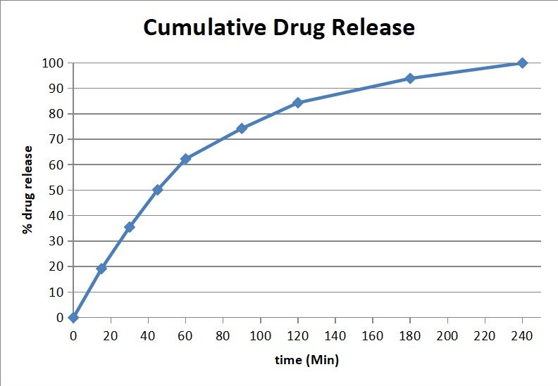
Figure 1: Cumulative drug release
This controlled release behaviour may be attributed to diffusion of phytoconstituents through the hydrated HPMC–PVP polymeric matrix, along with the permeation-enhancing effects of menthol and propylene glycol. The sustained release profile is advantageous for maintaining prolonged therapeutic levels and reducing application frequency. Similar diffusion-controlled release behaviour has been reported for polymer-based transdermal systems (21, 23).
Drug Release Kinetics
Kinetic modelling (Table 3-4) revealed that the drug release data best fitted the first-order model (figure 3) (R² = 0.9993), indicating concentration-dependent release. The Higuchi model (figure 4) also showed good linearity (R² = 0.9649), confirming diffusion-controlled drug release from the matrix. The Korsmeyer–Peppas model (figure 5) demonstrated excellent correlation (R² = 0.9999) with a release exponent (n) value of 0.8765, suggesting anomalous (non-Fickian) transport involving both diffusion and polymer relaxation mechanisms. The comparatively low R² value for the zero-order model (0.8193) indicates that release was not constant but dependent on concentration gradients (figure 2). Overall, the kinetic data confirm that the formulated patches provide controlled and sustained release governed predominantly by diffusion with matrix relaxation behaviour.
In-Vitro Anti-Inflammatory Activity
The anti-inflammatory activity evaluated by the egg albumin denaturation assay demonstrated concentration-dependent inhibition (table 5). At 1000 µg/mL, the extract exhibited 69% inhibition compared to 84.37% for diclofenac sodium. Although the activity of the extract was lower than the standard drug, it showed significant inhibition at higher concentrations,
Table 3: In vitro drug release data and transformed variables for kinetic modelling of Acalypha indica extract from herbal transdermal patches.
|
Time (min) |
% Cumulative Drug Release (Qt) |
% Drug Remaining (100-Qt) |
log % Drug Remaining (First Order) |
√Time (Higuchi) |
Log (Time) (K-P) |
Log (Qt) (K-P) |
|
0 |
0 |
100 |
2 |
0 |
||
|
15 |
19.19 |
80.81 |
1.907465 |
3.872983 |
1.176091 |
1.283075 |
|
30 |
35.54 |
64.46 |
1.80929 |
5.477226 |
1.477121 |
1.550717 |
|
45 |
50.18 |
49.82 |
1.697404 |
6.708204 |
1.653213 |
1.700531 |
|
60 |
62.32 |
37.68 |
1.576111 |
7.745967 |
1.778151 |
1.794627 |
|
90 |
74.28 |
25.72 |
1.410271 |
9.486833 |
1.954243 |
1.870872 |
|
120 |
84.38 |
15.62 |
1.193681 |
10.95445 |
2.079181 |
1.92624 |
|
180 |
93.93 |
6.07 |
0.783189 |
13.41641 |
2.255273 |
1.972804 |
|
240 |
100 |
0 |
- |
15.49193 |
2.380211 |
2 |
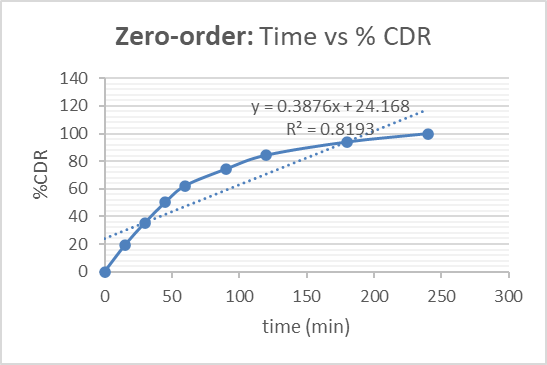
Figure 2: Zero-order release kinetics of Acalypha indica extract from transdermal patches.
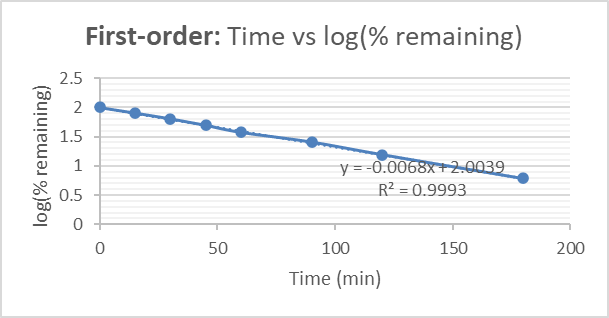
Figure 3: First-order release kinetics of Acalypha indica extract from transdermal patches.
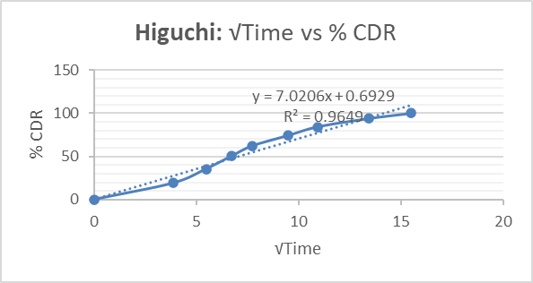
Figure 4: Higuchi diffusion model for Acalypha indica extract release from transdermal patches.
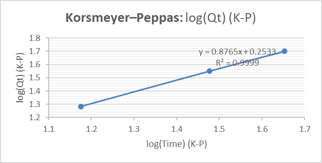
Figure 5: Korsmeyer–Peppas model for Acalypha indica extract release from transdermal patches.
Table 4: Kinetic model fitting parameters for in vitro release of Acalypha indica extract from herbal transdermal patches.
|
S. No |
Kinetic Model |
Equation |
Slope (k / n) |
R² value |
Interpretation |
|
1 |
Zero-order |
Qt = Q? + k?t |
0.3876 |
0.8193 |
Poor linearity |
|
2 |
First-order |
log(100−Qt) vs t |
−0.0068 |
0.9993 |
Best fit (concentration-dependent release) |
|
3 |
Higuchi model |
Qt = kH√t |
7.0206 |
0.9649 |
Diffusion-controlled release |
|
4 |
Korsmeyer–Peppas |
log Qt vs log t |
0.8765 (n) |
0.9999 |
Anomalous (non-Fickian) transport |
confirming its therapeutic potential. The observed activity may be attributed to flavonoids, phenolics, and tannins that stabilize proteins and inhibit inflammatory pathways. These findings are consistent with previous reports highlighting the anti-inflammatory potential of Acalypha indica (22, 24).
Table 5: In vitro Anti-inflammatory Protein Denaturation Assay
|
Conc. (µg/mL) |
Absorbance Extract |
Absorbance Diclofenac |
% Inhibition Extract |
% Inhibition Diclofenac |
|
1000 |
0.247 |
0.125 |
69% |
84.37% |
|
500 |
0.432 |
0.204 |
47% |
74.5% |
|
250 |
0.521 |
0.347 |
34% |
56.6% |
|
125 |
0.605 |
0.503 |
24.3% |
37% |
Stability Studies
Accelerated stability studies conducted as per ICH Q1A (R2) guidelines (40 ± 2°C / 75 ± 5% RH) for 90 days demonstrated no significant changes in physical appearance, flexibility, or integrity of the patches. The surface pH showed a slight decrease from 6.4 to 5.9 but remained within the acceptable physiological skin range (5.5–7.5), indicating non-irritant nature. No cracking, discoloration, or brittleness was observed, confirming compatibility of formulation components and stability of the polymeric matrix. These results suggest that the formulated herbal transdermal patches are stable under accelerated conditions and suitable for long-term storage. The results collectively demonstrate that the formulated Acalypha indica herbal transdermal patches possess satisfactory physicochemical properties, mechanical strength, stability, sustained drug release behaviour, and significant anti-inflammatory activity. The diffusion-controlled and anomalous release mechanism supports prolonged therapeutic action, aligning with the objective of achieving sustained anti-inflammatory efficacy. Although slightly less potent than diclofenac sodium, the herbal formulation offers the advantage of natural origin with potential for reduced adverse effects. Therefore, the developed transdermal patch system represents a promising alternative for controlled and effective anti-inflammatory therapy.
SUMMARY AND CONCLUSION
The present investigation was undertaken to formulate and evaluate herbal transdermal patches incorporating the methanolic extract of Acalypha indica with the objective of achieving sustained drug release and enhanced anti-inflammatory efficacy. Hydrophilic polymers (HPMC and PVP) were successfully employed to develop a flexible and stable polymeric matrix suitable for transdermal delivery. The formulated patches exhibited satisfactory physicochemical and mechanical properties, including uniform thickness (0.33 mm), minimal weight variation (0.174 g), high folding endurance (>500), and adequate percentage elongation (42%), indicating good flexibility and mechanical strength. Drug content uniformity (92%) confirmed efficient incorporation of the extract within the matrix, while acceptable moisture content (22.11%) and low moisture uptake (1.26%) supported stability under varying environmental conditions. Flatness (101%) and surface pH within the physiological range further demonstrated dimensional stability and skin compatibility. These findings comply with standard pharmaco-technical requirements for transdermal systems described in standard pharmaceutics literature (13, 15, 21).
UV–Visible spectroscopic analysis revealed characteristic absorption peaks with a prominent λmax at 603 nm, indicating the presence of flavonoids and phenolic constituents responsible for the therapeutic activity. The calibration curve showed good linearity (R² = 0.9737), validating the analytical method for quantitative estimation in accordance with principles described in pharmaceutical analysis (16). Accelerated stability studies performed according to ICH Q1A (R2) guidelines confirmed that the patches remained physically and chemically stable for 90 days without significant changes in appearance, flexibility, or pH, indicating good formulation integrity and storage stability (20).
The in vitro anti-inflammatory activity assessed by the protein denaturation assay demonstrated concentration-dependent inhibition, with 69% inhibition at 1000 µg/mL compared to 84.37% for diclofenac sodium. Although slightly lower than the standard drug, the extract exhibited substantial activity, supporting its traditional use in inflammatory conditions (22, 24). The in vitro skin permeation study using a Franz diffusion cell demonstrated sustained and controlled drug release, achieving complete release over 240 minutes. Kinetic modelling indicated that the release followed first-order kinetics with strong correlation to the Higuchi and Korsmeyer–Peppas models, suggesting diffusion-controlled and anomalous (non-Fickian) transport mechanisms (23, 25, 26).
In conclusion, the formulated herbal transdermal patches of Acalypha indica demonstrated satisfactory physicochemical characteristics, sustained release behaviour, stability under accelerated conditions, and significant in vitro anti-inflammatory activity. The developed system represents a promising natural alternative for transdermal anti-inflammatory therapy, offering advantages such as prolonged therapeutic action, improved patient compliance, and reduced dosing frequency. However, further in vivo studies and clinical investigations are necessary to substantiate the therapeutic efficacy, safety, and long-term applicability of the developed formulation.
ACKNOWLEDGEMENT
The authors are thankful to the Management of Arunai College of Pharmacy, Tiruvannamalai, Tamil Nadu, India for providing the necessary facilities to carry out this research work.
REFERENCES







Jaya Prakash S, Abinaya S, Abinaya V, Ajithkumar S, Anishfathima A, Anooth N And Senthilkumar S k., Formulation and Evaluation of Herbal Transdermal Patches of Acalypha indica for Anti-Inflammatory Activity., Int. J. of Pharm. Sci., 2026, Vol 4, Issue 3, 4080-4090, https://doi.org/10.5281/zenodo.19354212
 10.5281/zenodo.19354212