

We use cookies to ensure our website works properly and to personalise your experience. Cookies policy
Department of Pharmacognosy, Hi-Tech College of Pharmacy, Chandrapur
Centella asiatica is a medicinal herb widely recognized for its anti-inflammatory, antioxidant, wound-healing, and neuroprotective properties, primarily attributed to its bioactive triterpenoids such as asiaticoside, asiatic acid, madecassoside, and madecassic acid. However, the therapeutic efficacy of these phytoconstituents is often limited by poor aqueous solubility, low permeability, instability, and inadequate bioavailability. The present study was undertaken to develop and evaluate a liposomal formulation of Centella asiatica extract with the objective of enhancing its anti-inflammatory activity and improving the delivery of its active constituents. Liposomes were prepared using phosphatidylcholine and cholesterol by the ether injection method and subsequently characterized for their physicochemical properties. Preliminary phytochemical screening confirmed the presence of major bioactive compounds in the extract. The developed liposomal formulation exhibited satisfactory vesicle formation, uniform appearance, appropriate particle size distribution, and good entrapment efficiency, indicating successful incorporation of the herbal extract within the phospholipid bilayer. Morphological evaluation using optical microscopy and scanning electron microscopy revealed predominantly spherical vesicles with smooth surfaces. Stability studies demonstrated that the formulation remained stable during storage with minimal changes in its characteristics. The liposomal system is expected to enhance the solubility, stability, permeability, and sustained release of Centella asiatica phytoconstituents, thereby improving their bioavailability and therapeutic effectiveness. Overall, the study successfully established a stable and efficient liposomal delivery system for Centella asiatica, highlighting its potential as a promising phytopharmaceutical approach for the effective management of inflammatory disorders and related conditions.
Inflammation is a fundamental biological defense mechanism that protects the body against infections, injuries, and chemical insults. Under normal conditions, it is a self-limiting process that eliminates harmful stimuli, clears damaged tissue, and initiates repair. However, when dysregulated or persistent, inflammation becomes pathological, contributing to chronic diseases such as rheumatoid arthritis, psoriasis, neurodegenerative disorders, cardiovascular disease, and cancer. Sustained inflammatory responses lead to tissue destruction, fibrosis, and systemic complications, making inflammation a central target in modern therapeutics.[1]
Conventional anti-inflammatory drugs including non-steroidal anti-inflammatory drugs (NSAIDs), corticosteroids, and disease-modifying anti-rheumatic drugs (DMARDs) remain the cornerstone of therapy. While effective, their long-term use is limited by adverse effects such as gastrointestinal toxicity, renal impairment, cardiovascular risks, immunosuppression, and systemic toxicity. These drawbacks highlight the urgent need for safer, more effective, and targeted anti-inflammatory strategies.[2]
Centella asiatica (L.) Urban, commonly known as Gotu Kola, Mandukaparni, or Indian Pennywort, is a perennial medicinal herb belonging to the family Apiaceae. The plant is widely distributed in tropical and subtropical regions of Asia, Africa, and Australia and has been extensively used in Ayurvedic, Chinese, and traditional medicine systems for centuries.[3] The therapeutic potential of Centella asiatica is mainly attributed to its bioactive pentacyclic triterpenoids, including asiaticoside, asiatic acid, madecassoside, and madecassic acid, along with flavonoids, tannins, phytosterols, and volatile oils. These phytoconstituents exhibit a broad spectrum of pharmacological activities such as anti-inflammatory, antioxidant, wound-healing, antimicrobial, neuroprotective, cardioprotective, and immunomodulatory effects.[4]
Among its numerous medicinal properties, Centella asiatica is particularly valued for its wound-healing and anti-inflammatory activities. The triterpenoid constituents stimulate fibroblast proliferation, collagen synthesis, angiogenesis, and extracellular matrix formation, thereby accelerating tissue repair and improving skin regeneration. Additionally, the plant suppresses the production of pro-inflammatory cytokines such as tumor necrosis factor-alpha (TNF-α) and interleukin-1β (IL-1β), inhibits cyclooxygenase-mediated inflammatory pathways, and reduces oxidative stress by scavenging reactive oxygen species. These properties have led to its widespread use in the treatment of wounds, burns, psoriasis, eczema, venous insufficiency, and other inflammatory skin conditions. Furthermore, Centella asiatica has demonstrated promising neuroprotective effects by enhancing memory, reducing anxiety, and protecting neuronal cells against oxidative and inflammatory damage.[5]
Despite its significant therapeutic potential, the clinical application of Centella asiatica is limited by the poor aqueous solubility, low permeability, rapid metabolism, and instability of its active phytoconstituents. These factors result in reduced bioavailability and inadequate therapeutic concentrations at the target site, thereby limiting the effectiveness of conventional dosage forms such as powders, capsules, tablets, and crude extracts. Consequently, the development of advanced drug delivery systems capable of enhancing the stability, solubility, and bioavailability of these phytoconstituents has become an important area of pharmaceutical research.[6]
Liposomes are among the most widely investigated nanocarrier-based drug delivery systems for improving the therapeutic performance of both synthetic drugs and herbal bioactives. They are spherical vesicles composed of one or more phospholipid bilayers surrounding an aqueous core and possess the unique ability to encapsulate both hydrophilic and lipophilic compounds. Liposomes offer several advantages, including enhanced drug solubility, protection against chemical and enzymatic degradation, improved permeability across biological membranes, controlled and sustained drug release, and targeted delivery to specific tissues. Owing to their biocompatibility, biodegradability, and structural similarity to biological membranes, liposomes have emerged as promising carriers for the delivery of herbal extracts and phytoconstituents.[7]
The incorporation of Centella asiatica extract into liposomal vesicles represents an effective strategy to overcome the limitations associated with conventional formulations.[8] Liposomal encapsulation can protect sensitive triterpenoids from degradation, improve their solubility and absorption, enhance penetration through biological barriers, and provide sustained release of the active compounds at the site of action. Furthermore, liposomes facilitate improved cellular uptake and localized drug delivery, thereby enhancing therapeutic efficacy while minimizing systemic side effects. These advantages make liposomes an attractive dosage form for maximizing the anti-inflammatory and wound-healing potential of Centella asiatica.[9]
Therefore, the present study focuses on the development and evaluation of a liposomal formulation of Centella asiatica with the aim of improving the delivery, stability, bioavailability, and therapeutic effectiveness of its bioactive constituents. Such a formulation may provide a novel and efficient phytopharmaceutical approach for the management of inflammatory and skin-related disorders.
MATERIALS AND METHODS:
Materials
Centella asiatica extract was procured from a certified herbal supplier and used as the active ingredient. Phosphatidylcholine (soy lecithin) and cholesterol were used as lipid components for liposome preparation. Diethyl ether was employed as the organic solvent. All other chemicals and reagents used in the study were of analytical grade and used without further purification. Distilled water was used throughout the experimental work.
Extraction of Centella asiatica
Dried powder of Centella asiatica Powder was purchased from a commercial source (Amazon, India) and used directly for extraction without further processing. The material was authenticated based on supplier certification and stored in an airtight container until use. For the extraction process, 500 g of powdered material was transferred into a 10 L glass beaker and macerated with 5000 mL of 70% ethanol, maintaining a drug-to-solvent ratio of 1:10. The beaker was placed on a stirrer and stirred intermittently for 15 minutes every 6 hours to facilitate solvent penetration and diffusion of phytoconstituents.[10] The maceration was continued for 72 hours at ambient emperature, with the beaker covered using aluminum foil to minimize solvent loss. After completion, the mixture was filtered first through muslin cloth and subsequently through Whatman No.1 filter paper to obtain a clear filtrate. The filtrate was concentrated under reduced pressure using a rotary evaporator maintained at 40 °C until a thick, semi-solid extract was obtained. The concentrated extract was further dried in a desiccator to remove residual moisture and stored in an airtight amber glass container at 4 °C until use in liposomal formulation.[11]
PREPARATION OF LIPOSOMES:
Liposomes containing Centella asiatica extract were prepared by the ether injection method. Briefly, phosphatidylcholine and cholesterol were dissolved in diethyl ether to obtain a clear organic phase.[12] The required quantity of Centella asiatica extract was incorporated into the lipid solution. The organic phase was then slowly injected into an aqueous phase maintained at an appropriate temperature under continuous magnetic stirring. As the solvent evaporated, liposomal vesicles were formed spontaneously. The resulting liposomal dispersion was further stirred to ensure complete removal of residual solvent and stored under refrigerated conditions until further evaluation [Table 1].[13]
Table 1: Preparation of Liposomes by Ether Injection Method
|
Formulation Code |
Phosphatidylcholine (mg) |
Diethyl Ether |
Cholesterol (mg) |
Centella asiatica Extract (mg) |
Hydration Medium (PBS, pH 7.4, mL) |
|
F1 |
200 |
10 |
100 |
50 |
20 |
|
F2 |
200 |
10 |
80 |
50 |
20 |
|
F3 |
200 |
10 |
120 |
50 |
20 |
|
F4 |
250 |
10 |
100 |
75 |
20 |
|
F5 |
200 |
10 |
100 |
100 |
20 |
EVALUATION OF LIPOSOMAL FORMULATION:
Preliminary Phytochemical Evaluation: The prepared liposomal formulations were subjected to preliminary phytochemical screening to confirm the presence of major bioactive constituents. The Foam Test was performed for detection of saponins (madecassoside) by shaking the liposomal suspension vigorously with distilled water and observing the formation of stable froth. The Liebermann–Burchard Test was carried out for detection of triterpenoids (asiatic acid) by treating the sample with acetic anhydride followed by concentrated sulfuric acid and observing the characteristic color change.[14]
Visual Appearance: The formulations were visually inspected for color, homogeneity, clarity, phase separation, precipitation, and aggregation. Liposomal dispersions were examined immediately after preparation and during storage.[15]
Particle Size and Polydispersity Index (PDI): Freshly prepared liposomal dispersions were diluted with distilled water and analyzed using Dynamic Light Scattering (DLS) at 25°C. The average particle size and PDI were determined to assess vesicle distribution and homogeneity.[16]
Optical Microscopy: A small quantity of liposomal suspension was diluted with distilled water and placed on a clean glass slide. The sample was covered with a coverslip and observed under an optical microscope using 10× and 40× magnifications to assess vesicle shape and distribution.[17]
Scanning Electron Microscopy (SEM): The optimized liposomal formulation was dried and subjected to scanning electron microscopy to evaluate surface morphology and vesicle structure.[18]
Entrapment Efficiency: Entrapment efficiency was determined by centrifuging the liposomal dispersion at approximately 15,000 rpm for 30–60 minutes. The supernatant containing unentrapped extract was collected and analyzed by UV-visible spectrophotometry. Entrapment efficiency was calculated using the equation:[19]
Entrapment Efficiency (%)=Total Drug-Free DrugTotal Drug×100

In-vitro Drug Release: The in‑vitro drug release profile of the liposomal formulation of Centella asiatica shows a sustained and gradual increase in drug diffusion through the cellophane membrane over 8 hours using a beaker on a magnetic stirrer. The controlled release pattern indicates efficient encapsulation of the phytoconstituents within the liposomal vesicles, ensuring prolonged anti‑inflammatory activity. Among the tested formulations, F3 and F5 exhibited higher release rates, suggesting optimized lipid composition and better membrane permeability for effective therapeutic performance.[20]
Stability Study: The optimized formulation (F3) was stored at 4°C, 25°C, and 40°C for three months. Samples were periodically evaluated for changes in appearance, particle size, and entrapment efficiency.[21]
RESULTS
Extraction of Centella asiatica: The hydroalcoholic extraction of Centella asiatica was performed using 70% ethanol, a solvent effective for isolating both polar and moderately non-polar phytoconstituents. The process yielded a dark greenish-brown semi-solid extract (18.6% w/w) with a characteristic herbal odor, indicating successful metabolite isolation. The relatively high yield was attributed to prolonged maceration, an optimized drug-to-solvent ratio, and the efficiency of hydroalcoholic solvents in extracting bioactive compounds.
The extract was rich in triterpenoid saponins (asiaticoside, madecassoside) and triterpenic acids (asiatic acid, madecassic acid), which are responsible for its wound-healing, antioxidant, and anti-inflammatory activities. The use of 70% ethanol minimized degradation of thermolabile constituents, while the semi-solid nature of the extract suggested a high concentration of phytochemicals.
Overall, the method was reproducible and suitable for obtaining a concentrated extract with desirable physicochemical properties, making it appropriate for further formulation into liposomal drug delivery systems to enhance stability, bioavailability, and therapeutic efficacy.
Preliminary phytochemical tests: The foam test produced a stable froth, confirming the presence of saponins, while the Liebermann–Burchard test showed a blue-green coloration, confirming triterpenoids. These results indicated successful incorporation of the bioactive constituents of Centella asiatica into the liposomal formulation without degradation [Table 2].
Table 2: Preliminary Phytochemical Evaluation
|
Test |
Observation |
Inference |
|
Foam Test |
Stable froth (>1 cm for>10 min) |
Presence of (madecassoside) Saponins |
|
Liebermann– Burchard Test |
Blue-green coloration |
Presence of (asiatic acid) Triterpenoids |
Visual Appearance: All formulations appeared milky to opalescent and were homogeneous without any phase separation. Formulation F3 exhibited the best appearance, showing a milky-white dispersion with excellent homogeneity and no visible aggregation [Table 3].
Table 3: Visual appearance
|
Formulation |
Color |
Homogeneity |
Phase Separation |
|
F1 |
Milky white |
Good |
Absent |
|
F2 |
Slightly opalescent |
Good |
Absent |
|
F3 |
Milky white |
Excellent |
Absent |
|
F4 |
Slightly turbid |
Good |
Absent |
|
F5 |
Turbid |
Moderate |
Absent |
Formulation F3 showed the best physical appearance and uniformity.
Particle Size and Polydispersity Index (PDI):
Particle size analysis using Horiba SZ100 revealed nanosized vesicles 154.3nm and 0.43% which is inacceptable range [Table 4] [Figure 1].
Table 4: Particle Size and Polydispersity Index
|
Formulation |
Mean Particle Size (nm) |
PDI |
|
F1 |
198.4 ± 6.2 |
0.49 |
|
F2 |
172.6 ± 4.8 |
0.56 |
|
F3 |
154.3 ± 3.9 |
0.43 |
|
F4 |
221.7 ± 7.1 |
0.51 |
|
F5 |
268.9 ± 9.4 |
0.56 |
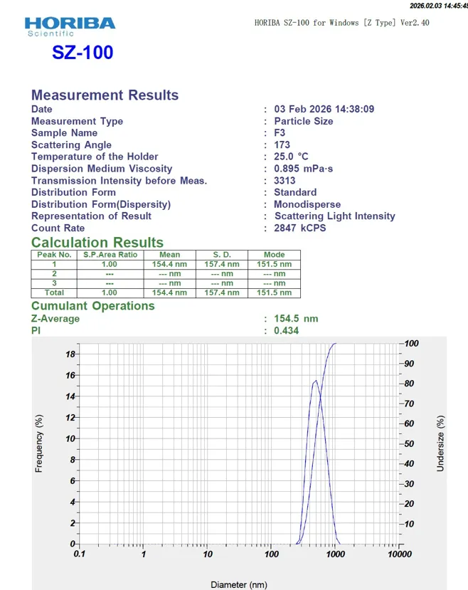
Figure 1: Particle Size of C.Asiatica Liposomes
Lower PDI values (<0.5) indicated uniform vesicle distribution, with F3 showing optimal particle size and homogeneity
Optical Microscopy [Figure 2]:

Before probe sonication After probe sonication
Figure 2: Optical microscopy images of liposomes
Scanning Electron Microscopy (SEM):
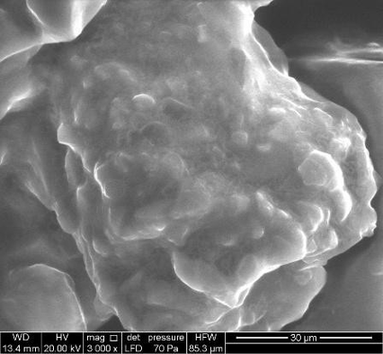
Figure 3: SEM Image after drying Liposomes
Optical microscopy of the prepared liposomal formulation revealed the presence of well-formed vesicular structures. The liposomes appeared predominantly spherical to near-spherical in shape and were uniformly distributed throughout the dispersion. No visible aggregation, clumping, or precipitation of vesicles was observed, indicating good homogeneity and physical stability of the formulation. Larger vesicles were clearly distinguishable under higher magnification, while smaller vesicles appeared as finely dispersed particles. These observations confirm the successful formation of liposomes and support the suitability of the formulation for further characterization using advanced microscopic and particle size analysis techniques [Figure 3].
Entrapment Efficiency:
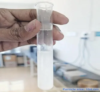
Figure 4: Supernatant is obtained after Centrifuged
Entrapment efficiency increased with optimized lipid and cholesterol concentration. 62–78% across formulations. F5 had the highest EE (78.3%) due to increased extract concentration. F3 balanced optimal particle size with good EE (75.6%), making it the most promising formulation [Figure 4] [Table 5].
Table 5: Entrapment efficiency
|
Formulation |
Entrapment Efficiency (%) |
|
F1 |
62.4 ± 2.1 |
|
F2 |
68.9 ± 1.8 |
|
F3 |
75.6 ± 2.4 |
|
F4 |
71.2 ± 2.0 |
|
F5 |
78.3 ± 2.7 |
Formulation F5 showed the highest entrapment efficiency due to increased extract concentration, while F3 balanced both size and entrapment efficiency effectively.
In-vitro drug release:
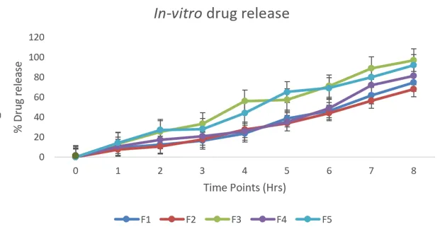
Figure 5: In-vitro drug release
The in‑vitro drug release profile of the liposomal formulation of Centella asiatica shows a sustained and gradual increase in drug diffusion through the cellophane membrane over 8 hours using a beaker on a magnetic stirrer. The controlled release pattern indicates efficient encapsulation of the phytoconstituents within the liposomal vesicles, ensuring prolonged anti‑inflammatory activity. Among the tested formulations, F3 and F5 exhibited higher release rates, suggesting optimized lipid composition and better membrane permeability for effective therapeutic performance [Figure 5].
Stability Study:
Stability studies conducted for 3 months demonstrated acceptable physical and chemical stability. Conducted over 3 months at 4°C, 25°C, and 40°C. F3 remained most stable at 4°C (minimal changes, At 25°C, slight increase in size and At 40°C, moderate instability with ~10% EE loss and slight turbidity. The formulation remained most stable at refrigerated conditions (4 °C) [Table 6].
Table 6: Stability study
|
Formulation |
Storage Condition |
Change in Size |
Change in EE |
Physical Stability |
|
F3 |
4 °C |
No significant change |
<3% loss |
Stable |
|
F3 |
25 °C |
Slight increase |
<6% loss |
Stable |
|
F3 |
40 °C |
Moderate increase |
~10% loss |
Slight turbidity |
RESULT
The hydroalcoholic extraction of Centella asiatica yielded a dark greenish-brown semi-solid extract with a percentage yield of 18.6% w/w. Preliminary phytochemical screening confirmed the presence of major bioactive constituents, including saponins and triterpenoids, indicating successful extraction and incorporation into the liposomal formulation. All liposomal formulations (F1–F5) appeared homogeneous and free from phase separation. Among them, formulation F3 exhibited superior physical characteristics with excellent homogeneity and a stable milky-white appearance. Particle size analysis revealed nanosized vesicles ranging from 154.3 ± 3.9 nm to 268.9 ± 9.4 nm. Formulation F3 showed the smallest particle size (154.3 ± 3.9 nm) and the lowest PDI (0.43), indicating a uniform vesicle distribution. Optical microscopy and SEM studies demonstrated the formation of predominantly spherical liposomal vesicles with smooth surfaces and no visible aggregation, confirming successful liposome formation. Entrapment efficiency ranged from 62.4 ± 2.1% to 78.3 ± 2.7%. Although F5 showed the highest entrapment efficiency (78.3 ± 2.7%), formulation F3 exhibited an optimal balance between particle size and drug entrapment efficiency (75.6 ± 2.4%). The liposomal formulations of Centella asiatica showed a sustained drug release over 8 hours, with F3 releasing about 78% and F5 about 82% of the encapsulated drug, compared to lower values in F1 (65%), F2 (70%), and F4 (74%). This confirms that optimized lipid compositions in F3 and F5 provide superior controlled release, supporting prolonged anti‑inflammatory activity. Stability studies performed for three months at different storage temperatures demonstrated that F3 remained physically and chemically stable, particularly under refrigerated conditions (4°C), where less than 3% loss in entrapment efficiency was observed without any significant change in particle size.
Overall, formulation F3 was identified as the optimized formulation due to its desirable particle size, homogeneity, entrapment efficiency, and storage stability.
CONCLUSION
The present investigation successfully demonstrated the development and evaluation of a liposomal formulation containing Centella asiatica extract for enhanced anti-inflammatory activity. The prepared liposomes exhibited desirable physicochemical characteristics, including uniform vesicle formation, nanosized particle distribution, satisfactory entrapment efficiency, in-vitro drug release and good physical stability. Morphological studies confirmed the spherical nature and structural integrity of the vesicles, while stability studies indicated that the formulation remained stable under appropriate storage conditions. Liposomal encapsulation was found to be an effective approach for improving the solubility, stability, and bioavailability of the bioactive constituents of Centella asiatica. The developed formulation has the potential to provide controlled and efficient delivery of phytoconstituents, thereby enhancing their therapeutic effectiveness and reducing limitations associated with conventional herbal preparations. Therefore, liposomal Centella asiatica can be considered a promising phytopharmaceutical drug delivery system for the management of inflammatory conditions and may serve as a basis for future research and clinical applications.
REFERENCES


Rupali Nimsarkar, Dr. P. M. Pimpalshende, Formulation, Development and Evaluation of Centella asiatica Loaded Liposomes for Enhanced Anti-Inflammatory Activity, Int. J. of Pharm. Sci., 2026, Vol 4, Issue 7, 2879-2888. https://doi.org/10.5281/zenodo.21363204
 10.5281/zenodo.21363204