
We use cookies to ensure our website works properly and to personalise your experience. Cookies policy
Dreamz College of Pharmacy, Himachal Pradesh Technical University, 175036
The present study aimed to formulate and evaluate sustained-release mucoadhesive microspheres of meloxicam using a combination of natural and synthetic polymers to enhance gastric retention, improve bioavailability, and achieve controlled drug release. Microspheres were prepared using the ionotropic gelation technique followed by polyelectrolyte complexation employing sodium alginate and chitosan, along with matrix modifiers such as HPMC and ethyl cellulose. A Quality by Design (QbD) approach using Box—Behnken design was implemented to optimize critical formulation variables. The prepared microspheres were evaluated for micromeritic properties, particle size, entrapment efficiency, mucoadhesion, and in vitro drug release. The optimized formulation (MM8) exhibited particle size of 528.6 ± 22 um, entrapment efficiency of 84.6 ± 1.7%, and mucoadhesion of 90.5 ± 1.8%. Drug release studies demonstrated sustained release up to 12 hours with 92.6 ± 2.0% release. Kinetic analysis revealed that drug release followed zero-order kinetics with a Korsmeyer—Peppas exponent (n 0.567), indicating non-Fickian diffusion. DSC and XRD studies confirmed partial amorphization without degradation. Stability studies showed no significant variation over three months. The study concluded that mucoadhesive microspheres of meloxicam provide a promising gastroretentive drug delivery system with enhanced therapeutic performance.
Among various GRDDS approaches, mucoadhesive systems offer a distinct advantage by adhering to the gastric mucosa, thereby ensuring prolonged retention and improved drug absorption. Mucoadhesive polymers such as sodium alginate and chitosan interact with mucin through hydrogen bonding and electrostatic interactions, leading to enhanced adhesion and drug retention. The incorporation of such polymers into drug delivery systems significantly improves the therapeutic efficacy of poorly soluble drugs (Thakur et al., 2026; Tiwari et al., 2026; Trinh et al., 2026; Tseng et al. , 2026). Microspheres represent a versatile multiparticulate drug delivery system capable of providing controlled and sustained drug release. Their small size and large surface area allow for uniform distribution in the gastrointestinal tract, enhancing drug absorption. When combined with mucoadhesive properties, microspheres can serve as an effective gastroretentive system, offering both prolonged retention and controlled drug release. The use of ionotropic gelation technique for microsphere preparation offers several advantages, including simplicity, cost-effectiveness, and avoidance of organic solvents. Sodium alginate, in the presence of calcium ions, forms a gel matrix that encapsulates the drug, while chitosan provides a mucoadhesive coating through polyelectrolyte complexation. The addition of synthetic polymers such as HPMC and ethyl cellulose further enhances the mechanical strength and release characteristics of the microspheres (Sharma et al., 2026; Szczepanowicz et al., 2026; Tanaka et al., 2026; Tang et al., 2026; Thakur et al., 2026; Tiwari et al., 2026; Trinh et al., 2026; Tseng et al., 2026).
Quality by Design (QbD) has become an integral part of pharmaceutical development, enabling systematic optimization of formulation variables and ensuring consistent product quality. The use of statistical tools such as Box—Behnken design allows for efficient evaluation of the effects of formulation variables on critical quality attributes such as entrapment efficiency, mucoadhesion, and drug release. The present study was therefore designed to develop and optimize mucoadhesive microspheres of meloxicam using a combination of natural and synthetic polymers. The objective was to enhance gastric retention, improve drug solubility, and achieve sustained drug release, thereby improving therapeutic efficacy and patient compliance.
MATERIALS AND METHODS
Materials

Preformulation Studies
Preformulation studies were conducted to evaluate the physicochemical properties of meloxicam and to ensure compatibility with selected excipients. The solubility of meloxicam was determined in distilled water and phosphate buffer (pH 6.8) using the shake flask method. The Imax of meloxicam was determined using UV-visible spectrophotometry in phosphate buffer (pH 6.8), which was found suitable for quantitative estimation. Drug—excipient compatibility was assessed using Fourier Transform Infrared (FTIR) spectroscopy, Differential Scanning Calorimetry (DSC), and X-ray Diffraction (XRD). FTIR spectra were recorded to identify potential interactions between drug and polymers, while DSC thermograms were used to analyze thermal behavior. XRD analysis was performed to evaluate the crystalline nature of the drug and its transformation in the formulated microspheres (Haimhoffer et al., 2021 ; Nicol et al., 2024; Ostr6ika-Cieélik, 2025; Steele & Austin, 2016).
Experimental Design (QbD Approach)
A Quality by Design approach was adopted to optimize the formulation variables. A threefactor, three-level Box—Behnken design was employed to evaluate the effect of independent variables on critical quality attributes (Haimhoffer et al., 2021; Nicol et al., 2024; Ostr6±kaCieélik, 2025; Steele & Austin, 2016).
Independent variables:
Dependent variables (responses):
A total of 17 experimental runs were generated, and formulations were coded as MM I to MM 17. Statistical analysis was performed using regression models and response surface methodology to identify optimal formulation conditions.
Preparation of Mucoadhesive Microspheres
Mucoadhesive microspheres of meloxicam were prepared using the ionotropic gelation technique followed by polyelectrolyte complexation. Accurately weighed meloxicam was dispersed in an aqueous solution of sodium alginate under continuous stirring to obtain a uniform dispersion. HPMC and ethyl cellulose were added to the mixture to enhance matrix formation and control drug release. The resulting dispersion was extruded dropwise through a syringe into a calcium chloride solution under constant stirring. Upon contact with the crosslinking solution, sodium alginate underwent instantaneous gelation, forming microspheres. The formed microspheres were allowed to cure for a specified time to ensure complete crosslinking. Subsequently, the microspheres were transferred to a chitosan solution to facilitate polyelectrolyte complexation. This step enhanced the mucoadhesive properties and mechanical strength of the microspheres. The microspheres were then filtered, washed with distilled water to remove excess reagents, and dried at room temperature (Abd El Hady et al., 2021; Ahmad et al., 2020; Baltzley et al., 2018; Essa et al., 2020; L6pez-Cebral et al., 2018; Mamona et al., 2025; Rath et al., 2025; Sharma et al., 2024; wang et al., 2025).
Evaluation of Microspheres
Micromeritic Properties
The flow properties of the prepared microspheres were evaluated by determining angle of repose, Carr's index, and Hausner ratio. These parameters provided insights into the handling and processing characteristics of the microspheres (Sapra et al., 2024; Sharma et al., 2024; Tripathi et al., 2024; J. Yang et al., 2024; X. Yang et al., 2024).
Particle Size Analysis

Entrapment Efficiency
Entrapment efficiency was determined by dissolving a known quantity of microspheres in phosphate buffer (pH 6.8) and analyzing the drug content using UV spectrophotometry. The percentage entrapment efficiency was calculated using the standard formula (Sapra et al. , 2024; Sharma et al., 2024; Tripathi et al., 2024; J. Yang et al., 2024; X. Yang et al., 2024).

Mucoadhesion Study
Mucoadhesive properties were evaluated using an ex vivo wash-off method. Freshly excised gastric mucosa was mounted on a suitable surface, and microspheres were applied. The setup was subjected to simulated gastric fluid under controlled conditions, and the percentage of microspheres retained over time was determined (Sapra et al., 2024; Sharma et al., 2024; Tripathi et al., 2024; J. Yang et al., 2024; X. Yang et al., 2024).
In Vitro Drug Release Study
Drug release studies were performed using a USP dissolution apparatus in phosphate buffer (pH 6.8). Samples were withdrawn at predetermined intervals and analyzed spectrophotometrically. The cumulative percentage drug release was calculated and plotted against time (Achmad et al., 2025; Pareek et al., 2024; Tripathi et al., 2024; X. Yang et al., 2024).
Drug Release Kinetics
The release data were fitted to various kinetic models, including zero-order, first-order, Higuchi, and Korsmeyer—Peppas models, to determine the mechanism of drug release.
Regression coefficients (R2) were calculated to identify the best-fit model.
Surface Morphology
Surface morphology of the microspheres was examined using scanning electron microscopy (SEM) to evaluate shape, surface characteristics, and structural integrity.
Thermal and Crystallinity Analysis
DSC and XRD analyses were conducted to assess the physical state of the drug within the microspheres and to detect any changes in crystallinity.
Stability Studies
Stability studies were performed on the optimized formulation under controlled conditions for a period of three months. Samples were evaluated at predetermined intervals for particle size, entrapment efficiency, mucoadhesion, and drug release. The stability of the formulation was assessed based on the absence of significant changes in these parameters (Achmad et al. , 2025; Pareek et al., 2024; Tripathi et al., 2024; X. Yang et al., 2024).
RESULTS
Calibration Curve of Meloxicam
The calibration curve of meloxicam in phosphate buffer (pH 6.8) demonstrated excellent linearity over the concentration range of 2—20 pg/mL. The regression equation obtained was y= 0.042x + 0.001, with a correlation coefficient ofR2 0.999, confirming the reliability of the analytical method.
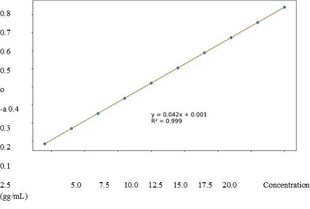
Figure 1: Calibration curve of meloxicam in phosphate buffer (pH 6.8).
Thermal and Crystallinity Analysis
DSC thermograms of meloxicam showed a sharp endothermic peak at —257—258 0 C, confirming its crystalline nature. In the microsphere formulation, a slight shift and broadening of the peak were observed, indicating partial amorphization of the drug.
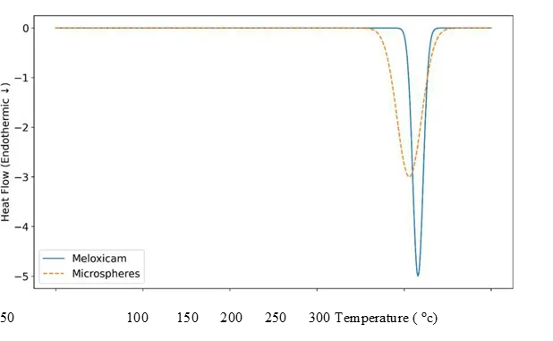
Figure 2: DSC thermograms of meloxicam and microspheres.
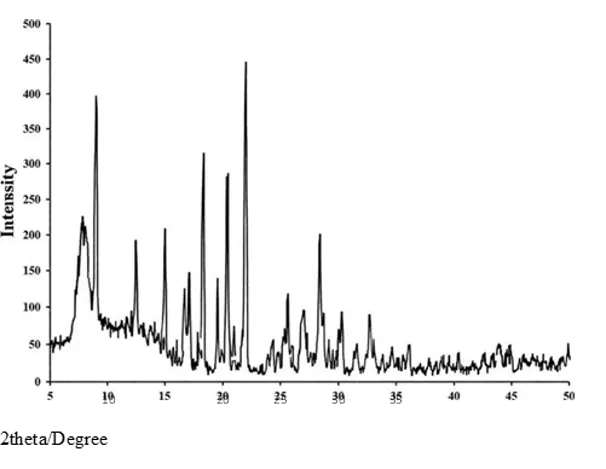
Figure 3: XRD pattern comparison.

The prepared microspheres exhibited acceptable flow properties.
Table 1: Micromeritic Properties of Microspheres
|
Batch |
Angle (0) |
Carr's Index (%) |
Hausner Ratio |
|
MMI |
30.1 ± 1.2 |
18.5 ±0.8 |
1.23 ±0.02 |
|
MM2 |
29.4 |
17.8 ±0.7 |
1.22 ±0.02 |
|
MM3 |
28.9 ± 1.1 |
17.2 ±0.6 |
1.21 ±O.OI |
|
MM4 |
27.6 |
15.8 |
1.19 |
|
MMS |
29.8 ±1.2 |
18.2 ±0.8 |
1.22 ±0.02 |
|
MM6 |
28.5 ± 1.0 |
16.9 ±0.7 |
1.20 |
|
MM7 |
27.9 ±0.8 |
15.6 ±0.6 |
1.18 |
|
MM8 |
26.8 ±0.9 |
14.3 |
1.16 |
|
MM9-MM17 |
(similar acceptable range) |
|
|
The results indicated good flowability, with MM8 showing superior properties.
Mucoadhesion Study
Table 2: Mucoadhesion Profile of Microspheres
|
Batch |
|
|
|
|
MMI |
78.2 ±2.3 |
70.5 ±2.1 |
62.4 ±2.o |
|
MM8 |
90.5 ± 1.8 |
84.2 ±1.9 |
76.8 ±1.8 |
|
MM17 |
89.2 ±1.9 |
82.6 ±2.o |
75.4 ± 1.9 |
All formulations showed gradual decrease in adhesion over time.

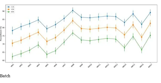
Figure 4: Mucoadhesion profile of all formulations (MMI—MMI 7).

In Vitro Drug Release Study
Table 3: Drug Release Profile
|
Batch |
QIh |
Q6h |
Q12h |
|
MMI |
32.5 ± 1.3 |
68.4 ±2.1 |
96.2 ±2.4 |
|
MM8 |
22.4 ± I |
55.6 ±1.8 |
92.6 ±2.o |
|
MM 17 |
23.1 ± 1.0 |
56.4 ± 1.9 |
91.5 ±2.1 |
The release pattern showed a biphasic profile with initial burst followed by sustained release.
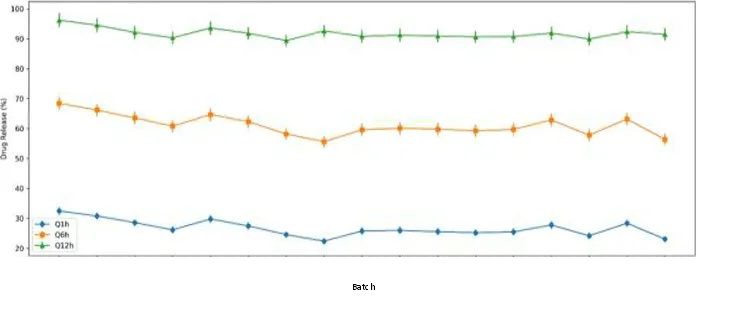
Figure 5: In vitro drug release profile of microspheres.
MM8 showed lowest burst release and controlled release behaviour.
Drug Release Kinetics
The optimized formulation (MM8) was analyzed for release kinetics. The R2 values indicated:
The Peppas exponent (n 0.567) confirmed non-Fickian diffusion, indicating combined diffusion and polymer relaxation mechanisms.
QbD Optimization
Response surface analysis showed the effect of formulation variables.
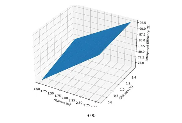
Figure 6: Response surface plot showing effect of alginate and chitosan on entrapment efficiency.
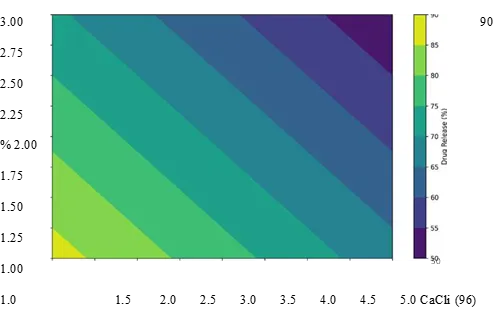
Figure 7: Contour plot showing effect of CaCb and alginate on drug release.
Stability Studies
Table 4: Stability Study Results
|
Time |
Particle Size (um) |
|
Mucoadhesion (%) |
Drug Release (%) |
|
O Month |
528.6 |
84.6 ± 1.7 |
90.5 ±1.8 |
92.6 ±2.o |
|
1 Month |
532.4 |
83.9 ± 1.8 |
89.6 ± 1.9 |
91.8 ±2.1 |
|
3 Months |
538.2 |
82.8 ±1.9 |
88.7 ±2.o |
90.9 ±2.2 |
No significant variation was observed, confirming stability.
DISCUSSION
The present study was designed to develop a robust gastroretentive mucoadhesive microsphere system of meloxicam capable of overcoming its inherent limitations such as poor aqueous solubility and variable bioavailability. The results obtained from formulation development, characterization, and optimization collectively demonstrated that the selected approach was scientifically sound and practically effective. The calibration curve established for meloxicam showed excellent linearity (R2 = 0.999), confirming the reliability of the analytical method employed throughout the study. This ensured accurate quantification of drug content during evaluation of entrapment efficiency and release studies. Preformulation analyses using FTIR, DSC, and XRD provided critical insights into drug—excipient compatibility. The absence of significant peak shifts in FTIR confirmed no chemical interaction, while DSC and XRD results indicated partial amorphization of meloxicam within the polymer matrix. This transformation from crystalline to partially amorphous form is advantageous, as it enhances drug dissolution and contributes to improved release characteristics.
The micromeritic evaluation revealed that all formulations exhibited acceptable flow properties, with angle of repose, Carr's index, and Hausner ratio within pharmacopeial limits. Among them, MM8 showed the best flow characteristics, which can be attributed to uniform particle size distribution and optimal polymer composition. Good flowability is essential not only for processing but also for ensuring reproducibility during large-scale production. Entrapment efficiency was significantly influenced by polymer concentration and cross-linking density. The optimized formulation (MM8) exhibited high entrapment efficiency (84.6 ± I . 7%), indicating effective drug encapsulation. The increase in sodium alginate concentration enhanced viscosity, leading to reduced drug diffusion into the external phase during preparation. Similarly, chitosan contributed to improved entrapment by forming a dense polyelectrolyte complex with alginate. Response surface analysis further confirmed that both polymers exerted a synergistic effect on entrapment efficiency, highlighting the importance of balanced formulation design.
Mucoadhesion studies demonstrated that all formulations adhered effectively to gastric mucosa, with a gradual decline over time. The optimized formulation exhibited maximum adhesion, which can be attributed to the presence of chitosan. The positively charged amino groups of chitosan interact with negatively charged mucin, resulting in strong electrostatic interactions. This prolonged adhesion ensures extended gastric residence time, which is critical for enhancing drug absorption and therapeutic efficacy. The in vitro drug release study revealed a biphasic release pattern characterized by an initial burst followed by sustained release. The initial release is attributed to surface-associated drug, while the sustained phase is governed by diffusion through the polymer matrix. The optimized formulation exhibited minimal burst release and controlled release up to 12 hours, indicating efficient matrix formation. The incorporation of both hydrophilic (HPMC) and hydrophobic (ethyl cellulose) polymers played a crucial role in modulating drug release. Kinetic analysis provided further insights into the mechanism of drug release. The high correlation with zero-order kinetics (R2 0.999) suggested a constant release rate independent of drug concentration. The Korsmeyer—Peppas model yielded an exponent value of 0.567, indicating non-Fickian diffusion. This confirms that drug release was governed by a combination of diffusion and polymer relaxation/swelling mechanisms, which is characteristic of well-designed sustained-release systems.
The QbD approach significantly strengthened the study by establishing a clear relationship between formulation variables and critical quality attributes. Response surface and contour plots demonstrated that increasing polymer concentration enhanced entrapment efficiency while reducing drug release due to increased cross-linking density. This systematic optimization ensured the selection of an ideal formulation with balanced properties. Stability studies confirmed that the optimized formulation remained stable over a period of three months, with no significant changes in particle size, entrapment efficiency, mucoadhesion, or drug release. The slight variations observed were within acceptable limits and can be attributed to minor polymer relaxation or environmental effects. Overall, the findings of this study strongly support the potential of mucoadhesive microspheres as an effective gastroretentive drug delivery system for meloxicam, offering controlled release, improved bioavailability, and enhanced therapeutic performance.
CONCLUSION
The present investigation successfully demonstrated the formulation and optimization of sustained-release mucoadhesive microspheres of meloxicam using a combination of natural and synthetic polymers. The ionotropic gelation technique, coupled with polyelectrolyte complexation, proved to be an effective and reproducible method for developing microspheres with desirable physicochemical and functional properties. The application of a Quality by Design approach enabled systematic optimization of formulation variables, ensuring a robust and efficient delivery system. Among all formulations, MM8 was identified as the optimized batch, exhibiting superior performance in terms of particle size (528.6 ± 22 pm), entrapment efficiency (84.6 ± 1.7%), and mucoadhesion (90.5 ± 1.8%). The formulation demonstrated a controlled and sustained drug release profile over 12 hours, with minimal initial burst release. Kinetic modeling confirmed that drug release followed zero-order kinetics and a non-Fickian diffusion mechanism (n 0.567), indicating the combined influence of diffusion and polymer relaxation. Thermal and crystallinity analyses (DSC and XRD) revealed partial amorphization of meloxicam within the polymer matrix, which is expected to enhance drug dissolution and bioavailability. Stability studies conducted over three months showed no significant changes in key parameters, confirming the physical and chemical stability of the optimized formulation. Overall, the developed mucoadhesive microspheres represent a promising gastroretentive drug delivery system capable of improving therapeutic efficacy, reducing dosing frequency, and enhancing patient compliance.
REFERENCES


Pooja Anjali, Dr. Puneet Kumar, Vijay Sharma, Neha, Naresh Kumar, Formulation, Development, and Evaluation of Sustained-Release Mucoadhesive Microspheres of Meloxicam Using Natural and Synthetic Polymers, Int. J. of Pharm. Sci., 2026, Vol 4, Issue 6, 1427-1439. https://doi.org/10.5281/zenodo.20564956
 10.5281/zenodo.20564956