
We use cookies to ensure our website works properly and to personalise your experience. Cookies policy
1Research Scholar, Shri Jagdishprasad Jhabarmal Tibrewala university, Vidyanagiri, Jhunjhunu – Churu road, Rajasthan- 6333010
2Guide, Shri Jagdishprasad Jhabarmal Tibrewala university, Vidyanagiri, Jhunjhunu – Churu road, Rajasthan- 6333010
3Co Guide, Shri Jagdishprasad Jhabarmal Tibrewala university, Vidyanagiri, Jhunjhunu – Churu road, Rajasthan- 6333010
Green nanotechnology has emerged as a promising and sustainable approach for the development of advanced biomedical and cosmeceutical formulations. Among various metallic nanomaterials, silver nanoparticles (AgNPs) have attracted considerable attention due to their remarkable antimicrobial, antioxidant, anti-inflammatory, wound healing, and skin-protective properties. Conventional methods used for nanoparticle synthesis often involve hazardous chemicals, high energy consumption, and toxic reducing agents, which may limit their biomedical applicability and environmental safety. Consequently, green synthesis approaches utilizing plant-derived phytochemicals as natural reducing and stabilizing agents have gained increasing importance in recent years. Crocus sativus flowers represent a highly valuable botanical resource for the green synthesis of silver nanoparticles because they are rich in biologically active compounds such as crocin, crocetin, picrocrocin, safranal, flavonoids, anthocyanins, and polyphenols. These phytochemicals play multifunctional roles during nanoparticle synthesis by facilitating the reduction of silver ions into metallic nanoparticles while simultaneously acting as natural capping and stabilizing agents. The resulting green-synthesized AgNPs exhibit enhanced biocompatibility, reduced toxicity, improved colloidal stability, and superior biological performance compared to chemically synthesized nanoparticles. This review comprehensively discusses the green synthesis of silver nanoparticles using saffron flower extracts and saffron floral waste, with particular emphasis on synthesis methodologies, reaction mechanisms, physicochemical factors affecting nanoparticle formation, and various characterization techniques including UV–Visible spectroscopy, Fourier Transform Infrared Spectroscopy (FTIR), X-ray Diffraction (XRD), Scanning Electron Microscopy (SEM), Transmission Electron Microscopy (TEM), Dynamic Light Scattering (DLS), and zeta potential analysis. Furthermore, the review highlights the antimicrobial, antioxidant, anti-inflammatory, wound healing, and anti-aging activities of saffron-mediated silver nanoparticles and their potential incorporation into skin care creams and topical cosmeceutical formulations. Additionally, the article discusses the advantages of green synthesis over conventional physical and chemical methods, including eco-friendliness, cost-effectiveness, reduced cytotoxicity, and enhanced biomedical compatibility. Current challenges associated with reproducibility, standardization, large-scale production, and regulatory approval are also critically analyzed. Overall, saffron-mediated green synthesized silver nanoparticles represent a highly promising strategy for the development of sustainable, multifunctional, and biologically safe nanocosmetic products with potential applications in advanced skin care therapies and herbal cosmeceutical industries.
In recent decades, the rapid advancement of nanotechnology has revolutionized multiple scientific disciplines, including medicine, pharmaceutics, biotechnology, and cosmetic science. Among these emerging interdisciplinary fields, nanocosmeceuticals have gained exceptional attention due to their ability to enhance the efficacy, stability, and delivery of active compounds in topical formulations. Conventional cosmetic preparations frequently suffer from major limitations such as poor skin penetration, inadequate stability of active ingredients, low bioavailability, rapid degradation upon exposure to light and oxygen, and reduced therapeutic efficiency. The human skin, particularly the stratum corneum, acts as a formidable barrier that restricts the permeation of many bioactive compounds. To overcome these limitations, nanotechnology-based delivery systems have been developed to improve the physicochemical and biological performance of cosmetic products. Nanotechnology involves the engineering of materials within the nanoscale range of approximately 1–100 nm, where materials exhibit unique optical, chemical, thermal, and biological properties due to their increased surface-area-to-volume ratio and quantum effects. At this nanoscale dimension, nanoparticles possess superior reactivity, enhanced penetration ability, controlled release characteristics, and improved interaction with biological membranes. Various nanosystems such as nanoemulsions, liposomes, solid lipid nanoparticles (SLNs), nanostructured lipid carriers (NLCs), dendrimers, and metallic nanoparticles are increasingly utilized in modern cosmetics and dermatological formulations. These systems not only protect sensitive active ingredients from oxidation and degradation but also improve localized retention, prolong therapeutic action, and enhance overall formulation performance. Consequently, nanotechnology has become a cornerstone in the development of next-generation cosmetic and skin care products. Simultaneously, there has been a substantial global shift toward herbal, sustainable, and environmentally friendly cosmetic products. Increasing awareness regarding the adverse effects of synthetic chemicals, preservatives, and artificial additives has encouraged consumers to seek safer and naturally derived alternatives. Conventional cosmetic ingredients such as parabens, sulfates, synthetic antioxidants, and chemical surfactants have been associated with allergic reactions, skin irritation, endocrine disruption, and environmental toxicity. As a result, herbal cosmeceuticals and green cosmetics have emerged as promising alternatives that combine cosmetic enhancement with therapeutic benefits. Plant-derived formulations are particularly valued because they contain a diverse range of bioactive phytochemicals including flavonoids, phenolic acids, alkaloids, tannins, terpenoids, carotenoids, and glycosides. These naturally occurring compounds exhibit strong antioxidant, anti-inflammatory, antimicrobial, anti-aging, and photoprotective activities. Unlike synthetic compounds that usually act through a single mechanism, herbal ingredients provide synergistic multi-target effects that enhance skin health while maintaining superior biocompatibility and reduced toxicity. Therefore, the integration of herbal ingredients into nanotechnology-based cosmetic formulations has opened a new avenue for the development of highly effective, safe, and eco-friendly nanocosmeceuticals. Although nanotechnology offers remarkable advantages, conventional methods used for nanoparticle synthesis present serious environmental and toxicological concerns. Traditional physical methods such as laser ablation, evaporation-condensation, and high-energy milling require sophisticated instrumentation, high energy consumption, and expensive operational conditions. Similarly, chemical synthesis methods depend heavily on hazardous reducing agents and stabilizers such as sodium borohydride, hydrazine, and various organic solvents. Residual traces of these toxic chemicals may remain adsorbed on nanoparticle surfaces, potentially causing cytotoxicity, oxidative stress, inflammation, and environmental contamination. Such drawbacks significantly limit the suitability of chemically synthesized nanoparticles for biomedical and cosmetic applications, especially for sensitive topical formulations.
To address these limitations, green synthesis of nanoparticles has emerged as an environmentally sustainable and biologically safe alternative. Green synthesis utilizes biological systems such as plants, bacteria, fungi, algae, and natural biomolecules as reducing and stabilizing agents for nanoparticle production. Among these approaches, plant-mediated synthesis has gained particular importance because it is simple, cost-effective, scalable, non-toxic, and environmentally benign. Plant extracts contain abundant phytochemicals capable of reducing metal ions into nanoparticles while simultaneously stabilizing them through natural capping mechanisms. This approach eliminates the need for hazardous chemicals, reduces energy consumption, minimizes waste generation, and produces highly biocompatible nanoparticles suitable for pharmaceutical and cosmetic applications. Among various metallic nanoparticles, silver nanoparticles (AgNPs) have attracted considerable attention owing to their extraordinary physicochemical and biological properties. Silver nanoparticles possess unique optical characteristics due to localized surface plasmon resonance (LSPR), along with remarkable antimicrobial, antioxidant, anti-inflammatory, and wound healing activities. AgNPs exhibit potent broad-spectrum antimicrobial activity against bacteria, fungi, and viruses through multiple mechanisms, including disruption of microbial cell membranes, generation of reactive oxygen species, leakage of intracellular components, and interference with microbial DNA replication. These properties make AgNPs highly valuable in skin care formulations intended for acne management, wound healing, skin protection, and preservation against microbial contamination. Furthermore, silver nanoparticles have demonstrated significant antioxidant and anti-inflammatory activities that contribute to the reduction of oxidative stress and inflammatory skin conditions. Due to their nanoscale size, AgNPs can penetrate deeper skin layers, improve local drug retention, and provide sustained therapeutic effects. Consequently, silver nanoparticles are increasingly incorporated into creams, gels, lotions, wound dressings, sunscreens, and anti-aging formulations. However, the biological performance and safety of AgNPs largely depend upon the synthesis method employed. Green synthesized silver nanoparticles are generally considered more stable, less toxic, and more biocompatible than chemically synthesized counterparts because they are naturally coated with plant-derived biomolecules. In this context, Crocus sativus, commonly known as saffron, represents an exceptionally promising botanical candidate for the green synthesis of silver nanoparticles. Saffron has been widely used since ancient times in traditional medicine, cosmetics, and food preparations due to its unique therapeutic and aromatic properties. The flowers and stigmas of saffron are rich in biologically active phytochemicals such as crocin, crocetin, safranal, picrocrocin, flavonoids, carotenoids, and phenolic compounds. These constituents exhibit powerful antioxidant, antimicrobial, anti-inflammatory, anti-aging, and skin-brightening activities, making saffron highly valuable in dermatological and cosmetic applications. The phytochemicals present in saffron flowers play a dual role during nanoparticle synthesis. Functional groups such as hydroxyl, carbonyl, and glycosidic moieties act as natural reducing agents that convert silver ions (Ag⁺) into metallic silver nanoparticles (Ag⁰). Simultaneously, these biomolecules adsorb onto the nanoparticle surface and function as stabilizing and capping agents, preventing aggregation and enhancing nanoparticle stability. As a result, saffron-mediated silver nanoparticles possess enhanced biological functionality and improved compatibility with topical formulations. In addition to their antimicrobial action, these nanoparticles may contribute synergistically to skin rejuvenation, reduction of hyperpigmentation, prevention of photoaging, and enhancement of overall skin health. The incorporation of saffron flower-mediated silver nanoparticles into skin care creams represents an innovative and sustainable strategy in modern cosmeceutical development. Such formulations combine the therapeutic benefits of herbal phytochemicals with the advanced delivery capabilities of nanotechnology, resulting in multifunctional products with enhanced efficacy and reduced toxicity. Moreover, the use of agricultural floral materials for nanoparticle synthesis supports sustainable utilization of plant resources and aligns with global efforts toward green chemistry and environmentally responsible manufacturing practices. Therefore, the present review aims to comprehensively discuss the green synthesis of silver nanoparticles using saffron flowers and their potential applications in skin care creams. The review focuses on the principles and advantages of green nanoparticle synthesis, phytochemical constituents of saffron flowers involved in nanoparticle formation, synthesis methodologies, characterization techniques, biological activities, skin care applications, safety considerations, and future prospects in herbal nanocosmeceutical research. The aim of the present review article is to comprehensively evaluate the green synthesis of silver nanoparticles using Crocus sativus flowers and to explore their potential applications in skin care cream formulations. The review focuses on the role of saffron flower phytochemicals in the reduction and stabilization of silver nanoparticles, various synthesis methodologies, physicochemical characterization techniques, biological activities, and their therapeutic significance in cosmetic and dermatological applications. Additionally, the article aims to discuss the advantages of green nanotechnology over conventional synthesis approaches, along with safety considerations, current research trends, challenges, and future prospects in the field of herbal nanocosmeceuticals.
Overview Of Silver Nanoparticles
Silver nanoparticles (AgNPs) are metallic nanostructures composed predominantly of silver atoms with dimensions generally ranging between 1 and 100 nm. Nanoparticles are considered ultrafine particles because their nanoscale size imparts unique physicochemical properties that differ significantly from bulk materials. Due to their extremely small dimensions, AgNPs possess a remarkably high surface area-to-volume ratio, resulting in increased surface reactivity and enhanced interaction with biological systems. These distinctive properties have attracted considerable attention in pharmaceutical, biomedical, and nanotechnological fields. AgNPs are particularly recognized for their unique optical, electronic, thermal, catalytic, magnetic, antimicrobial, antiviral, antifungal, anti-inflammatory, and anticancer properties, making them highly valuable in drug delivery, wound healing, diagnostics, biosensing, tissue engineering, and therapeutic applications. The size and morphology of silver nanoparticles are highly dependent on the synthesis method employed, including physical, chemical, and biological approaches. Generally, AgNPs exhibit particle sizes within the range of 1–100 nm, although controlled synthesis techniques can precisely regulate their dimensions according to specific applications. Biological and plant-mediated synthesis methods frequently produce stable and well-dispersed nanoparticles with sizes predominantly ranging from 5 to 60 nm. Parameters such as temperature, pH, silver nitrate concentration, reducing agents, reaction duration, and stabilizing materials significantly influence the final particle size and distribution. The nanoscale dimensions critically affect the biological activity, catalytic efficiency, optical characteristics, and cellular uptake of AgNPs, with smaller particles usually demonstrating enhanced reactivity and antimicrobial effectiveness due to their larger exposed surface area. Morphologically, silver nanoparticles display remarkable structural diversity. Although spherical nanoparticles are the most commonly synthesized and thermodynamically stable forms, AgNPs can also be fabricated into nanorods, nanowires, nanoplates, triangular structures, cubes, disks, and polygonal shapes depending on the synthesis conditions and preparation techniques. The morphology of AgNPs plays a crucial role in determining their physicochemical and biological behavior, as particle shape strongly influences optical properties, plasmonic activity, cellular interactions, and antimicrobial performance. Certain anisotropic structures such as triangular and rod-shaped nanoparticles exhibit superior optical and electronic properties compared to conventional spherical particles because of their unique surface arrangements and electron oscillation characteristics. The surface properties of silver nanoparticles are equally important in determining their stability, dispersibility, reactivity, and biological interactions. One of the most significant characteristics of AgNPs is their exceptionally high surface area-to-volume ratio, which increases the proportion of reactive atoms exposed on the particle surface. This enhanced surface reactivity contributes substantially to their catalytic efficiency and potent antimicrobial activity through improved interaction with microbial membranes and intracellular components. However, due to their high surface energy, nanoparticles possess an inherent tendency to aggregate or agglomerate. To overcome this limitation, various capping or stabilizing agents such as plant phytochemicals, polymers, surfactants, proteins, and other biomolecules are incorporated during synthesis. These agents provide steric hindrance and electrostatic repulsion, thereby maintaining colloidal stability and preventing particle aggregation. Another important surface-related property of AgNPs is Surface Plasmon Resonance (SPR), which represents the collective oscillation of conduction electrons on the nanoparticle surface upon interaction with incident light. SPR produces characteristic absorption bands in the visible region and is responsible for the distinctive color variations observed in AgNP suspensions, ranging from pale yellow to dark brown depending on particle size, shape, and surrounding medium. This optical phenomenon is widely utilized in UV–Visible spectroscopic characterization and plays a significant role in biosensing, imaging, and diagnostic applications. In addition, zeta potential is commonly used to evaluate the surface charge and colloidal stability of AgNPs. High positive or negative zeta potential values generate strong electrostatic repulsive forces between nanoparticles, thereby preventing aggregation, reducing sedimentation, and ensuring long-term formulation stability.
Silver nanoparticles (AgNPs) possess unique physicochemical properties that distinguish them from bulk silver and make them highly valuable in biomedical, pharmaceutical, cosmetic, and industrial applications. Among these properties, their remarkable optical behavior is considered one of the most important characteristics and is primarily attributed to the phenomenon known as Localized Surface Plasmon Resonance (LSPR). LSPR occurs when metallic nanoparticles are significantly smaller than the wavelength of incident light. Upon exposure to light, the conduction electrons present on the surface of AgNPs undergo coherent collective oscillation against the metallic lattice under the influence of the electromagnetic field. This resonant oscillation generates intense absorption and scattering of light within the visible region. The wavelength and intensity of the absorption peak are highly dependent on particle size, morphology, surface chemistry, aggregation state, and the surrounding dielectric environment. As a result, AgNP dispersions exhibit characteristic color changes ranging from pale yellow to deep brown. UV–Visible spectrophotometry is commonly employed to monitor this optical behavior, where spherical silver nanoparticles typically exhibit a strong absorption peak between 400 and 450 nm, confirming successful nanoparticle synthesis and stability. Another highly significant property of silver nanoparticles is their exceptional broad-spectrum antimicrobial activity against Gram-positive and Gram-negative bacteria, fungi, and viruses, including multidrug-resistant microbial strains. Unlike conventional antimicrobial agents that target a single biochemical pathway, AgNPs exert their antimicrobial effects through multiple simultaneous mechanisms, thereby minimizing the possibility of microbial resistance development. Due to their nanoscale size and high surface area, AgNPs can readily attach to microbial cell walls and membranes, causing structural alterations and increased membrane permeability. This interaction leads to leakage of essential intracellular constituents and eventual rupture of the microbial cell membrane. Furthermore, AgNPs continuously release silver ions (Ag⁺), which possess a strong affinity for sulfur- and phosphorus-containing biomolecules. These ions interact with respiratory enzymes and membrane proteins, disrupting ATP production and cellular metabolism. In addition, AgNPs induce the formation of reactive oxygen species (ROS) and free radicals, resulting in oxidative stress that damages lipids, proteins, and nucleic acids within microbial cells. Silver ions also interact directly with microbial DNA, causing condensation of the DNA structure, inhibition of replication processes, and arrest of cell division, ultimately leading to microbial death. In addition to antimicrobial activity, biologically synthesized or green-synthesized silver nanoparticles exhibit considerable antioxidant properties. During green synthesis, plant extracts or natural biomolecules function both as reducing agents and stabilizing agents, forming a protective outer coating or corona around the nanoparticles. This natural capping layer is rich in phytoconstituents such as polyphenols, flavonoids, tannins, and glycosides, which possess strong antioxidant potential. These phytochemicals contain active hydroxyl groups capable of donating electrons or hydrogen atoms to neutralize harmful free radicals and reactive oxygen species. Consequently, green-synthesized AgNPs demonstrate efficient free radical scavenging activity against superoxide radicals, hydroxyl radicals, and hydrogen peroxide. This antioxidant capability contributes significantly to cellular protection against oxidative stress, UV-induced skin damage, premature aging, and tissue degradation, thereby enhancing their therapeutic value in dermatological and wound-healing applications. Silver nanoparticles also exhibit potent anti-inflammatory properties that further support their biomedical and therapeutic applications. Recent nanomedicine and nanocosmeceutical studies have demonstrated that AgNPs can effectively modulate inflammatory signaling pathways and reduce excessive inflammatory responses. AgNPs suppress the overexpression of major pro-inflammatory mediators and transcription factors, thereby reducing the production of inflammatory cytokines such as tumor necrosis factor-alpha (TNF-α), interleukin-6 (IL-6), and interleukin-1 beta (IL-1β). In addition, they downregulate inflammatory enzymes including cyclooxygenase-2 (COX-2) and inducible nitric oxide synthase (iNOS), resulting in reduced edema, erythema, pain, and nitric oxide-mediated cellular injury. AgNPs further contribute to tissue repair by limiting the infiltration of inflammatory cells such as mast cells and neutrophils into damaged tissues. This modulation of the inflammatory microenvironment promotes transition from the inflammatory phase to the proliferative phase of healing, thereby accelerating granulation tissue formation, collagen deposition, re-epithelialization, and restoration of the skin barrier. Owing to these combined antimicrobial, antioxidant, and anti-inflammatory properties, silver nanoparticles have emerged as highly promising multifunctional agents in wound healing, tissue engineering, drug delivery, and advanced therapeutic formulations. Silver nanoparticles (AgNPs) have attracted enormous scientific and industrial attention because of their exceptional physicochemical, antimicrobial, optical, catalytic, and surface-modification properties. Their nanoscale dimensions, high surface area-to-volume ratio, tunable surface chemistry, and ability to interact effectively with biological systems have enabled their extensive application in pharmaceutical, biomedical, cosmetic, and dermatological fields. Owing to these multifunctional characteristics, AgNPs are increasingly being explored as advanced therapeutic agents, drug delivery carriers, diagnostic tools, antimicrobial coatings, and functional ingredients in nanocosmeceutical formulations.
In pharmaceutical sciences, silver nanoparticles play a significant role in the development of advanced drug delivery systems and novel therapeutic formulations. Many conventional drugs, especially hydrophobic compounds and biological macromolecules, suffer from poor aqueous solubility, instability, low bioavailability, and rapid metabolic degradation. AgNPs function as efficient nanocarriers capable of encapsulating, adsorbing, or conjugating therapeutic molecules onto their reactive surfaces, thereby protecting them from premature degradation and enhancing drug stability. Surface functionalization of AgNPs using polymers, ligands, surfactants, or plant-derived biomolecules allows precise modulation of drug release kinetics and targeted delivery to specific tissues or pathological sites. Stimuli-responsive AgNP-based systems can release therapeutic agents selectively under conditions such as acidic pH, elevated temperature, or inflammatory microenvironments, thereby improving therapeutic efficacy while minimizing systemic toxicity and adverse effects. Silver nanoparticles are also widely investigated as potent antimicrobial agents for combating multidrug-resistant (MDR) microorganisms. Unlike conventional antibiotics that typically target a single microbial pathway, AgNPs exert antimicrobial effects through multiple simultaneous mechanisms including disruption of microbial membranes, generation of reactive oxygen species, protein denaturation, and inhibition of DNA replication. This multifaceted mode of action significantly reduces the possibility of microbial resistance development. Furthermore, AgNPs exhibit strong synergistic effects when combined with standard antibiotics such as penicillin, ampicillin, erythromycin, and ciprofloxacin. These nanoparticle-antibiotic conjugates enhance bacterial susceptibility, lower the minimum inhibitory concentration (MIC) of antibiotics, and restore the effectiveness of drugs against resistant bacterial strains. In the biomedical field, silver nanoparticles have emerged as highly promising materials for anticancer therapy, medical device coatings, biosensing, and tissue engineering applications. AgNPs demonstrate selective cytotoxicity toward cancer cells primarily through oxidative stress-mediated mechanisms. Upon entering cancer cells, AgNPs induce excessive intracellular reactive oxygen species (ROS) generation, leading to mitochondrial dysfunction, DNA damage, cytochrome c release, and activation of apoptotic signaling pathways. Since cancer cells generally possess compromised antioxidant defense systems and abnormal metabolic activity, they are more susceptible to oxidative injury induced by AgNPs. In addition, silver nanoparticles exhibit anti-angiogenic properties by suppressing vascular endothelial growth factor (VEGF) and other angiogenic mediators, thereby inhibiting the formation of new blood vessels required for tumor growth and metastasis. Another major biomedical application of AgNPs involves infection control through antimicrobial coatings on medical devices and healthcare materials. Bacterial colonization and biofilm formation on catheters, prosthetic implants, orthopedic devices, heart valves, and surgical instruments represent significant causes of hospital-acquired infections. AgNP-coated surfaces effectively prevent microbial adhesion, disrupt biofilm formation, and inhibit bacterial proliferation on medical devices. As a result, silver nanoparticle coatings are increasingly incorporated into wound dressings, catheters, surgical tools, masks, hospital textiles, and implantable devices to maintain sterile surfaces and reduce the risk of nosocomial infections. Moreover, the unique optical properties of AgNPs, particularly Localized Surface Plasmon Resonance (LSPR), have enabled their application in biosensing and diagnostic technologies. Small changes in the nanoparticle surface environment produce measurable shifts in optical absorption spectra, allowing the development of highly sensitive diagnostic assays for detecting pathogens, biomarkers, genetic mutations, and disease-associated molecules. In cosmetic and dermatological applications, silver nanoparticles have become important components of modern nanocosmeceutical formulations because of their combined antimicrobial, antioxidant, anti-inflammatory, and wound-healing properties. AgNPs significantly accelerate wound healing by reducing microbial contamination, suppressing excessive inflammatory responses, and promoting tissue regeneration. They inhibit pro-inflammatory cytokines such as tumor necrosis factor-alpha (TNF-α) and interleukin-6 (IL-6), thereby minimizing chronic inflammation and facilitating progression toward the proliferative phase of healing. AgNPs also stimulate fibroblast proliferation, keratinocyte migration, granulation tissue formation, collagen synthesis, and re-epithelialization, ultimately improving wound closure and skin regeneration. Furthermore, silver nanoparticles help regulate collagen alignment during tissue remodeling, thereby reducing hypertrophic scarring and improving the structural integrity and appearance of healed skin. Silver nanoparticles are also extensively utilized in anti-acne and therapeutic skincare formulations. Acne vulgaris is primarily associated with the proliferation of microorganisms such as Cutibacterium acnes and Staphylococcus epidermidis within sebaceous follicles. Conventional acne therapies often rely on antibiotics and harsh chemicals that may cause irritation, dryness, erythema, and bacterial resistance. Biologically synthesized or green AgNPs provide a safer and more biocompatible alternative due to their targeted antimicrobial activity and reduced cytotoxicity. Plant-mediated AgNPs capped with bioactive phytochemicals offer additional anti-inflammatory and antioxidant benefits, helping to soothe irritated skin, reduce oxidative stress, control sebum oxidation, and minimize inflammatory lesions without causing excessive skin dryness. In addition, silver nanoparticles are increasingly explored as natural preservatives in cosmetic products. Traditional preservatives such as parabens, formaldehyde releasers, and phenoxyethanol have raised concerns regarding skin irritation, allergic reactions, and possible endocrine-disrupting effects. Due to their broad-spectrum antimicrobial activity, AgNPs can effectively inhibit microbial contamination in cosmetic formulations, thereby reducing or eliminating the need for synthetic chemical preservatives. Furthermore, the tunable optical properties of AgNPs resulting from Surface Plasmon Resonance enable their application as stable decorative pigments in cosmetics such as lipsticks, eye shadows, and beauty products, where they provide long-lasting color stability and enhanced aesthetic appeal. Collectively, these diverse pharmaceutical, biomedical, and cosmetic applications highlight the enormous potential of silver nanoparticles as multifunctional nanomaterials in advanced healthcare and consumer product development.
Green Synthesis Of Silver Nanoparticles
Green synthesis is an environmentally friendly, sustainable, and non-toxic approach for the fabrication of nanoparticles using naturally available biological resources instead of hazardous synthetic chemicals. In nanoparticle synthesis, this method has emerged as an important alternative to conventional physical and chemical techniques because it minimizes environmental hazards while enhancing biocompatibility and safety. Green synthesis primarily utilizes biological systems such as plant extracts, microorganisms including bacteria, fungi and yeast, algae, and natural biopolymers as reducing and stabilizing agents during nanoparticle formation. Among these, plant-mediated synthesis has gained particular attention due to its simplicity, rapidity, cost-effectiveness, and ability to produce stable nanoparticles without the need for complex laboratory conditions. In the synthesis of silver nanoparticles (AgNPs), biological extracts contain diverse phytochemicals and bioactive metabolites such as flavonoids, polyphenols, alkaloids, proteins, terpenoids, glycosides, tannins, and sugars that actively participate in nanoparticle formation. These biomolecules donate electrons to reduce silver ions (Ag⁺) derived from silver nitrate (AgNO₃) into metallic silver atoms (Ag⁰), leading to nucleation and formation of nanoparticles. Simultaneously, these biological compounds adsorb onto the nanoparticle surface and act as capping or stabilizing agents, preventing aggregation and maintaining colloidal stability through steric and electrostatic interactions.
The concept of green synthesis strongly aligns with the fundamental principles of Green Chemistry, which focus on minimizing environmental pollution, reducing hazardous waste generation, and promoting safer chemical processes. One of the primary objectives of green nanoparticle synthesis is waste prevention through the elimination or substantial reduction of toxic chemical byproducts commonly generated during conventional synthesis. Unlike chemical methods that rely on hazardous reducing agents such as sodium borohydride, hydrazine hydrate, and toxic organic solvents, green synthesis employs safe, biodegradable, and non-toxic biological materials under aqueous conditions. Water serves as the primary solvent medium in most biological synthesis protocols, thereby avoiding the use of harmful organic solvents and ensuring environmental compatibility. Another important principle incorporated into green synthesis is energy efficiency. Conventional physical methods such as thermal decomposition, laser ablation, evaporation-condensation, and irradiation techniques require high temperatures, high pressures, expensive instrumentation, and substantial electrical energy consumption. In contrast, green synthesis reactions are generally performed under mild conditions, including room temperature, atmospheric pressure, and physiological pH, significantly reducing energy requirements and operational costs. Furthermore, the use of renewable feedstocks such as medicinal plants, agricultural residues, fruit peels, and naturally cultivable microorganisms ensures sustainability and renewability of raw materials for large-scale nanoparticle production. Compared to conventional physical and chemical synthesis approaches, green synthesis offers several significant advantages in terms of toxicity, safety, cost-effectiveness, scalability, and biomedical applicability. Traditional chemical synthesis often leaves residual toxic chemicals, surfactants, and synthetic stabilizers adsorbed onto the nanoparticle surface, which may induce cytotoxicity, oxidative stress, and adverse biological reactions when applied in pharmaceutical or biomedical systems. In contrast, green-synthesized silver nanoparticles are naturally coated with a biologically active organic corona composed of plant-derived phytochemicals and biomolecules, which enhances their biocompatibility and reduces the risk of secondary toxicity. This natural capping layer also contributes additional therapeutic properties such as antioxidant, anti-inflammatory, and antimicrobial activities, creating a synergistic effect between the metallic silver core and the bioactive surface molecules. Economically, green synthesis is highly advantageous because it utilizes inexpensive and readily available natural resources such as plant leaves, flowers, seeds, fruit wastes, and microbial cultures, thereby reducing the dependency on costly synthetic chemicals and sophisticated equipment. The process is simple, rapid, and can be performed using basic laboratory infrastructure without the need for advanced vacuum systems or high-energy machinery. Another important advantage of green synthesis is its suitability for large-scale industrial production. Scaling up conventional chemical synthesis often generates enormous quantities of hazardous waste requiring expensive treatment and disposal procedures, whereas green synthesis follows relatively straightforward one-step or few-step reaction pathways that are easier to scale sustainably with minimal environmental burden. Additionally, the mild synthesis conditions preserve the stability and functional integrity of the nanoparticles, making them particularly suitable for pharmaceutical, biomedical, food, agricultural, and cosmeceutical applications. Due to their enhanced stability, reduced toxicity, eco-friendly nature, and multifunctional therapeutic properties, green-synthesized silver nanoparticles are increasingly considered superior alternatives to conventionally synthesized nanoparticles for advanced nanomedicine and sustainable nanotechnology applications. The synthesis of silver nanoparticles (AgNPs) can be broadly classified into three major methodologies, namely physical methods, chemical methods, and biological or green synthesis methods. These approaches differ significantly in their synthesis mechanisms, energy requirements, environmental impact, particle stability, scalability, and biomedical applicability. The selection of a suitable synthesis method plays a critical role in determining the physicochemical characteristics, morphology, surface chemistry, toxicity profile, and functional performance of the resulting nanoparticles. Over the years, increasing attention has been directed toward the development of environmentally sustainable and biocompatible synthesis approaches, particularly green synthesis, due to the limitations associated with conventional physical and chemical methods. Physical methods of nanoparticle synthesis generally follow a top-down approach in which bulk metallic materials are mechanically or physically reduced into nanosized particles. Commonly employed physical techniques include laser ablation, ion sputtering, evaporation-condensation, thermal decomposition, ball milling, and physical vapor deposition. These methods are advantageous because they avoid the direct use of toxic reducing chemicals and minimize chemical contamination in the final nanoparticle product. As a result, physically synthesized nanoparticles often exhibit relatively pure metallic surfaces. However, physical synthesis methods possess several important drawbacks. One of the major challenges is the high tendency of nanoparticles to undergo agglomeration due to the absence of effective stabilizing agents during synthesis. Particle aggregation significantly reduces the surface area, uniformity, and functional efficiency of the nanoparticles. In addition, physical methods require sophisticated instrumentation, high vacuum systems, elevated temperatures, and enormous energy inputs, making them economically expensive and operationally complex. The requirement for specialized equipment and harsh operating conditions also limits their practicality for large-scale industrial production and biomedical applications. Chemical methods represent one of the most widely utilized strategies for nanoparticle fabrication and generally operate through a bottom-up approach, where nanoparticles are assembled atom-by-atom or molecule-by-molecule from metallic precursor salts. Common chemical synthesis techniques include chemical reduction, sol-gel processing, microemulsion methods, electrochemical synthesis, and photochemical reduction. In silver nanoparticle synthesis, reducing agents such as sodium borohydride, hydrazine hydrate, citrate, and ascorbic acid are frequently employed to convert silver ions (Ag⁺) from precursor salts such as silver nitrate (AgNO₃) into metallic silver atoms (Ag⁰). Chemical synthesis methods provide excellent control over nanoparticle size, morphology, distribution, crystallinity, and surface characteristics, enabling the production of highly uniform and customizable nanoparticles for various industrial applications. Despite these advantages, chemical synthesis is associated with considerable limitations related to toxicity and environmental safety. The process commonly requires hazardous reducing agents, toxic organic solvents, and synthetic stabilizers, which can generate dangerous chemical byproducts and environmental pollutants. Furthermore, traces of these toxic chemicals may remain adsorbed on the nanoparticle surface even after purification, thereby increasing cytotoxicity and limiting their direct application in pharmaceutical, biomedical, food, and agricultural systems. The disposal of chemical waste generated during large-scale synthesis also poses serious environmental and regulatory challenges. To overcome the limitations associated with physical and chemical synthesis approaches, biological or green synthesis methods have emerged as environmentally friendly, sustainable, and biocompatible alternatives for nanoparticle production. Green synthesis generally follows a bottom-up mechanism and utilizes natural biological resources such as plants, bacteria, fungi, yeast, algae, and biopolymers for nanoparticle fabrication. Among these biological systems, plant-mediated synthesis is the most extensively explored due to its simplicity, rapidity, cost-effectiveness, and ability to produce highly stable nanoparticles. Various plant parts including leaves, flowers, fruits, seeds, peels, roots, rhizomes, and bark extracts are rich in bioactive phytochemicals such as flavonoids, polyphenols, terpenoids, alkaloids, proteins, sugars, enzymes, and glycosides, which actively participate in nanoparticle synthesis. These biomolecules perform a dual role during the synthesis process. First, they act as reducing agents by donating electrons to reduce silver ions (Ag⁺) into metallic silver nanoparticles (Ag⁰). Second, they function as natural capping and stabilizing agents by forming a protective organic coating around the nanoparticle surface, thereby preventing aggregation and enhancing colloidal stability without the need for synthetic stabilizers.
Green synthesis offers numerous advantages over conventional synthesis methods. The process is highly eco-friendly because it eliminates the use of hazardous chemicals, toxic solvents, and dangerous reducing agents. As a result, green-synthesized nanoparticles exhibit improved biocompatibility, lower cytotoxicity, and enhanced suitability for pharmaceutical, biomedical, cosmetic, and agricultural applications. Another major advantage is the mild reaction conditions under which synthesis occurs. Unlike physical methods that require extreme temperatures and high energy consumption, biological synthesis reactions are typically completed rapidly under ambient temperature, atmospheric pressure, and aqueous conditions, thereby significantly reducing operational costs and energy requirements. Additionally, the phytochemical corona surrounding green-synthesized nanoparticles imparts additional therapeutic properties such as antioxidant, anti-inflammatory, antimicrobial, and wound-healing activities, creating functional synergy between the metallic silver core and the natural biomolecular coating. These properties enhance nanoparticle bioavailability, stability, and biological effectiveness against pathogenic microorganisms and biofilm-producing bacteria. Despite these significant advantages, green synthesis methods also face certain challenges, particularly regarding reproducibility and standardization. Since the composition of plant extracts and biological materials can vary depending on geographic location, climatic conditions, harvesting season, plant age, and extraction methods, achieving uniform nanoparticle size, morphology, and consistency across different production batches remains difficult. This variability can affect the scalability and commercial standardization of green synthesis protocols. Nevertheless, continuous advancements in nanotechnology, phytochemical characterization, and process optimization are progressively improving the reproducibility and industrial feasibility of green nanoparticle synthesis. Overall, biological or green synthesis has emerged as one of the most promising approaches for the sustainable production of silver nanoparticles due to its eco-friendly nature, low toxicity, cost-effectiveness, energy efficiency, and enhanced biomedical compatibility. Green synthesis, particularly plant-mediated synthesis of silver nanoparticles (AgNPs), has emerged as a highly promising alternative to conventional physical and chemical synthesis methods due to its eco-friendly nature, economic feasibility, reduced toxicity, and enhanced biocompatibility. Traditional nanoparticle synthesis methods often involve hazardous chemicals, sophisticated instrumentation, high energy consumption, and environmentally unsafe procedures, which limit their biomedical applicability and sustainability. In contrast, green synthesis utilizes naturally available biological resources such as plant extracts, microorganisms, algae, and biopolymers to fabricate nanoparticles under mild and environmentally benign conditions. The bioactive phytochemicals present in these natural sources not only reduce metallic ions into nanoparticles but also stabilize the particles through natural capping mechanisms, thereby providing multiple functional advantages over conventional synthesis pathways. One of the most important advantages of green synthesis is its eco-friendly and sustainable nature. Conventional physical synthesis methods require extreme operational conditions such as high temperatures, high pressures, vacuum environments, and intensive energy input, resulting in elevated operational costs and substantial environmental burden. Similarly, chemical synthesis approaches rely heavily on toxic reducing agents, hazardous organic solvents, and synthetic stabilizers that generate dangerous chemical waste and environmental pollutants. Green synthesis fundamentally overcomes these limitations by replacing toxic chemicals with natural biological extracts and aqueous solvent systems. In most plant-mediated synthesis protocols, water acts as the primary solvent medium, thereby eliminating the use of harmful organic solvents and minimizing ecotoxicological risks. Furthermore, green synthesis reactions are generally completed rapidly under ambient temperature, atmospheric pressure, or mild heating conditions, significantly reducing energy consumption and carbon footprint. The process produces minimal or no hazardous byproducts, making it an environmentally sustainable and safer alternative for nanoparticle production. Another major benefit of green synthesis is its remarkable cost-effectiveness. Conventional nanoparticle synthesis often requires expensive precursor chemicals, sophisticated purification systems, high-energy instruments, and specialized manufacturing facilities, which collectively increase production costs. In contrast, green synthesis employs inexpensive, renewable, and widely available biological materials such as medicinal plants, agricultural residues, fruit peels, weeds, and microbial cultures. Plant species such as Moringa oleifera, Azadirachta indica, Terminalia arjuna, and Eugenia roxburghii have been extensively investigated as economical biological sources for nanoparticle synthesis. Additionally, the phytochemicals present in plant extracts perform dual functions by acting simultaneously as reducing agents and stabilizing or capping agents. This eliminates the requirement for separate synthetic stabilizers, surfactants, or polymeric capping materials that are commonly required in chemical synthesis methods. The synthesis process itself is relatively simple, rapid, and does not require sophisticated high-pressure reactors, vacuum chambers, or advanced industrial machinery, thereby significantly reducing equipment and operational expenses. Consequently, green synthesis offers a highly economical and scalable approach suitable for large-scale industrial production. Reduced toxicity is another critical advantage associated with green-synthesized silver nanoparticles. Chemically synthesized nanoparticles often retain residual toxic chemicals, solvents, surfactants, and reducing agents adsorbed onto their surfaces even after purification. These residual contaminants may induce oxidative stress, inflammation, cytotoxicity, genotoxicity, and adverse biological responses when applied in pharmaceutical or biomedical systems. In contrast, green synthesis avoids the use of hazardous chemical reagents, resulting in nanoparticles with cleaner and biologically safer surfaces. The natural phytochemical coating surrounding green-synthesized nanoparticles significantly reduces the risk of secondary toxicity and improves their compatibility with living systems. Comparative biological studies have demonstrated that green-synthesized nanoparticles produce lower inflammatory responses and reduced cellular stress compared to conventionally synthesized nanoparticles. Furthermore, in vitro safety assessments, including hemolysis and cytotoxicity studies, have shown that biologically synthesized AgNPs exhibit potent antimicrobial activity while maintaining minimal or negligible toxicity toward healthy mammalian cells and tissues. This reduced toxicity profile makes green nanoparticles particularly suitable for biomedical, pharmaceutical, cosmetic, and therapeutic applications. Biocompatibility represents one of the most valuable features of green-synthesized nanoparticles. In plant-mediated synthesis, the nanoparticle surface becomes naturally coated with biologically active phytochemicals such as flavonoids, polyphenols, terpenoids, proteins, alkaloids, and glycosides. This natural organic corona stabilizes the nanoparticles and creates a biologically compatible interface that closely resembles natural cellular environments. As a result, green-synthesized nanoparticles can interact more safely and efficiently with biological tissues, reducing the likelihood of immune rejection, irritation, or adverse cellular reactions. The phytochemical capping layer also contributes additional therapeutic properties such as antioxidant, anti-inflammatory, wound-healing, and antimicrobial activities, thereby creating functional synergy between the metallic silver core and the surrounding biological molecules. This synergistic interaction enhances therapeutic efficiency and improves targeted biological performance in applications such as drug delivery, wound healing, tissue engineering, anticancer therapy, and antimicrobial treatment. Moreover, green-synthesized silver nanoparticles exhibit enhanced surface bioavailability and strong activity against multidrug-resistant pathogens and bacterial biofilms while causing minimal damage to surrounding healthy tissues. Collectively, these advantages establish green synthesis as a highly effective, sustainable, and biocompatible strategy for the development of advanced silver nanoparticle-based biomedical and pharmaceutical systems.
Saffron Flowers As A Biological Reducing Agent
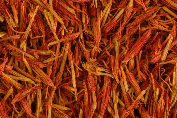

Fig 1: Botanical Profile of Crocus sativus (Saffron)
Crocus sativus, commonly known as saffron or “red gold,” is a perennial, stemless, bulbous herb belonging to the family Iridaceae and is considered one of the world’s most valuable medicinal and culinary plants. The plant is highly prized for its bright scarlet-red stigmas, which are used as a spice, coloring agent, fragrance component, and therapeutic material in traditional and modern medicine. Despite its enormous commercial value, saffron is regarded as a low-yielding crop because only the stigmas are economically useful and must be harvested manually with extreme care. During processing, large quantities of floral biomass waste such as petals, tepals, stamens, and styles are generated. Recent scientific investigations have revealed that these byproducts possess considerable phenolic, antioxidant, antimicrobial, and anti-inflammatory potential due to the presence of bioactive phytochemicals including flavonoids, polyphenols, anthocyanins, and glycosides.
Taxonomically, Crocus sativus belongs to the kingdom Plantae, division Magnoliophyta or Spermatophyta, class Liliopsida (Monocotyledonae), order Liliales, and family Iridaceae. The genus is Crocus and the species is Crocus sativus L. Morphologically, the plant is characterized by an underground storage organ known as a corm or cormus, which functions as the primary vegetative and nutrient storage structure. The plant generally grows up to approximately 30 cm in height and produces long, narrow, linear green leaves. The aerial floral structures consist of sepals, petals, stamens, style, and stigmas. The flowers are cup-shaped with a characteristic purple or parma coloration. The commercially important spice is obtained from the three dried scarlet-red stigmas attached to the yellow style. Biomass distribution within the flower is highly uneven, with the stigmas accounting for only about 7% of the total floral mass, while the remaining 93% consists of sepals, petals, stamens, and styles. Consequently, nearly 30 kg of floral waste, mainly tepals, is produced during the preparation of 1 kg of commercial saffron spice. Crocus sativus is cultivated extensively in several regions across Asia, Europe, and the Mediterranean basin. Iran is the largest producer of saffron globally, while in India its cultivation is mainly confined to the Kashmir region due to favorable climatic conditions. Other saffron-producing countries include Spain, Greece, Italy, France, Azerbaijan, Turkey, Morocco, China, Egypt, and Mexico. The plant thrives best under semi-arid and arid environmental conditions characterized by cold winters, moderate precipitation during spring and winter, and dry summers with minimal rainfall. Saffron cultivation requires well-drained soil and specific climatic conditions for optimal flowering and stigma development. The harvesting process is extremely labor-intensive because the delicate flowers must be manually collected and the tiny stigmas carefully separated by hand. This meticulous harvesting process, combined with the low yield of stigmas per flower, contributes significantly to saffron’s status as one of the most expensive and valuable botanical commodities in the world. The pharmacological significance, characteristic sensory attributes, and therapeutic efficacy of Crocus sativus are primarily attributed to its rich and diverse phytochemical composition. The plant contains a complex array of secondary metabolites including carotenoids, apocarotenoids, flavonoids, anthocyanins, glycosides, and phenolic compounds that collectively contribute to its medicinal, antioxidant, anti-inflammatory, antimicrobial, neuroprotective, and anticancer properties. Although the commercially valuable stigmas are particularly rich in specialized apocarotenoids such as crocin, picrocrocin, and safranal, the non-stigmatic floral parts including petals, tepals, and leaves also represent important reservoirs of polyphenolic bioactive compounds with considerable pharmaceutical and nanotechnological importance. Among the major phytoconstituents of saffron, crocin is regarded as one of the most significant bioactive compounds. Chemically, crocin is a rare water-soluble carotenoid derivative identified as a glycosyl ester of crocetin linked with gentiobiose sugars. Unlike most naturally occurring carotenoids that are highly lipophilic, the high glycosyl content of crocin imparts remarkable hydrophilic properties, allowing it to dissolve readily in aqueous media. Crocin constitutes approximately 6–16% of the dry weight of saffron stigmas and is primarily responsible for the characteristic golden-yellow coloration of saffron. Pharmacologically, crocin exhibits potent antioxidant activity through effective free radical scavenging and inhibition of oxidative stress-induced cellular damage. Numerous studies have demonstrated its neuroprotective, anti-inflammatory, hepatoprotective, cardioprotective, and antitumor properties. Crocin has also shown promising effects in improving memory, reducing neuronal degeneration, and protecting tissues against oxidative injury. Picrocrocin is another major apocarotenoid constituent responsible for the characteristic bitter taste of saffron. Chemically, it is a monoterpene glycoside with the molecular formula C₁₆H₂₆O₇ and constitutes approximately 3.7–4% of saffron stigma dry weight. Picrocrocin plays a crucial role as a precursor molecule in the formation of safranal during post-harvest processing. Under conditions such as drying, heat exposure, acidic or alkaline hydrolysis, and enzymatic degradation, picrocrocin undergoes cleavage to produce glucose and an intermediate aglycone known as 4-hydroxy-β-cyclocitral. This intermediate subsequently undergoes dehydration to generate safranal, the principal volatile aromatic compound of saffron. Safranal is a low-molecular-weight monoterpene aldehyde chemically identified as 2,6,6-trimethyl-1,3-cyclohexadiene-1-carboxaldehyde (C₁₀H₁₄O). It represents nearly 60% of the volatile fraction of saffron essential oil and is mainly responsible for the distinct sweet, hay-like, and slightly iodoform aroma of saffron. Interestingly, freshly harvested saffron flowers contain very low concentrations of safranal, and its formation largely depends on post-harvest processing methods, particularly drying and storage conditions that facilitate the degradation of picrocrocin. In addition to its sensory significance, safranal possesses considerable biological activities including antioxidant, anticonvulsant, antidepressant, anxiolytic, anti-inflammatory, and antimicrobial properties.
Flavonoids constitute one of the most abundant classes of phytochemicals present in saffron petals and leaves, which together account for the major proportion of floral biomass generated during saffron processing. Important flavonoids identified in saffron include kaempferol and its glycosidic derivatives, quercetin, catechin, rutin, and anthocyanins such as delphinidin. Among these, kaempferol glycosides are recognized as the predominant secondary metabolites in saffron petals. These flavonoids exhibit strong antioxidant and free radical scavenging activities, thereby protecting cells against oxidative damage and inflammation. In recent years, saffron-derived flavonoids have gained substantial attention in green nanotechnology because they can effectively function as natural reducing and capping agents during the green synthesis of metallic nanoparticles. Their polyhydroxylated structures facilitate electron donation for metal ion reduction while simultaneously stabilizing the synthesized nanoparticles through surface adsorption. Saffron flowers are also rich sources of phenolic compounds and non-flavonoid polyphenols that contribute significantly to their biological activities. Polar solvent extracts such as methanol, aqueous ethanol, and ethyl acetate extracts have been reported to contain high total phenolic content. These phenolic constituents exhibit potent antioxidant, anti-inflammatory, hepatoprotective, renoprotective, antimicrobial, and chemoprotective properties by inhibiting lipid peroxidation and reducing oxidative stress-mediated tissue damage. The hydroxyl functional groups present in these phenolic compounds are highly reactive and capable of donating electrons, making them highly valuable in green synthesis processes for the reduction of metallic salts into stable and biocompatible nanoparticles. Consequently, saffron-derived phenolic compounds and flavonoids have attracted increasing interest for applications in nanomedicine, pharmaceutical formulations, wound healing systems, and advanced cosmeceutical products. Phytochemicals derived from Crocus sativus floral biomass and agricultural residues play a crucial multifunctional role in the green synthesis of metallic nanoparticles, particularly silver nanoparticles (AgNPs). In plant-mediated nanotechnology, biological extracts replace hazardous synthetic chemicals by functioning simultaneously as reducing agents, stabilizing agents, and capping agents during nanoparticle formation. Saffron petals, leaves, and other non-stigmatic floral wastes are exceptionally rich in bioactive phytochemicals such as crocin, flavonoids, polyphenols, anthocyanins, glycosides, proteins, amino acids, and terpenoids, which collectively regulate the nucleation, growth, morphology, stability, and biological performance of nanoparticles. These phytochemicals not only facilitate nanoparticle synthesis under eco-friendly conditions but also impart enhanced therapeutic and biomedical properties to the resulting nanostructures. One of the most fundamental roles of phytochemicals during nanoparticle synthesis is their function as reducing agents during the bioreduction phase. The synthesis of metallic nanoparticles initially requires the conversion of oxidized metallic precursor ions, such as silver ions (Ag⁺) from silver nitrate (AgNO₃) or gold ions (Au³⁺) from chloroauric acid (HAuCl₄), into their zero-valent metallic forms (Ag⁰ or Au⁰). Saffron-derived phytochemicals, especially polyphenols and flavonoids such as kaempferol and quercetin, contain multiple hydroxyl (-OH) functional groups attached to aromatic structures that possess strong electron-donating capacity. During the reduction process, these hydroxyl groups undergo oxidation while simultaneously transferring electrons or hydrogen atoms to the metallic ions. As a result, the metal ions are reduced into neutral metallic atoms that subsequently aggregate to form nanoscale nuclei. The generalized reduction pathway may be represented as follows:
Mn++Phytochemical-OH→M0+Phytochemical=O+nH+

As the concentration of zero-valent metal atoms increases in the reaction medium, supersaturation occurs, leading to rapid nucleation and nanoparticle formation. The antioxidant strength, concentration, and chemical composition of the phytochemicals directly influence nucleation kinetics, particle size distribution, morphology, and homogeneity of the synthesized nanoparticles. Therefore, phytochemical composition plays a decisive role in controlling nanoparticle characteristics during green synthesis. Following nanoparticle formation, phytochemicals serve as stabilizing agents that prevent nanoparticle aggregation and maintain colloidal stability. Newly formed metallic nanoparticles possess extremely high surface energy and naturally tend to agglomerate through van der Waals attractive forces, resulting in larger inactive aggregates. To prevent this phenomenon, various saffron-derived biomolecules such as polysaccharides, proteins, crocin glycosides, anthocyanins, and flavonoids adsorb spontaneously onto the nanoparticle surface. These molecules stabilize nanoparticles through both electrostatic stabilization and steric hindrance mechanisms. The adsorption of polar and ionizable functional groups onto the metallic surface imparts a surface charge to the nanoparticles, typically generating negative zeta potential values. This electrostatic charge creates repulsive forces between adjacent nanoparticles, thereby preventing particle clustering and maintaining dispersion stability. Simultaneously, bulky phytochemical structures physically surround the nanoparticle core and create steric barriers that restrict close particle-to-particle interactions. This steric hindrance effectively isolates nanoparticles from one another, preventing aggregation and sedimentation without the need for synthetic stabilizers such as polyvinylpyrrolidone (PVP) or polyethylene glycol (PEG). Consequently, saffron-derived phytochemicals ensure long-term colloidal stability under environmentally benign conditions. Another highly important function of phytochemicals in green synthesis is their role in nanoparticle capping and surface functionalization. The capping mechanism involves the formation of chemical interactions between specific functional groups of phytochemicals and the outer surface atoms of the metallic nanoparticle core. Fourier-transform infrared spectroscopy (FTIR) analyses of green-synthesized nanoparticles frequently reveal the presence of functional groups such as hydroxyl (-OH), carbonyl (=O), carboxyl (-COOH), amide (-NH–C=O), and amino (-NH₂) groups on the nanoparticle surface. These groups originate from flavonoids, phenolic acids, proteins, amino acids, terpenoids, picrocrocin derivatives, and safranal intermediates present within saffron extracts. Through coordination bonding and surface adsorption, these functional groups establish a stable organic coating around the nanoparticles. This capping layer not only protects nanoparticles from oxidation and aggregation but also regulates nanoparticle growth by selectively occupying active crystal facets. Such thermodynamic control influences crystal growth direction and surface energy distribution, enabling controlled synthesis of nanoparticles with different morphologies including spherical, rod-shaped, triangular, and anisotropic structures. Importantly, the phytochemical capping layer also enhances the biological and therapeutic performance of the synthesized nanoparticles. Since the nanoparticle surface remains coated with naturally occurring bioactive compounds such as crocin, kaempferol, and other antioxidant phytochemicals, the resulting nanoconjugates exhibit improved biocompatibility and safer interaction with mammalian cells. These phytochemical-coated nanoparticles demonstrate synergistic biological effects including enhanced antioxidant activity, anti-inflammatory effects, antimicrobial potency, antiproliferative activity against cancer cells, and stronger disruption of bacterial biofilms. Furthermore, the presence of natural phytochemical coronas improves cellular uptake, reduces cytotoxicity, and enhances nanoparticle bioavailability in pharmaceutical and biomedical applications. Therefore, phytochemicals not only drive nanoparticle synthesis but also critically determine the structural stability, therapeutic efficiency, and biomedical compatibility of green-synthesized metallic nanoparticles.
Synthesis Process Of Silver Nanoparticles Using Saffron Flowers
The preparation of saffron flower extract represents a crucial preliminary step in the green synthesis of silver nanoparticles (AgNPs), as the efficiency of nanoparticle formation depends strongly on the concentration and stability of phytochemicals present within the botanical extract. In the case of Crocus sativus, non-stigmatic floral components such as petals, sepals, stamens, and leaves serve as abundant reservoirs of bioactive metabolites including flavonoids, polyphenols, anthocyanins, glycosides, crocin derivatives, proteins, and phenolic acids. These compounds act as natural reducing and stabilizing agents during nanoparticle synthesis. Therefore, careful processing of saffron floral waste is essential to preserve the structural integrity and biological activity of these sensitive phytochemicals. The preparation process begins with the systematic collection of saffron floral biomass generated as agricultural waste during commercial saffron harvesting. Since only the scarlet stigmas are utilized for spice production, nearly 90% of the flower, including petals and other floral structures, remains unused. These discarded floral materials are collected directly from saffron cultivation fields and subjected to thorough cleaning procedures to remove soil particles, dust, microbial contaminants, and other environmental impurities. Initially, the flowers are washed several times with running tap water followed by repeated rinsing with distilled or double-distilled water to ensure maximum purity before extraction. Following washing, the floral material undergoes controlled drying to prevent degradation of temperature-sensitive phytochemicals. Shade drying is the most commonly employed method, where the flowers are spread in thin layers within a clean, well-ventilated environment protected from direct sunlight. This drying process is generally performed at ambient room temperatures ranging between 25°C and 30°C. Exposure to direct ultraviolet radiation is strictly avoided because it may induce photo-oxidation and degradation of important bioactive constituents such as crocin, anthocyanins, flavonoids, and phenolic compounds. In some laboratory protocols, controlled oven drying at mild temperatures between 37°C and 40°C is alternatively used until constant weight is achieved. Once the material becomes completely dehydrated and brittle, it is mechanically pulverized using a laboratory grinder to obtain a fine, homogeneous powder. Pulverization disrupts plant cellular structures and increases the surface area available for solvent penetration, thereby enhancing the efficiency of phytochemical extraction. The powdered material is subsequently stored in airtight containers under cool and dry conditions to prevent moisture absorption and chemical degradation. The extraction stage is primarily designed to isolate and transfer maximum quantities of intracellular phytochemicals into the liquid phase while maintaining their structural integrity. The most widely reported approach for saffron flower waste is aqueous decoction or solid-liquid extraction. In this method, a measured quantity of dried saffron flower powder, typically ranging from 1 g to 10 g, is mixed with a defined volume of solvent, commonly between 50 mL and 100 mL, in a clean Erlenmeyer flask. The mixture is subjected to controlled heating with continuous magnetic stirring on a thermostatic hotplate. Extraction temperatures generally range from 50°C to 80°C for durations of approximately 15–30 minutes. Mild heating facilitates disruption of plant cell walls and improves dissolution of bioactive metabolites into the solvent while minimizing decomposition of thermolabile glycosides and phenolic compounds essential for nanoparticle reduction. After extraction, the crude mixture is allowed to cool to room temperature and subsequently undergoes filtration and clarification processes. Initially, the extract is filtered through analytical filter papers such as Whatman No. 1 to remove coarse plant debris and suspended particles. To further eliminate ultrafine particulates and microfibers that may interfere with subsequent spectroscopic characterization techniques such as UV-Visible spectroscopy, the filtrate is often centrifuged at speeds of approximately 4000–5000 rpm for about 10 minutes. This step produces a clear and homogeneous phytochemical-rich extract suitable for nanoparticle synthesis. The purified extract is then transferred into sterilized amber-colored glass containers and stored under refrigerated conditions at approximately 4°C. Cold storage minimizes microbial contamination, enzymatic degradation, and oxidation of sensitive phytochemicals prior to use in nanoparticle synthesis reactions. Solvent selection plays a decisive role in determining the composition, extraction efficiency, and reducing capability of saffron flower extracts. In most green synthesis studies involving saffron floral biomass, distilled or deionized water is selected as the primary extraction solvent due to its excellent compatibility with green chemistry principles. Water effectively dissolves highly polar phytochemicals such as crocin derivatives, kaempferol glycosides, anthocyanins, and phenolic acids because these compounds contain multiple hydroxyl and sugar moieties that confer strong hydrophilicity. Furthermore, the use of water completely eliminates toxic organic solvents from the synthesis process, thereby enhancing the safety, eco-friendliness, and biomedical suitability of the resulting nanoparticles. Although certain experimental studies employ aqueous ethanol or methanol mixtures to improve extraction of less polar flavonoids and aglycones, purely aqueous extracts remain preferred for direct nanoparticle synthesis because residual organic solvents may destabilize colloidal suspensions and introduce safety concerns in pharmaceutical and cosmetic applications. Another important factor influencing extract performance is solvent pH. Saffron aqueous extracts are naturally slightly acidic because of dissolved phenolic acids and organic metabolites. The pH of the extraction medium directly affects the ionization state of phenolic hydroxyl groups, thereby altering their electron-donating ability and redox potential during nanoparticle synthesis. Consequently, pH variations significantly influence nucleation kinetics, particle size distribution, morphology, and colloidal stability of the synthesized silver nanoparticles. Therefore, careful control of extraction conditions, solvent composition, and storage parameters is essential for obtaining highly active and reproducible saffron flower extracts suitable for efficient green nanoparticle synthesis. The green synthesis of silver nanoparticles (AgNPs) using Crocus sativus floral waste involves a carefully controlled sequence of experimental procedures designed to maximize nanoparticle formation while preserving the bioactivity of plant-derived phytochemicals. The synthesis process relies on the reduction of silver ions (Ag⁺) from silver nitrate (AgNO₃) into zero-valent metallic silver (Ag⁰) through the action of naturally occurring reducing biomolecules present within saffron flower extracts. Comparative analyses of studies conducted by Baran et al. and Bagherzade et al. demonstrate both common principles and methodological variations in nanoparticle synthesis protocols. The synthesis procedure begins with the preparation of silver nitrate solution, which serves as the metallic precursor source for nanoparticle formation. In the study conducted by Baran et al. (2023), highly pure silver nitrate salt (99.8% purity) obtained from Sigma Aldrich was dissolved in distilled water to prepare the reaction solution. Similarly, Bagherzade et al. (2017) used analytical-grade silver nitrate procured from Merck Company. Different precursor concentrations and extract ratios were systematically evaluated to optimize nanoparticle formation efficiency, and the optimal concentration was determined to be 2 mmol/L silver nitrate solution. To ensure sterility and prevent microbial interference during subsequent antibacterial evaluations, the prepared silver nitrate solution was passed through a sterile 0.45 μm membrane filter prior to use. Proper preparation of the silver nitrate solution is essential because the concentration of silver ions directly influences nucleation kinetics, nanoparticle size distribution, and colloidal stability during synthesis. Mixing conditions represent another critical determinant of nanoparticle morphology and synthesis efficiency. In the Baran et al. protocol, the aqueous extract prepared from dried purple saffron flowers was directly mixed with the silver nitrate solution under environmentally friendly and low-cost reaction conditions designed for rapid nanoparticle production. In contrast, Bagherzade et al. employed a more controlled and precise mixing methodology. In this procedure, 5 mL of saffron floral waste extract was added dropwise into 20 mL of the optimized 2 mmol/L silver nitrate solution. Instead of conventional magnetic stirring, ultrasonic irradiation was applied throughout the reaction process. Ultrasound energy promotes rapid dispersion of reactants, enhances mass transfer, and improves nucleation homogeneity by generating localized cavitation effects within the reaction medium. This technique facilitates uniform nanoparticle growth and contributes significantly to achieving narrow particle size distributions and improved structural consistency. Reaction parameters such as temperature, reaction duration, storage conditions, and purification methods play decisive roles in nanoparticle crystallization and stability. In the Baran et al. study, the synthesis protocol was optimized for extremely rapid nanoparticle formation, with successful synthesis occurring within only 15 minutes of reaction time. The resulting nanoparticles exhibited remarkable colloidal stability, maintaining consistent UV-Visible absorption characteristics even after one month of storage at room temperature. In comparison, Bagherzade et al. utilized prolonged ultrasonic irradiation for approximately 3 hours to ensure complete reduction and nanoparticle maturation. UV-Visible spectroscopic analysis demonstrated progressive increases in absorbance intensity for up to 5 hours without significant shifts in the characteristic surface plasmon resonance wavelength, indicating stable nanoparticle formation. To preserve sensitive phytochemicals prior to synthesis, saffron extracts were initially prepared and maintained under dark conditions at room temperature for 24 hours. Furthermore, all reaction glassware was sterilized by heating at 160°C for 2 hours to eliminate contamination. Following synthesis, the nanoparticles were isolated through centrifugation at 8000 rpm for 10 minutes, allowing separation of nanoparticle pellets from unreacted phytochemicals and residual plant debris.
One of the most important visual indicators of nanoparticle synthesis is the characteristic color change occurring during the reduction of silver ions into metallic nanoparticles. Initially, silver nitrate solutions appear colorless or pale. Upon addition of saffron flower extract, a distinct transformation to yellowish-brown or deep brown coloration is observed, signifying nanoparticle formation. This phenomenon occurs because of Surface Plasmon Resonance (SPR), where conduction electrons on the nanoparticle surface undergo collective oscillation upon interaction with light. In the Baran et al. study, nanoparticle formation was confirmed within 15 minutes by the appearance of a strong SPR absorption peak at approximately 405.68 nm, indicating the formation of small and highly stable silver nanoparticles. The rapid synthesis was attributed to the presence of abundant phytochemicals such as flavonoids, phenols, alkaloids, and terpenoids capable of rapidly reducing Ag⁺ ions into Ag⁰ nanoparticles. In the Bagherzade et al. study, the reaction mixture gradually developed a deep brown color corresponding to nanoparticle formation, and UV-Visible spectroscopic analysis revealed a characteristic SPR peak centered around 450 nm. The shift in absorption wavelength suggested slight differences in nanoparticle size and morphology compared to the Baran et al. protocol. Comparative characterization studies demonstrated that the synthesis procedures produced nanoparticles with distinct structural properties. The Baran et al. method yielded highly stable nanoparticles with rapid synthesis kinetics and an SPR peak around 405 nm, indicative of fine particle dispersion and smaller nanoparticle size. In contrast, the Bagherzade et al. approach generated crystalline, predominantly spherical silver nanoparticles with average particle sizes of approximately 15 nm and a distribution range between 12 and 20 nm. These nanoparticles were naturally capped and stabilized by saffron-derived phytochemicals, which prevented aggregation and enhanced colloidal stability. Overall, both studies demonstrate that saffron floral waste serves as an efficient, sustainable, and biocompatible biological source for the green synthesis of stable silver nanoparticles suitable for pharmaceutical, biomedical, antimicrobial, and cosmeceutical applications. The green synthesis of silver nanoparticles (AgNPs) using Crocus sativus extracts is strongly influenced by several physicochemical parameters including pH, temperature, precursor concentration, extract concentration, and reaction time. These variables directly regulate the reduction kinetics of silver ions (Ag⁺), nucleation rate, nanoparticle growth behavior, morphology, particle size distribution, colloidal stability, and final biological activity of the synthesized nanoparticles. Comparative studies involving saffron-mediated synthesis methods demonstrate that even slight variations in reaction conditions can significantly alter nanoparticle characteristics and synthesis efficiency. Among the most influential factors, pH plays a critical role because it governs the ionization state and surface charge of phytochemicals present in saffron extracts. Plant metabolites such as flavonoids, phenolic acids, anthocyanins, terpenoids, and glycosides contain multiple hydroxyl and carboxyl functional groups whose protonation state changes depending on pH conditions. These alterations directly affect the electron-donating ability of the phytochemicals and consequently their efficiency in reducing Ag⁺ ions into metallic Ag⁰ nanoparticles. In the reported studies, aqueous extraction methods were employed without extensive artificial pH adjustment, thereby maintaining the naturally slightly acidic to neutral pH characteristic of saffron floral extracts. This mild pH environment helps preserve delicate secondary metabolites, particularly anthocyanins and phenolic compounds, which serve dual functions as reducing and capping agents. Under these balanced conditions, nanoparticle formation proceeds smoothly without rapid coagulation, precipitation, or destabilization of the colloidal suspension. The pH environment also influences electrostatic stabilization by affecting nanoparticle surface charge and zeta potential, thereby contributing to long-term colloidal stability.
Temperature is another highly important parameter because it determines the kinetic energy available for nucleation and nanoparticle growth. Elevated temperatures generally accelerate molecular movement and increase the rate of reduction reactions, whereas mild temperatures help preserve thermolabile phytochemicals and promote controlled nanoparticle formation. In the synthesis procedure reported by Baran et al., the reduction process was successfully performed under ambient room-temperature conditions without requiring excessive thermal energy. The high reducing capability of the saffron flower phytochemicals enabled rapid conversion of silver ions into nanoparticles within a very short duration while maintaining excellent stability. In contrast, Bagherzade et al. implemented ultrasonic irradiation instead of conventional thermal heating during nanoparticle synthesis. Although the extraction stage was carried out at room temperature, all glassware was sterilized at 160°C for 2 hours before synthesis to eliminate contamination. Ultrasonic irradiation generated localized acoustic cavitation and microscopic hotspots within the solution, supplying sufficient localized energy to promote homogeneous nucleation and controlled crystal growth. This approach resulted in highly crystalline, spherical nanoparticles with an average diameter of approximately 15 nm. Therefore, both thermal and non-thermal energy inputs can significantly influence nanoparticle morphology, crystallinity, and size distribution. The concentration ratio between the silver nitrate precursor and the saffron extract is another decisive parameter affecting nanoparticle synthesis efficiency and stability. Proper concentration balance ensures adequate availability of reducing biomolecules for complete reduction of silver ions while simultaneously providing sufficient capping molecules to stabilize the newly formed nanoparticles. In the study conducted by Bagherzade et al., systematic optimization experiments were performed using different extract volumes ranging from 1 to 10 mL combined with varying silver nitrate concentrations. The optimal synthesis condition was identified as 5 mL of saffron wastage extract mixed with 20 mL of 2 mmol/L AgNO₃ solution. Under these conditions, the concentration of phytochemicals such as flavonoids, antioxidants, aldehydes, ketones, and phenolic compounds was sufficient to efficiently reduce Ag⁺ ions while simultaneously capping the nanoparticle surfaces. This natural capping prevented excessive particle aggregation by generating steric hindrance and electrostatic repulsion between nanoparticles. Insufficient extract concentration can lead to incomplete reduction and poor stabilization, whereas excessive extract concentration may cause excessive surface coating, particle enlargement, or agglomeration. Therefore, optimization of precursor-to-extract ratio is essential for achieving uniform particle size and stable colloidal systems. Reaction time also critically influences nanoparticle synthesis because it determines the progression from initial ion reduction to complete nanoparticle nucleation, growth, and stabilization. Different synthesis methods exhibit significantly different kinetic profiles depending on the efficiency of phytochemical reduction and physical agitation techniques. In the Baran et al. study, the saffron flower extract demonstrated exceptionally rapid reduction kinetics, producing stable nanoparticles within only 15 minutes. Successful nanoparticle formation was confirmed through the appearance of a characteristic Surface Plasmon Resonance (SPR) absorption peak at approximately 405.68 nm. In contrast, the ultrasonic-assisted synthesis method employed by Bagherzade et al. required approximately 3 hours of continuous ultrasonic irradiation to complete nanoparticle synthesis. During this process, UV-Visible spectroscopic monitoring revealed a progressive increase in absorbance intensity up to the fifth hour of observation, indicating continuous nanoparticle production over time. Importantly, the SPR absorption peak remained fixed near 450 nm throughout the reaction period without wavelength shifting, demonstrating that nanoparticle size remained highly uniform despite increasing nanoparticle concentration. Long-term stability studies further confirmed that the synthesized colloidal nanoparticles retained identical SPR profiles even after one month of storage, indicating excellent colloidal stability and resistance to aggregation. Overall, the synthesis of silver nanoparticles using saffron extracts is a highly sensitive process controlled by interconnected physicochemical factors including pH, temperature, concentration ratios, and reaction duration. Careful optimization of these parameters is essential to achieve stable, uniformly distributed, biocompatible, and highly active nanoparticles suitable for pharmaceutical, biomedical, antimicrobial, and nanocosmeceutical applications.
Characterization Of Silver Nanoparticles
The characterization of silver nanoparticles (AgNPs) is an essential step in confirming successful nanoparticle synthesis and understanding their physicochemical properties, including particle size, morphology, crystalline structure, elemental composition, and surface chemistry. In saffron-mediated green synthesis, the nanoparticles produced using Crocus sativus flower extracts and saffron processing waste were comprehensively characterized using various spectroscopic, microscopic, and diffraction-based analytical techniques. These characterization studies confirmed the successful formation of stable, crystalline, and biologically capped silver nanoparticles suitable for pharmaceutical, antimicrobial, biomedical, and cosmeceutical applications.
Uv–Visible Spectroscopy: UV–Visible spectroscopy was employed as the primary and most rapid analytical technique to confirm the formation of silver nanoparticles through the phenomenon of Surface Plasmon Resonance (SPR). SPR occurs when incident electromagnetic radiation excites the collective oscillation of free conduction electrons present on the surface of metallic nanoparticles. This interaction generates a characteristic absorption peak in the visible region, which serves as direct evidence for nanoparticle formation. The first indication of successful synthesis in both studies was the visible color transformation of the reaction mixture. In the study conducted by Bagherzade et al., the initially colorless silver nitrate solution changed into a deep brown colloidal suspension after treatment with saffron wastage extract. Similarly, Baran et al. observed a yellowish-brown to brown coloration upon interaction between the saffron purple flower extract and silver nitrate solution. These visual color changes confirmed the reduction of silver ions (Ag⁺) into metallic silver nanoparticles (Ag⁰). Spectral analysis further validated nanoparticle formation. Baran et al. reported a sharp and intense SPR absorption peak at 405.68 nm, which appeared within only 15 minutes of reaction time, indicating extremely rapid nanoparticle synthesis. In contrast, Bagherzade et al. monitored the reaction under ultrasonic irradiation and identified a stable SPR peak centered around 450 nm. The absorbance intensity progressively increased up to five hours without any significant shift in the peak position, suggesting increased nanoparticle yield while maintaining uniform particle size distribution. Long-term UV–Visible monitoring demonstrated excellent colloidal stability, as the synthesized nanoparticles retained identical absorption profiles even after one month of room-temperature storage.
Fourier Transform Infrared Spectroscopy (Ftir): Fourier Transform Infrared Spectroscopy (FTIR) was utilized to identify the phytochemical constituents and functional groups involved in the reduction and stabilization of silver nanoparticles. FTIR analysis provided strong evidence that naturally occurring biomolecules present in saffron extracts acted simultaneously as reducing agents and capping agents during nanoparticle synthesis. In the study by Bagherzade et al., FTIR spectra revealed a broad absorption peak at 3414 cm⁻¹, corresponding to O–H stretching vibrations of hydroxyl groups derived from phenolic compounds and flavonoids. A sharp peak at 1622 cm⁻¹ indicated the presence of carbonyl (C=O) stretching or secondary amine groups. Additional peaks at 1413 cm⁻¹ and 1075 cm⁻¹ were associated with C–N and C–O stretching vibrations, respectively, confirming the involvement of carbohydrates, glycosides, and bioactive constituents such as crocin, crocetin, picrocrocin, and safranal in nanoparticle stabilization. Similarly, Baran et al. identified several important functional groups in the saffron purple flower extract. Major absorption bands were observed at 3287 cm⁻¹ corresponding to O–H and N–H stretching vibrations of proteins and phenolic compounds, while the peak at 1633 cm⁻¹ represented C=O stretching of amide groups or aromatic compounds. Additional peaks at 2924 cm⁻¹, 1379 cm⁻¹, and 1017 cm⁻¹ confirmed the presence of terpenes, alkaloids, flavonoids, and phenolic biomolecules attached to the nanoparticle surface. These phytochemicals formed a natural protective capping layer around the silver core, preventing aggregation and enhancing colloidal stability.
Scanning Electron Microscopy (Sem) And Edx Analysis: Scanning Electron Microscopy (SEM) and Field Emission Scanning Electron Microscopy (FE-SEM) were employed to evaluate the external morphology, surface topology, and distribution pattern of the synthesized nanoparticles. These imaging techniques revealed that the saffron-mediated silver nanoparticles were predominantly spherical in shape and exhibited highly uniform distribution. FE-SEM imaging conducted by Bagherzade et al. demonstrated that the nanoparticles synthesized using saffron wastage extract were exceptionally well dispersed and monodispersed with minimal aggregation. Likewise, SEM analysis performed by Baran et al. showed dense and compact arrangements of spherical nanoparticles synthesized from saffron purple flower extracts. The nanoparticles remained structurally separated due to the presence of phytochemical capping agents surrounding the metallic core, which effectively minimized van der Waals-mediated agglomeration. Energy-Dispersive X-ray (EDX) spectroscopy was further used to determine the elemental composition and purity of the synthesized nanoparticles. Both studies identified a strong characteristic silver signal between 3 keV and 3.4 keV, confirming the successful formation of metallic silver nanoparticles. Baran et al. additionally reported the presence of carbon, oxygen, chlorine, and potassium in the EDX spectrum, which directly supported the existence of a bio-organic capping layer derived from saffron phytochemicals. The elemental analysis revealed approximately 51.52% silver content in the synthesized nanoparticles.
Transmission Electron Microscopy (Tem): Transmission Electron Microscopy (TEM) was employed as the definitive high-resolution imaging technique to determine the exact particle size, morphology, and nanoscale distribution of the synthesized silver nanoparticles. TEM imaging provided direct visual evidence of nanoparticle formation and structural uniformity. According to Bagherzade et al., TEM micrographs confirmed that the silver nanoparticles synthesized through ultrasonic-assisted green synthesis were predominantly spherical and highly uniform in distribution. Detailed particle size analysis demonstrated a narrow size range between 12 nm and 20 nm, with an average particle diameter of approximately 15 nm. The clear separation observed between adjacent nanoparticles confirmed the effectiveness of the naturally occurring phytochemical capping agents in preventing aggregation and maintaining colloidal stability.
X–Ray Diffraction (Xrd): X-ray Diffraction (XRD) analysis was carried out to determine the crystalline structure, phase purity, and long-range atomic arrangement of the synthesized silver nanoparticles. The diffraction profiles obtained from both saffron-mediated synthesis studies confirmed the formation of highly crystalline silver nanoparticles possessing a face-centered cubic (FCC) lattice structure. Distinct Bragg diffraction peaks were observed at characteristic 2θ values corresponding to metallic silver crystal planes. Peaks appearing at 38.2°, 44.3°, 64.5°, and 77.5° corresponded to the (111), (200), (220), and (311) crystallographic planes, respectively. Among these, the intense diffraction peak associated with the (111) plane indicated preferential crystalline orientation and high crystallinity of the nanoparticles. Importantly, no additional impurity peaks or secondary crystalline phases were detected in the XRD spectra, confirming the complete reduction of silver ions into pure, crystalline, zero-valent metallic silver nanoparticles. The absence of contaminating peaks further demonstrated the effectiveness of saffron-derived phytochemicals in mediating clean and eco-friendly nanoparticle synthesis.
Biological Activities Of Saffron-Mediated Silver Nanoparticles
Antimicrobial Activity
Activity against Skin Pathogens: The green synthesis of silver nanoparticles utilizing saffron (Crocus sativus L.) extracts has emerged as a powerful biomedical strategy, exhibiting exceptional antimicrobial prowess against a diverse array of pathogenic microorganisms. Comprehensive investigations show that saffron-mediated silver nanoparticles (AgNPs or SNPs) demonstrate potent bactericidal and fungicidal profiles at remarkably low minimum inhibitory concentrations (MIC). These nanostructures possess broad-spectrum efficacy, vigorously suppressing virulent Gram-positive skin and systemic threats like Staphylococcus aureus and Streptococcus pneumoniae, alongside notorious Gram-negative pathogens including Pseudomonas aeruginosa, Klebsiella pneumoniae, Escherichia coli, Shigella flexneri, and Acinetobacter species. Furthermore, saffron-stabilized nanoparticles display significant anticandidal activity, rendering them highly effective against opportunistic fungal agents such as Candida albicans, which frequently compromise cutaneous and mucosal barrier integrity. Notably, research underscores that the overall antimicrobial efficacy of these nanoparticles is highly size-dependent; as the particle dimensions contract down to the 12–20 nm range, the relative surface-area-to-volume ratio increases, allowing a vastly heightened contact interface with microbes and drastically elevating their lethality against resilient strains. Mechanism of Antimicrobial Action: The antimicrobial mechanisms governing saffron-mediated silver nanoparticles are multi-tiered and rely fundamentally on both physical disruption and chemical toxicity. Structurally, the toxicity and efficiency of these biogenic nanoparticles are heavily dictated by their precise physical dimensions and the steady rate of silver ion ($Ag^+$) emission over time. These nanoparticles interact dynamically with the complex three-dimensional cell walls and lipid membranes of Gram-positive and Gram-negative bacteria. Due to their ultra-small, spherical morphology (frequently averaging around 15 nm), they easily attach to and penetrate the cellular envelopes. Once localized at or within the bacterial cell wall, they initiate severe physical damage, destabilizing structural components and causing membrane perforation, which triggers the leakage of vital intracellular constituents. Concurrently, the constant release of silver ions from the internalized nanoparticles disrupts metabolic machinery. These ions bind tightly to cellular sulfur- and phosphorus-rich biomolecules like proteins and nucleic acids, ultimately halting DNA replication, inactivating essential respiratory enzymes, and inducing catastrophic oxidative stress that leads swiftly to programmed cell death.
Antioxidant Activity
Free Radical Scavenging: The outstanding biological functionality of saffron-mediated silver nanoparticles is deeply intertwined with the rich phytochemical cocktail native to the Crocus sativus plant matrix. Saffron wastages, petals, and floral parts contain substantial reservoirs of secondary metabolites belonging to the flavonoid and carotenoid families. Fourier Transform Infrared Spectroscopy (FTIR) characterization consistently confirms that the synthesized silver cores are naturally capped and stabilized by these plant secondary metabolites. Prominent among these active components are valuable pigments like anthocyanins (such as delphinidin, petunidin, and malvidin), flavonols (including kaempferol, quercetin, and isorhamnetin), and primary therapeutic carotenoids like crocin, picrocrocin, and safranal. When integrated onto the surface of the silver nanoparticles, these natural compounds retain their innate hydrogen-donating and electron-transfer capabilities. This dense decoration enables the biogenic nanoparticles to operate as highly active free radical scavengers, efficiently neutralizing reactive oxygen species (ROS) and volatile free radicals in the immediate biological microenvironment. Protection against Oxidative Stress: By functioning as an advanced antioxidant delivery platform, saffron-mediated silver nanoparticles offer robust protective shields against environmental and metabolic oxidative stress. In dermatological and cellular contexts, unmitigated oxidative stress causes widespread lipid peroxidation, severely damages structural proteins like collagen and elastin, and induces genetic mutations that accelerate skin aging and cellular degradation. The unique synergy between the inert metallic core and the external antioxidant saffron shell creates a sustained defense line. The surrounding carotenoids and flavonoids efficiently absorb radiant energy and deactivate singlet oxygen molecules, while the overall nanoparticle matrix steadily breaks down damaging peroxides. This collective activity prevents the onset of oxidative chain reactions, protects surrounding dermal cells from toxin-induced necrosis, and maintains cellular homeostasis, validating the formulation's viability as a premium therapeutic ingredient in protective skincare and advanced anti-aging matrices.
Anti-Inflammatory Activity
Reduction of Skin Inflammation: Saffron-mediated silver nanoparticles possess pronounced anti-inflammatory properties that are crucial for managing acute and chronic cutaneous disorders. Cutaneous inflammation is typically driven by an overproduction of pro-inflammatory cytokines, chemokines, and nitric oxide synthesized by infiltrating immune cells. The biological compounds naturally present on these engineered green nanoparticles—specifically crocin and quercetin derivatives—act as regulatory agents that help modulate complex cellular signaling cascades. By interrupting these pathways, the nanoparticles actively diminish the expression of key inflammatory mediators. Concurrently, the silver nanoparticle core limits local fluid accumulation and controls cellular migration to injured sites. This dual mechanical and chemical approach leads to a measurable reduction in tissue swelling, localized heat, erythema, and discomfort, effectively soothing irritated dermal layers. Dermatological Significance: From a clinical and dermatological perspective, the anti-inflammatory capacity of saffron-synthesized silver nanoparticles offers vast potential for product development. Managing persistent skin conditions like eczema, psoriasis, acne vulgaris, and contact dermatitis requires therapeutic agents that can simultaneously control secondary microbial infections while calming aggressive immune responses. Saffron-mediated nanoparticles satisfy both requirements within a single biogenic platform. Their eco-friendly, non-toxic manufacturing path ensures that they do not introduce hazardous stabilizing chemical residues (such as sodium borohydride or hydrazine), which are notorious for causing secondary skin irritation or allergic contact hypersensitivity. Consequently, these biogenic nanoparticles provide a safer, more biocompatible alternative for topical dermatological applications, offering a gentler therapeutic profile for sensitive and inflamed skin barriers.
Wound Healing Potential
Tissue Regeneration: The therapeutic utility of silver ions and advanced nanostructured silver has long been documented in treating complex cutaneous wounds, including deep abrasions, lacerations, and severe burn injuries. Saffron-mediated silver nanoparticles markedly accelerate tissue regeneration by orchestrating a structured sequence of cellular activities. During the proliferative phase of wound healing, these nanoparticles stimulate the migration and multiplication of dermal fibroblasts and keratinocytes. This cellular surge is essential for building a robust new extracellular matrix and accelerating re-epithelialization across open wound beds. Furthermore, the presence of the silver core encourages controlled angiogenesis—the formation of new capillary networks—ensuring that the rapidly regenerating tissue receives an optimal supply of oxygen and vital nutrients necessary to sustain metabolic growth and fast-track recovery. Skin Repair Mechanisms: The exact skin repair mechanisms governed by saffron-mediated silver nanoparticles operate via a comprehensive, multi-stage therapeutic approach. Initially, the nanoparticles establish a highly sterile wound environment; their exceptional antimicrobial properties successfully eliminate virulent opportunistic pathogens that contaminate burn surfaces and wound preservative solutions, thereby preventing prolonged inflammation and chronic infection delays. As the healing process transitions into the remodeling phase, the nanoparticles help regulate the deposition of collagen fibers, ensuring they lay down in an organized, parallel matrix rather than a chaotic alignment. This careful structural regulation dramatically improves the tensile strength of the newly repaired skin while minimizing the formation of hypertrophic scars or keloids. By concurrently mitigating local oxidative stress, suppressing excessive inflammatory responses, and preventing cellular plugging, saffron-mediated silver nanoparticles optimize every stage of the natural healing cascade, achieving rapid, clean, and cosmetically favorable skin repair.
Applications In Skin Care Creams
Green-synthesized silver nanoparticles utilizing saffron (Crocus sativus) extract (CS-AgNPs) serve as multifunctional cosmetic bioactive systems with significant dermatological potential. The synthesis process depends on naturally occurring phytochemicals present in saffron floral waste, including crocin, picrocrocin, safranal, flavonoids, anthocyanins, kaempferol, and other phenolic compounds. These bioactive constituents perform dual roles during nanoparticle synthesis by acting as reducing agents that convert silver ions (Ag⁺) into metallic silver nanoparticles (Ag⁰), while simultaneously functioning as capping and stabilizing agents that prevent nanoparticle aggregation and maintain colloidal stability. The resulting nanoparticles exhibit enhanced biological compatibility and therapeutic effectiveness suitable for topical cosmetic applications. In anti-acne cosmetic formulations, CS-AgNPs demonstrate remarkable antimicrobial activity against acne-associated microorganisms, particularly Gram-positive bacterial strains such as Staphylococcus aureus. Studies indicate that these nanoparticles exhibit effective antimicrobial action even at very low minimum inhibitory concentrations (MICs), highlighting their strong bactericidal efficiency. The antimicrobial mechanism is primarily associated with the interaction of the metallic silver core with sulfur-containing thiol groups and phosphorus-rich membrane components of microbial cells. This interaction causes disruption of bacterial cell walls, membrane permeability alterations, respiratory chain inhibition, leakage of intracellular materials, and eventual microbial death. Additionally, the phytochemical capping layer contributes synergistically to antimicrobial performance, enhancing the overall effectiveness against resistant skin pathogens. CS-AgNPs also possess strong anti-aging potential because of their antioxidant-rich phytochemical coating. The saffron-derived capping molecules, including quercetin, kaempferol, anthocyanins, and other flavonoids, efficiently scavenge reactive oxygen species (ROS) generated during oxidative stress and photoaging. Excessive ROS production contributes significantly to collagen degradation, elastin damage, wrinkle formation, and premature skin aging. The antioxidant network present on the nanoparticle surface helps neutralize these radicals and protects dermal structures from oxidative injury. Furthermore, the naturally capped nanoparticles exhibit enhanced biocompatibility and safer interaction with cellular pathways, making them highly suitable for prolonged cosmetic use. In skin-brightening formulations, saffron-mediated silver nanoparticles provide additional cosmetic benefits through their influence on melanogenesis pathways. Certain saffron phytochemicals, particularly flavonols and carotenoid derivatives, exhibit inhibitory activity against tyrosinase, the key enzyme involved in melanin synthesis. By suppressing tyrosinase activity, these nanoparticles may reduce excessive pigmentation and promote a more uniform skin tone. The nanoscale dimensions of the particles also improve the distribution of active compounds across the skin surface, thereby enhancing optical uniformity and brightness. Saffron-mediated silver nanoparticles provide several advantages in topical drug and cosmetic delivery systems due to their nanoscale dimensions, high surface activity, and biologically functionalized surfaces. Their unique physicochemical properties significantly improve penetration, controlled release, and formulation stability. One major advantage is enhanced penetration across the skin barrier. Due to their extremely small particle size and high surface-area-to-volume ratio, CS-AgNPs establish greater interaction with the stratum corneum and underlying dermal layers. Experimental findings reported particle dimensions ranging from approximately 15 nm for spherical nanoparticles to nearly 67.9 nm for cylindrical structures, dimensions that are considered favorable for improved topical diffusion and skin interaction. The reduced size facilitates close contact with biological membranes and improves the delivery of active compounds into target tissues. Controlled release behavior is another important characteristic of these nanoparticles. The organic phytochemical capping layer surrounding the metallic silver core functions as a natural diffusion-regulating barrier. Functional groups such as hydroxyl, carbonyl, amide, ketone, and carboxylic acid moieties slow the release of active silver ions (Ag⁺) into the surrounding environment. This sustained release profile minimizes sudden burst release, reduces the possibility of local cytotoxicity, prolongs antimicrobial activity, and enhances therapeutic duration on the skin surface. Improved colloidal stability is also a defining advantage of saffron-mediated nanoparticles. The phytochemical capping molecules generate electrostatic and steric stabilization around the metallic core. Negatively charged phenolic and carbonyl-containing groups create repulsive forces between neighboring particles, preventing aggregation and precipitation. Simultaneously, bulky organic molecules surrounding the nanoparticles produce steric hindrance that physically separates the particles from each other. UV–Visible spectroscopic evaluations demonstrated long-term stability of the synthesized colloids, where characteristic Surface Plasmon Resonance (SPR) peaks between approximately 405.68 nm and 450 nm remained unchanged over extended storage periods without bathochromic shifts or peak broadening, confirming resistance to agglomeration. The successful incorporation of saffron-mediated silver nanoparticles into cosmetic and topical formulations requires careful optimization of formulation variables, including cream base selection, excipient compatibility, pH control, and long-term stability assessment.
Selection of the cream base significantly influences the stability and therapeutic performance of the nanoparticles. Oil-in-water (O/W) emulsions are generally preferred for anti-acne and skin-brightening products because they provide a lighter, non-greasy feel and allow easier dispersion of hydrophilic nanoparticles within the aqueous external phase. In contrast, water-in-oil (W/O) emulsions are more suitable for anti-aging and dry-skin applications because they provide enhanced occlusivity and prolonged moisturization. The hydrophilic nature of the phytochemical-coated nanoparticles, particularly due to abundant hydroxyl and carbonyl functional groups, facilitates their uniform distribution in aqueous formulation systems and minimizes phase separation. Compatibility studies are essential to preserve nanoparticle integrity throughout formulation development. The surfactants selected for stabilization should ideally be non-ionic or mildly anionic in nature to avoid disruption of the protective phytochemical capping layer. Strong ionic interactions may destabilize the nanoparticles and induce aggregation or precipitation. Maintaining an appropriate formulation pH is equally critical. Topical preparations should ideally remain within the physiological skin pH range of approximately 5.0–5.5. Significant deviations from this range may alter the ionization state of the carboxyl and hydroxyl groups present on the nanoparticle surface, thereby compromising electrostatic stabilization and inducing aggregation. Fourier Transform Infrared Spectroscopy (FTIR) serves as an important analytical tool during compatibility evaluation. FTIR analysis can confirm whether the characteristic functional groups associated with the nanoparticle surface remain intact after incorporation into the formulation matrix. Key absorption bands associated with hydroxyl stretching vibrations (approximately 3342–3453 cm⁻¹) and amide or carbonyl groups (approximately 1640–1653 cm⁻¹) are commonly monitored to assess chemical compatibility between nanoparticles and excipients. Stability testing is another critical aspect of formulation development. UV–Visible spectroscopy is routinely employed to monitor the characteristic SPR peak of the nanoparticles over prolonged storage periods. Maintenance of the SPR peak within the range of approximately 405.68–450 nm without peak shifting or broadening confirms structural stability and absence of nanoparticle aggregation. Formulations are also subjected to accelerated thermal and mechanical stress conditions, including freeze–thaw cycling, elevated temperature storage (40–50°C), and centrifugation studies to evaluate phase stability and precipitation tendencies. An additional advantage of saffron-mediated silver nanoparticles lies in their intrinsic antimicrobial activity, which may reduce the requirement for high concentrations of synthetic preservatives. Since these nanoparticles demonstrate strong inhibitory activity against bacterial and fungal organisms, including Candida albicans, they can contribute to self-preserving formulation systems while maintaining microbiological stability throughout product shelf life.
Safety, Toxicity And Regulatory Aspects
Silver nanoparticles (AgNPs) are widely recognized for their broad-spectrum antimicrobial properties against bacteria, fungi, and viruses. Owing to their exceptionally high surface-area-to-volume ratio, nanoscale dimensions, and unique physicochemical behavior, AgNPs have gained extensive applications in wound healing systems, antimicrobial coatings, drug delivery platforms, cosmetics, food packaging, and biomedical therapeutics. In recent years, green synthesis approaches utilizing biological materials such as saffron (Crocus sativus) extracts have emerged as environmentally friendly alternatives to conventional chemical synthesis routes. In these methods, plant-derived phytochemicals act simultaneously as reducing, stabilizing, and capping agents during the conversion of silver ions (Ag⁺) into metallic silver nanoparticles (Ag⁰). Compared to conventional reducing agents like sodium borohydride and hydrazine, saffron-mediated synthesis offers reduced environmental toxicity, improved biocompatibility, and enhanced biological functionality. However, despite these advantages, the increased biological activity of AgNPs also raises important concerns regarding their cytotoxicity, cellular interactions, and long-term safety profiles. The biological activity and toxicity of silver nanoparticles are closely associated with their physicochemical properties, particularly particle size, morphology, surface chemistry, and rate of silver ion release. The same mechanisms responsible for the destruction of pathogenic microorganisms can also influence healthy mammalian cells if nanoparticle exposure exceeds safe biological limits. One of the primary mechanisms of toxicity involves direct interaction between AgNPs and cellular membranes. Silver nanoparticles exhibit strong affinity toward sulfur-containing proteins and thiol groups present in membrane-associated enzymes and structural proteins. Upon contact, nanoparticles accumulate on the cell surface and induce structural destabilization, membrane deformation, increased permeability, and eventual membrane rupture. This process leads to leakage of intracellular contents and disruption of normal cellular integrity. Following membrane penetration, AgNPs can enter intracellular compartments where they interact with phosphorus-containing biomolecules, particularly nucleic acids such as DNA. Binding of silver nanoparticles or released silver ions to DNA interferes with replication processes, transcriptional activities, and cellular division. Simultaneously, AgNPs impair mitochondrial and respiratory functions by disrupting proton gradients and damaging membrane-bound respiratory enzymes. This results in inhibition of ATP synthesis, oxidative imbalance, and eventual cellular apoptosis or necrosis. Reactive oxygen species (ROS) generation further intensifies cellular stress, producing oxidative damage to lipids, proteins, and genetic material. Consequently, the antimicrobial effectiveness and cytotoxicity of AgNPs are fundamentally interconnected through these shared cellular disruption pathways. The toxicity profile of silver nanoparticles is highly size-dependent. Smaller nanoparticles possess a dramatically increased surface-area-to-volume ratio, enabling greater interaction with biological membranes and more efficient penetration into cellular compartments. Additionally, reduced particle dimensions accelerate the release of silver ions, which further amplifies biological activity. Nanoparticles within the smaller nanoscale range therefore exhibit stronger antimicrobial potency but may simultaneously demonstrate increased cytotoxic potential if concentration control is inadequate. Shape and morphology also contribute to toxicity behavior, as spherical nanoparticles generally display more uniform cellular interaction patterns compared with irregularly shaped or aggregated particles.
The sensitivity of microorganisms toward AgNPs varies significantly between Gram-positive and Gram-negative bacterial species due to differences in cell wall architecture. Gram-negative bacteria such as Escherichia coli, Pseudomonas aeruginosa, Klebsiella pneumoniae, and Shigella flexneri possess a complex dual-membrane structure containing lipopolysaccharides (LPS), phospholipids, and porin channels. This rigid outer membrane can function as a partial protective barrier, limiting the immediate penetration of nanoparticles and reducing sensitivity in some cases. In contrast, Gram-positive bacteria such as Staphylococcus aureus, Bacillus subtilis, and Bacillus cereus possess a thick peptidoglycan-rich cell wall but lack the additional outer lipopolysaccharide membrane. The absence of this external protective layer often makes Gram-positive organisms more vulnerable to silver nanoparticle penetration and ion diffusion, although susceptibility may vary depending on nanoparticle size, concentration, and synthesis conditions. An important feature of green synthesis is the role of plant-derived capping agents in modulating nanoparticle stability and biological compatibility. During saffron-mediated synthesis, various secondary metabolites—including flavonoids, phenols, anthocyanins, tannins, and terpenoids—adsorb onto the nanoparticle surface and form a protective organic coating. Compounds such as kaempferol, delphinidin, petunidin, and malvidin contribute to this natural capping phenomenon. The organic shell generated by these phytochemicals prevents uncontrolled nanoparticle aggregation and stabilizes the colloidal suspension through electrostatic and steric hindrance mechanisms. More importantly, this biologically active coating modifies the surface chemistry and interfacial interactions of the nanoparticles with living tissues. Compared with chemically synthesized nanoparticles lacking biocompatible coatings, phytochemical-capped AgNPs generally demonstrate improved dispersion, reduced aggregation, and potentially lower nonspecific toxicity. From a medical and dermatological perspective, silver nanoparticles possess significant therapeutic value due to their ability to control microbial infections and accelerate wound sterilization. AgNP-based systems are widely investigated for managing burns, cuts, abrasions, chronic wounds, acne lesions, and skin infections. Their antimicrobial activity helps prevent secondary infection cascades and supports cleaner wound environments. However, their deep penetrative ability and membrane-disruptive characteristics also necessitate strict dose optimization to avoid excessive tissue irritation, inflammatory responses, or damage to healthy cells. Long-term or high-dose exposure may produce adverse biological effects, particularly if nanoparticles accumulate within tissues or induce prolonged oxidative stress. Consequently, although saffron-mediated silver nanoparticles offer substantial biomedical promise due to their enhanced antimicrobial efficacy, antioxidant functionality, and biocompatible green synthesis pathway, comprehensive toxicological evaluation remains essential. Optimization of nanoparticle concentration, particle size, release kinetics, and surface chemistry is critical for balancing therapeutic effectiveness with biological safety. Future research should therefore focus on standardized toxicity profiling, controlled dosage systems, in vivo biocompatibility assessment, and long-term safety studies to ensure the safe clinical translation of biogenic silver nanoparticle technologies in pharmaceutical, cosmetic, and dermatological applications. Safety evaluation is a crucial component in the development and commercialization of engineered nanomaterials, particularly silver nanoparticles (AgNPs) intended for biomedical, pharmaceutical, and cosmetic applications. Although green synthesis methods using saffron (Crocus sativus) extracts offer improved biocompatibility and reduced environmental toxicity compared with conventional chemical methods, comprehensive toxicological assessment remains essential before clinical or commercial implementation. Safety studies are broadly categorized into in vitro and in vivo investigations, both of which provide complementary information regarding nanoparticle interactions with biological systems. In vitro studies, meaning “in glass,” are laboratory-based experiments performed outside living organisms using isolated cells, tissues, or biological components cultured under controlled conditions. These studies serve as the first screening platform for evaluating the cytotoxicity and biological interactions of silver nanoparticles. They provide important information regarding cellular uptake, oxidative stress generation, membrane damage, inflammatory responses, and genotoxicity caused by nanoparticle exposure. In the context of topical or nanocosmetic applications, in vitro studies are commonly conducted using skin fibroblasts, keratinocytes, macrophages, or epithelial cell lines to determine whether the nanoparticles induce irritation, apoptosis, or abnormal cellular responses. These experimental systems allow researchers to directly observe nanoparticle–cell interactions without the complexity of full-body physiological systems, making them highly useful for preliminary toxicity screening and formulation optimization. In contrast, in vivo studies are performed within living organisms such as rodents or other animal models. These investigations provide systemic biological information that cannot be fully replicated through in vitro approaches. In vivo studies evaluate pharmacokinetics and biodistribution, including the absorption, distribution, metabolism, and excretion (ADME) of nanoparticles throughout the body. They also help determine whether nanoparticles accumulate within critical organs such as the liver, kidneys, spleen, lungs, or brain after repeated exposure. Additionally, in vivo models are essential for assessing systemic toxicity, immunotoxicity, allergenicity, inflammatory responses, and long-term biological compatibility under acute or chronic exposure conditions. Such studies are particularly important for topical nanocosmetic systems because nanoparticles with extremely small dimensions may potentially penetrate beyond superficial skin layers and interact with deeper tissues. As nanotechnology becomes increasingly integrated into cosmetic and dermatological formulations, specialized regulatory frameworks have been developed to ensure product safety and consumer protection. Cosmetic regulatory authorities and international agencies establish guidelines governing the production, labeling, characterization, and commercialization of nanomaterial-containing products. Regulatory systems generally require explicit disclosure of nano-scale ingredients on product labels, frequently by adding the designation “[nano]” after the ingredient name. Furthermore, nanomaterials associated with persistent bioaccumulation, unknown long-term toxicity, or hazardous biological behavior are often restricted or prohibited from consumer use.
Before commercialization, nanocosmetic formulations must undergo comprehensive safety assessment procedures supported by a complete scientific dossier. One of the primary requirements is detailed physicochemical characterization of the nanoparticles. Parameters such as particle size, morphology, surface area, crystal structure, colloidal stability, and surface charge must be carefully documented because these characteristics strongly influence biological behavior and toxicity profiles. Exposure assessment is another critical requirement and involves evaluating the intended route of administration—whether dermal, inhalational, or oral—as well as determining the extent of nanoparticle penetration into the stratum corneum and deeper skin layers. Toxicological profiling is also mandatory to confirm that the formulation does not induce dermal irritation, sensitization, phototoxicity, systemic toxicity, or adverse immunological effects under normal conditions of use. The saffron-mediated silver nanoparticles described in the reviewed literature demonstrate several characteristics relevant to nanocosmetic safety evaluation. During green synthesis, naturally occurring saffron phytochemicals—including flavonoids, anthocyanins, terpenoids, and phenolic compounds—act simultaneously as reducing agents and capping stabilizers. Functional groups such as hydroxyl (–OH) and carbonyl (C=O) moieties reduce silver ions (Ag⁺) into metallic silver nanoparticles (Ag⁰) while forming a protective organic coating around the nanoparticle surface. This phytochemical capping layer improves colloidal stability, minimizes uncontrolled aggregation, and enhances overall biocompatibility compared with chemically synthesized nanoparticles lacking natural stabilizers. Physicochemical characterization of saffron-derived AgNPs demonstrated characteristic Surface Plasmon Resonance (SPR) peaks typically ranging between approximately 380 nm and 450 nm in UV–Visible spectroscopic analysis, confirming successful nanoparticle synthesis and colloidal stability. Electron microscopy and diffraction studies revealed predominantly spherical or cylindrical nanoparticles with average dimensions ranging from approximately 12 nm to 68 nm depending on synthesis conditions such as standard stirring or ultrasonic irradiation. Since nanoparticle size directly influences biological penetration and toxicity, accurate size control is considered a critical safety parameter during formulation development.
The reviewed studies also highlighted the broad-spectrum antimicrobial efficacy of saffron-mediated AgNPs against numerous clinically relevant microorganisms, including Escherichia coli, Pseudomonas aeruginosa, Klebsiella pneumoniae, Staphylococcus aureus, Shigella flexneri, Bacillus subtilis, and Candida albicans. Antimicrobial activity was strongly size-dependent, with smaller nanoparticles exhibiting greater penetration efficiency and stronger interaction with intracellular sulfur- and phosphorus-containing biomolecules, ultimately disrupting microbial respiration and DNA replication. However, this same enhanced biological activity also underscores the importance of careful safety optimization because excessive penetration and ion release may potentially damage healthy mammalian cells if improperly controlled. Differences in bacterial sensitivity were also associated with variations in cell wall architecture. Gram-negative bacteria possess a rigid outer lipopolysaccharide membrane that may partially hinder nanoparticle penetration, whereas Gram-positive bacteria lack this additional outer membrane and may therefore exhibit greater susceptibility to silver ion interaction. Understanding these interaction mechanisms is essential for balancing antimicrobial efficacy with biological safety in therapeutic and cosmetic formulations. Overall, saffron-mediated silver nanoparticles represent a promising class of green nanomaterials with significant biomedical and cosmetic potential due to their combined antimicrobial, antioxidant, and biocompatible properties. Nevertheless, comprehensive safety evaluation through standardized in vitro and in vivo testing, detailed physicochemical characterization, regulatory compliance, and long-term toxicological assessment remains indispensable to ensure safe human application and successful clinical translation.
Current Research Trends And Recent Advancements
Herbal nanocosmetics represent a rapidly emerging field in dermatological and cosmetic science, integrating the therapeutic potential of medicinal plants with the enhanced delivery efficiency of nanoscale systems. In recent years, plant-mediated nanotechnology has attracted significant attention because plant extracts naturally contain a wide range of bioactive secondary metabolites such as flavonoids, phenols, tannins, glycosides, terpenoids, anthocyanins, and carotenoids. These compounds not only function as reducing agents during nanoparticle synthesis but also act as natural capping and stabilizing agents that prevent aggregation and improve nanoparticle stability. In saffron (Crocus sativus)-mediated synthesis, phytochemicals including crocin, picrocrocin, safranal, kaempferol, quercetin, and anthocyanins contribute significantly to the formation and stabilization of silver nanoparticles. The resulting biogenic nanoparticles exhibit improved antioxidant, antimicrobial, anti-inflammatory, and anti-aging properties, making them highly suitable for advanced cosmetic formulations. Unlike chemically synthesized nanoparticles, herbal nanocosmetics provide excellent biocompatibility, lower toxicity, and enhanced skin compatibility while avoiding the introduction of hazardous synthetic stabilizers or reducing chemicals. Sustainable nanotechnology, commonly referred to as green synthesis, has become one of the most important modern advancements in bionanotechnology. Conventional nanoparticle synthesis techniques often require toxic reducing agents such as sodium borohydride, hydrazine, or organic solvents, along with high-energy physical processes that generate environmental hazards. Green synthesis approaches overcome these limitations by utilizing biological resources including plant extracts, microorganisms, agricultural waste materials, and natural polymers. In saffron-mediated synthesis, the hydroxyl (-OH), carbonyl (C=O), amide, and phenolic functional groups present in floral extracts efficiently reduce silver ions (Ag⁺) into metallic silver nanoparticles (Ag⁰) under mild aqueous conditions. An important trend in sustainable nanotechnology is the valorization of agricultural waste. In saffron cultivation, only the red stigmas possess major commercial value, while large quantities of purple petals and floral residues are discarded. Recent studies successfully repurpose these waste materials as economical and eco-friendly biofactories for nanoparticle synthesis. Such strategies not only reduce environmental waste burden but also create high-value biomedical nanomaterials from low-cost natural resources. Furthermore, green synthesis methods demonstrate rapid reaction kinetics, with nanoparticle formation occurring within minutes to a few hours at room temperature. Advanced methods such as ultrasonic irradiation additionally improve nanoparticle uniformity, yielding highly monodispersed spherical nanostructures with enhanced crystallinity and long-term stability.
Another major advancement in current bionanotechnology is the development of smart topical delivery systems. Nanoparticles synthesized through green methods possess exceptionally small dimensions, generally ranging from approximately 12–68 nm, which provide an enormous surface-area-to-volume ratio. This nanoscale architecture enables intimate interaction with biological membranes and skin barriers, improving penetration and localized therapeutic activity. Saffron-mediated silver nanoparticles function as advanced antimicrobial delivery systems capable of targeting pathogenic microorganisms through multiple mechanisms. The nanoparticles attach to bacterial cell walls, disrupt membrane integrity, interfere with sulfur- and phosphorus-containing biomolecules, inhibit respiration, and damage DNA replication pathways. Because of these targeted actions, nanosilver-based topical systems are increasingly investigated for wound dressings, burn management, anti-acne formulations, and skin infection therapies. Beyond dermatological applications, smart nanosystems are also being explored in agricultural preservation technologies, where they suppress microbial contamination and extend the shelf-life of fruits, flowers, and food products by preventing vascular blockage and microbial spoilage. Combination herbal nanoparticle systems constitute another highly significant research trend. These systems combine the intrinsic medicinal properties of herbal phytochemicals with the enhanced bioactivity of metallic nanoparticles, creating synergistic therapeutic effects superior to either component alone. The successful synthesis and stabilization of these systems are confirmed using multiple analytical characterization techniques. UV–Visible spectroscopy identifies Surface Plasmon Resonance (SPR) peaks generally appearing within the 380–450 nm region, confirming nanoparticle formation. Fourier Transform Infrared Spectroscopy (FTIR) detects functional groups such as hydroxyl, carbonyl, and amide bonds associated with plant-derived capping molecules. X-Ray Diffraction (XRD) analysis confirms the crystalline face-centered cubic (fcc) lattice structure of metallic silver through characteristic Bragg reflection planes including (111), (200), (220), and (311). Transmission Electron Microscopy (TEM) and Field Emission Scanning Electron Microscopy (FE-SEM) reveal predominantly spherical and highly monodispersed nanoparticles ranging approximately from 15–67 nm in size. The antimicrobial performance of these herbal nanoparticle systems is exceptionally strong against a broad spectrum of pathogenic microorganisms. Gram-positive bacteria such as Staphylococcus aureus, Bacillus subtilis, and Bacillus cereus often demonstrate high sensitivity due to the absence of an outer lipopolysaccharide membrane, allowing easier nanoparticle penetration. Gram-negative organisms including Escherichia coli, Pseudomonas aeruginosa, Klebsiella pneumoniae, and Shigella flexneri are also effectively inhibited despite their complex outer membrane structures. Additionally, strong antifungal activity has been reported against opportunistic pathogens such as Candida albicans. The synergistic mechanism combines the membrane-disrupting action of the metallic silver core with the antioxidant, anti-inflammatory, and antimicrobial activities of the phytochemical coating layer. This multifaceted mode of action reduces required therapeutic doses, enhances efficacy, minimizes toxicity, and decreases the likelihood of microbial resistance development. Overall, current research trends in green bionanotechnology strongly emphasize sustainability, multifunctionality, biocompatibility, and targeted therapeutic efficiency. The integration of herbal phytochemistry with nanoscale engineering has opened new opportunities in nanomedicine, nanocosmetics, wound healing systems, antimicrobial therapies, and smart topical drug delivery. Saffron-mediated silver nanoparticles serve as an excellent model of this modern interdisciplinary approach, demonstrating how agricultural waste resources can be transformed into highly valuable biomedical nanomaterials with broad pharmaceutical and cosmetic applications.
FUTURE PERSPECTIVES
Green nanotechnology utilizing saffron (Crocus sativus) has emerged as an eco-friendly, economical, and highly promising strategy for the synthesis of silver nanoparticles (AgNPs). The phytochemical constituents naturally present in saffron floral parts—including flavonoids, phenols, terpenoids, anthocyanins, carotenoids, crocin, and safranal—act as powerful reducing and stabilizing agents that convert silver ions (Ag⁺) into stable metallic silver nanoparticles (Ag⁰). These biogenic nanoparticles exhibit excellent antimicrobial, antioxidant, anti-inflammatory, and wound-healing properties, making them highly attractive for biomedical, pharmaceutical, agricultural, and cosmeceutical applications. Furthermore, the utilization of saffron floral waste materials, particularly purple petals discarded during saffron harvesting, provides an important sustainable approach for agricultural waste valorization. Despite these promising advancements, several major scientific, technological, and regulatory limitations continue to hinder the transition of saffron-mediated silver nanoparticles from laboratory research to large-scale clinical and industrial applications. One of the most significant research gaps is the limited availability of clinical and in vivo studies. Most currently available literature focuses predominantly on in vitro antimicrobial investigations against pathogenic microorganisms such as Staphylococcus aureus, Bacillus subtilis, Escherichia coli, Pseudomonas aeruginosa, Klebsiella pneumoniae, and Candida albicans. These studies consistently demonstrate strong antimicrobial activity at relatively low minimum inhibitory concentrations (MICs). However, the biological behavior of nanoparticles within complex living systems remains insufficiently explored. There is a critical lack of extensive animal-model investigations and human clinical trials evaluating therapeutic performance, pharmacokinetics, tissue penetration, immune response, wound-healing efficacy, and long-term therapeutic outcomes. Without comprehensive in vivo validation, it remains difficult to establish the real biomedical applicability of saffron-derived silver nanoparticles. Another important limitation is the lack of long-term toxicity and safety evaluation. Although green synthesis methods avoid the use of hazardous reducing chemicals such as sodium borohydride and hydrazine, the nanoparticles themselves may still exhibit toxicity depending on particle size, morphology, concentration, and silver ion release kinetics. Smaller nanoparticles possess higher surface-area-to-volume ratios, which significantly increases their biological interaction and penetration potential. While this improves antimicrobial efficiency, it may also increase cytotoxicity toward healthy mammalian cells. Presently, very limited information exists regarding chronic toxicity, organ accumulation, biodistribution, nephrotoxicity, hepatotoxicity, genotoxicity, reproductive toxicity, and long-term environmental impact. Additionally, the biological behavior of the organic phytochemical capping layer under prolonged exposure conditions remains poorly understood. These gaps emphasize the urgent need for comprehensive toxicological profiling before commercial or clinical approval can be achieved. Scale-up and industrial production also represent major engineering challenges. Most saffron-mediated nanoparticle syntheses are currently optimized only for small laboratory-scale conditions involving benchtop centrifugation, flask-based reactions, and localized ultrasonic irradiation. Translating these carefully controlled microscale synthesis conditions into industrial-scale manufacturing systems is highly complicated. Maintaining consistent nanoparticle size distribution, monodispersity, morphology, crystallinity, capping efficiency, and reaction reproducibility during large-batch production remains difficult. Variability in saffron phytochemical composition due to geographical origin, harvesting season, and extraction methods can further complicate standardization. Therefore, advanced process engineering approaches such as continuous-flow synthesis systems, automated reactors, and industrial bioprocess optimization are required to achieve reliable large-scale manufacturing. Stability-related limitations also remain insufficiently addressed. Existing studies indicate that saffron-mediated silver nanoparticles maintain relatively stable UV–Visible Surface Plasmon Resonance (SPR) peaks within approximately 405–450 nm for short-term storage durations. However, comprehensive long-term stability studies under variable environmental conditions are lacking. Factors such as temperature fluctuations, humidity, light exposure, pH variations, ionic strength, and oxidation can significantly influence nanoparticle aggregation, silver ion release, and degradation of the phytochemical capping layer. Understanding the long-term structural and physicochemical stability of these nanoparticles is essential for developing commercially viable pharmaceutical and cosmetic products with acceptable shelf lives. Another important gap concerns limited dermatological and skin-compatibility evaluation. Saffron-mediated silver nanoparticles possess considerable potential in topical formulations including anti-acne creams, wound-healing gels, anti-aging products, and antimicrobial dressings. Nevertheless, detailed studies examining their interactions with human skin are still scarce. Important parameters such as penetration depth through the stratum corneum, irritation potential, allergenicity, phototoxicity, sensitization, effects on the native skin microbiome, and chronic dermal exposure safety have not yet been comprehensively investigated. Since silver nanoparticles possess strong membrane-disrupting capabilities, establishing safe concentration limits and biocompatibility profiles is essential for dermatological applications.
Future research perspectives strongly emphasize the integration of advanced computational technologies, smart delivery systems, and sustainable biomedical engineering approaches. Artificial intelligence (AI) and machine learning (ML) are expected to play transformative roles in nanoparticle optimization and predictive formulation design. By analyzing experimental variables such as precursor concentration, extract composition, pH, temperature, reaction time, and ultrasonic frequency, AI-based models can accurately predict nanoparticle size, morphology, stability, and antimicrobial performance. Such computational tools can significantly reduce trial-and-error experimentation while improving synthesis reproducibility and industrial scalability. Targeted nanocosmetic and topical delivery systems also represent a major future direction. Saffron-derived nanoparticles uniquely combine the antimicrobial properties of metallic silver with the antioxidant and anti-inflammatory activities of saffron phytochemicals such as crocin, safranal, kaempferol, and quercetin. This multifunctional nature makes them highly attractive for advanced skincare systems, including anti-aging formulations, acne treatments, wound dressings, and skin-brightening products. Future delivery systems may incorporate these nanoparticles into nanogels, liposomes, nanoemulsions, polymeric carriers, and smart transdermal systems to improve controlled release, site-specific targeting, and skin penetration while minimizing toxicity. The development of biodegradable nanoformulations is another promising advancement. Embedding saffron-mediated silver nanoparticles within biodegradable polymers, hydrogels, nanofibers, or wafer systems can provide controlled and sustained silver ion release while reducing environmental accumulation and systemic toxicity risks. Such biodegradable carrier systems would improve therapeutic efficiency while ensuring safe degradation into non-toxic byproducts after use. Commercial cosmeceutical and biomedical applications are also expected to expand considerably in the future. The massive quantities of saffron floral waste generated annually provide a highly economical and renewable raw material source for green nanoparticle synthesis. Optimized large-scale production technologies could enable the development of premium eco-certified cosmetics, antimicrobial preservatives, advanced wound-care materials, biomedical coatings, and smart therapeutic systems. However, achieving this commercial transition will require strict regulatory standardization, validated safety profiles, reproducible manufacturing protocols, and multidisciplinary collaboration among nanotechnologists, pharmaceutical scientists, dermatologists, toxicologists, and biomedical engineers. Overall, saffron-mediated green synthesis of silver nanoparticles represents a highly promising intersection of nanotechnology, phytochemistry, sustainability, and biomedical science. Although current studies demonstrate substantial antimicrobial and therapeutic potential, significant gaps related to toxicity evaluation, clinical validation, long-term stability, industrial scalability, and regulatory approval remain unresolved. Future advancements integrating artificial intelligence, biodegradable smart delivery systems, precision nanocosmetics, and sustainable manufacturing technologies are expected to overcome these barriers and establish saffron-derived silver nanoparticles as valuable next-generation biomedical and cosmeceutical materials.
CONCLUSION
The integration of green nanotechnology and phytochemistry represents a major advancement in the development of sustainable and high-performance cosmeceuticals. The green synthesis of silver nanoparticles (AgNPs) using saffron (Crocus sativus) floral waste offers a unique approach that combines environmental sustainability with advanced biomedical and dermatological applications. This strategy not only minimizes environmental burden through agricultural waste utilization but also generates multifunctional nanomaterials possessing remarkable antimicrobial, antioxidant, anti-inflammatory, and wound-healing properties. The convergence of nanotechnology with plant-based bioactive compounds therefore establishes a promising platform for the future development of safe, ecofriendly, and therapeutically efficient topical formulations. Conventional physical and chemical methods used for nanoparticle synthesis often involve expensive instrumentation, high energy consumption, and hazardous reducing agents such as sodium borohydride and hydrazine. These toxic chemicals may leave residual contaminants on nanoparticle surfaces, potentially causing cytotoxicity, environmental pollution, and skin irritation. In contrast, green synthesis provides a safer and more economical alternative by utilizing natural plant extracts as both reducing and stabilizing agents. The process generally operates under mild reaction conditions using aqueous solvents and ambient temperatures, thereby reducing the overall environmental impact and eliminating toxic byproducts. Furthermore, the phytochemicals present in saffron extracts perform dual functions by reducing silver ions (Ag⁺) into metallic silver nanoparticles (Ag⁰) while simultaneously forming a protective capping layer around the nanoparticle surface. This natural organic coating enhances colloidal stability through steric hindrance and electrostatic repulsion, preventing aggregation and improving long-term nanoparticle stability. Saffron floral biomass, particularly the discarded purple petals and floral residues generated during saffron processing, serves as a valuable renewable source of biologically active phytochemicals. These floral components are rich in flavonoids, phenols, anthocyanins, carotenoids, glycosides, and terpenoids, including compounds such as kaempferol, quercetin, crocin, crocetin, picrocrocin, and safranal. These phytochemicals contain hydroxyl and carbonyl functional groups capable of efficiently donating electrons for nanoparticle reduction. Beyond their reducing activity, these compounds remain adsorbed on the nanoparticle surface as biocompatible capping agents. This phytochemical coating contributes additional therapeutic benefits including antioxidant, anti-inflammatory, anti-aging, and skin-brightening effects. Consequently, saffron-mediated silver nanoparticles function as multifunctional therapeutic systems that combine the intrinsic biological activity of saffron phytochemicals with the potent antimicrobial activity of nanosilver. The incorporation of saffron-derived silver nanoparticles into topical cream formulations addresses several limitations associated with conventional cosmetic products, including poor penetration, instability of active ingredients, and limited localized bioavailability. Due to their nanoscale dimensions, typically ranging between approximately 12 nm and 68 nm, these nanoparticles possess enhanced surface-area-to-volume ratios and improved interaction with the skin barrier. Their small particle size facilitates deeper penetration through the stratum corneum, enabling sustained and localized therapeutic delivery. Within topical systems, these nanoparticles exhibit strong broad-spectrum antimicrobial activity against important skin pathogens such as Staphylococcus aureus and acne-associated bacteria through mechanisms including membrane disruption, reactive oxygen species generation, and inhibition of DNA replication. In addition, saffron-mediated AgNPs demonstrate significant anti-inflammatory and tissue-repair properties by regulating inflammatory mediators, reducing oxidative stress, and supporting collagen organization during wound healing. Their intrinsic antimicrobial nature also offers the potential to reduce dependence on synthetic preservatives such as parabens and formaldehyde releasers, thereby supporting the development of cleaner and safer cosmetic formulations.
The future potential of saffron-mediated silver nanoparticles in herbal nanocosmetics and biomedical applications is highly promising. The utilization of natural botanical waste materials aligns closely with global green chemistry principles, sustainable manufacturing strategies, and increasing consumer demand for eco-certified products. However, successful commercialization requires overcoming several important challenges. Standardization of plant extract composition across different geographical regions, cultivation conditions, and harvesting seasons is essential to ensure reproducible nanoparticle synthesis and consistent product quality. Comprehensive in vitro and in vivo toxicological investigations are also necessary to establish long-term safety profiles, dermal compatibility, dosage limitations, and pharmacokinetic behavior. Additionally, regulatory frameworks specific to nanocosmetics and nanomedicine must be carefully addressed through detailed physicochemical characterization, safety assessment, and clinical validation. Overall, saffron flower-mediated silver nanoparticles represent an innovative and environmentally responsible advancement in green bionanotechnology. Their multifunctional biological properties, combined with sustainable synthesis methods and promising dermatological applications, position them as valuable candidates for next-generation herbal nanocosmetics, advanced wound-healing systems, antimicrobial formulations, and smart topical delivery platforms. With continued interdisciplinary research, process optimization, and rigorous safety evaluation, saffron-based silver nanoparticles have the potential to become a cornerstone of future ecofriendly pharmaceutical and cosmetic technologies.
REFERENCES

Remya Bernard*, Dr. Ankit Singh, Dr. Raslamol. K., Green Synthesis of Silver Nanoparticles Using Saffron Flowers for Skin Care Applications: A Review, Int. J. of Pharm. Sci., 2026, Vol 4, Issue 6, 5044-5085. https://doi.org/10.5281/zenodo.20761742
 10.5281/zenodo.20761742