

We use cookies to ensure our website works properly and to personalise your experience. Cookies policy
1,2PharmD intern, Swamy Vivekanandha College of Pharmacy
3Asst. Professor, Swamy Vivekanandha College of Pharmacy
Haemoglobin E-?-thalassemia (HbE/?-thalassemia) is a common hereditary hemoglobinopathy in South and Southeast Asia, with symptoms ranging from mild anaemia to severe transfusion-dependent disease. We describe the case of a 10-year-old girl who had intermittent jaundice and recurring abdominal pain for two years. A clinical examination indicated hepatosplenomegaly, moderate icterus, and noticeable pallor. A peripheral smear revealed severe microcytic hypochromic anaemia with a haemoglobin level of 4.3 g/dL, anisopoikilocytosis, target cells, teardrop cells, and nucleated red blood cells, all of which were indicative of chronic haemolytic anaemia, according to preliminary laboratory tests. Iron tests were within normal bounds, ruling out iron deficiency anaemia, while biochemical analysis revealed primarily indirect hyperbilirubinemia. HbE/?-thalassemia was validated by haemoglobin electrophoresis, which showed HbE 44.5%, HbF 52.2%, and HbA 3.3%. Echocardiography did not identify any structural cardiac abnormalities, but abdominal ultrasonography did demonstrate hepatosplenomegaly with gallbladder sludge. One unit (200 mL) of packed red blood cells, folic acid supplements, and supportive care were used to treat the patient. The haemoglobin level rose to 9.4 g/dL after the transfusion, and there were no transfusion-related side effects and a noticeable clinical improvement. The significance of taking into account HbE/?-thalassemia in children who exhibit severe microcytic anaemia, jaundice, and hepatosplenomegaly is demonstrated by this instance. In order to minimize disease-related complications, enhance quality of life, and maximize long-term clinical outcomes for patients with transfusion-dependent HbE/?-thalassemia, early diagnosis through haemoglobin electrophoresis, prompt initiation of transfusion therapy, and routine long-term follow-up are crucial.
One of the most frequent severe hereditary hemoglobin disorders in the world, haemoglobin E–β-thalassemia (HbE/β-thalassemia) is especially common in Southeast Asia and the Indian subcontinent, where it significantly increases childhood morbidity and healthcare costs. A β-thalassemia allele and a structural haemoglobin E (HbE) variant resulting from a mutation in the β-globin gene (HBB) co-occur in this condition. From moderate anaemia that requires little to no intervention to severe transfusion-dependent illness that resembles β-thalassemia major, the clinical picture is extremely diverse. The kind of β-thalassemia mutation, fetal haemoglobin (HbF) production, genetic modifiers, and environmental variables all impact this heterogeneity[1].
An imbalance between α- and β-globin chains results from faulty β-globin synthesis, which is the fundamental pathophysiology of HbE/β-thalassemia. Reduced red blood cell survival, persistent haemolysis, and inefficient erythropoiesis are the results of excess unpaired α-globin chains precipitating within erythroid precursor cells. As a result, afflicted individuals experience growth retardation, splenomegaly, hepatomegaly, compensatory bone marrow expansion, and persistent anaemia. In addition to raising the likelihood of pigment gallstones and other problems, ongoing haemolysis also causes indirect hyperbilirubinemia[2]. Severe microcytic hypochromic anaemia, anisopoikilocytosis, target cells, nucleated red blood cells, and distinctive hemoglobin fractions shown by hemoglobin electrophoresis or high-performance liquid chromatography (HPLC), which continue to be the mainstay for diagnosis, are typical laboratory findings[3].
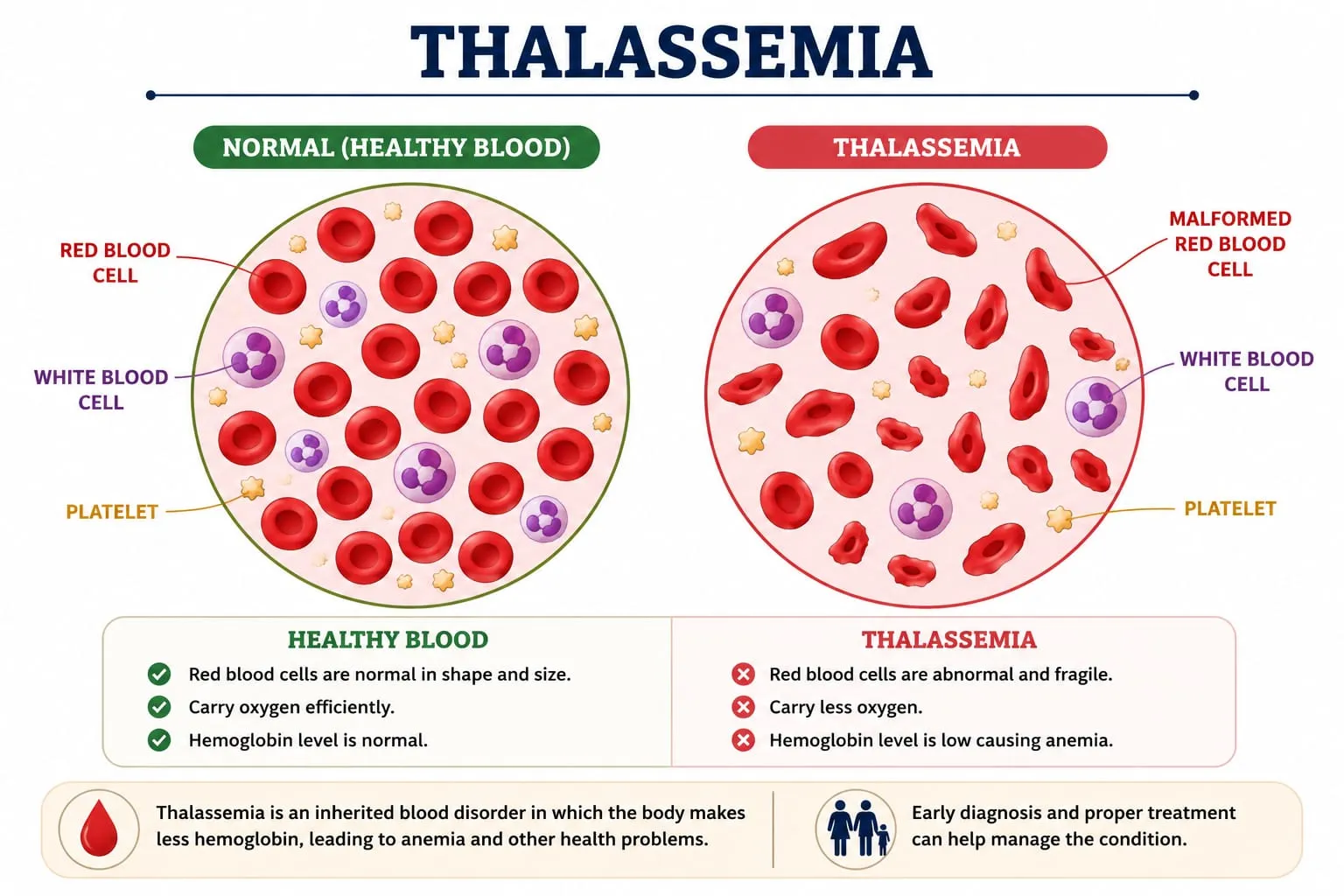
Fig 1 : Morphological changes in Red Blood Cells in Thalassemia.
The goal of managing transfusion-dependent HbE/β-thalassemia is to maintain sufficient hemoglobin concentrations with frequent transfusions of packed red blood cells (PRBCs), which will reduce ineffective erythropoiesis, encourage normal growth, and enhance quality of life[4]. However, iron overload, transfusion-transmitted infections, alloimmunization, and delayed haemolytic transfusion reactions are significant side effects of long-term transfusion therapy. An overall red blood cell alloimmunization prevalence of about 11% was reported in a systematic review involving more than 9,000 transfusion-dependent thalassemia patients. Antibodies were primarily directed against Rh and Kell antigens, underscoring the significance of appropriate blood compatibility testing and extended antigen matching whenever possible. As a result, long-term care necessitates a multidisciplinary strategy that includes thorough transfusion support, iron chelation therapy, routine haematological monitoring, and organ failure surveillance. Reducing disease-related morbidity and improving long-term survival need early diagnosis and prompt beginning of suitable treatment. Newborn screening, carrier finding, genetic counselling, and prenatal diagnosis continue to be crucial preventative measures that can greatly lower the burden of disease in nations with a high prevalence of hemoglobinopathies. Additionally, new developments in gene-editing technologies and hematopoietic stem cell transplantation present intriguing therapeutic alternatives for certain patients, but many people in settings with limited resources are still unable to access these methods[5].
This case reports on a 10-year-old child with transfusion-dependent HbE/β-thalassemia who had hepatosplenomegaly and severe chronic haemolytic anaemia. The case emphasizes the usefulness of hemoglobin electrophoresis for diagnosis, the significance of thorough clinical and laboratory evaluation, and the positive short-term reaction to prompt packed red blood cell transfusion. In order to reduce disease-related complications and improve patient outcomes, it also highlights the necessity of early detection and organized long-term follow-up[6].
CASE DESCRIPTION
A 10-year-old girl, complained of intermittent jaundice and recurring abdominal pain for about two years when she arrived at the pediatric department. The child was 121.5 cm tall and weighed 19.4 kg. She had no prior history of being admitted to a neonatal intensive care unit and was born at term by a lower segment cesarean section. In accordance with the national immunization schedule, she was fully immunized. There were no notable prenatal or perinatal difficulties reported, and she was the child of a non-consanguineous marriage. Any history of drug allergies was disputed by the family. She had been evaluated by a paediatrician two years prior to the current admission due to persistent pallor and jaundice. The paediatrician recommended blood transfusion at that time, but the parents refused and instead chose Ayurvedic treatment, which lasted for about a year and a half before being stopped three months before admission. The child's symptoms continued after treatment, necessitating hospitalization for additional assessment and care.
The child's vital signs were stable at admission, and he or she was conscious, aware, and focused. Her blood pressure was 90/60 mmHg, her body temperature was 98.1°F, her respiration rate was 20 breaths per minute, her pulse rate was 80 beats per minute, and her oxygen saturation was 98% on room air. A general examination showed mild icterus and significant pallor. A respiratory examination found normal bilateral air entry, while a cardiovascular examination revealed normal first and second heart sounds without murmurs. An examination of the abdomen revealed considerable hepatosplenomegaly, with the spleen palpable almost 8 cm below the left costal edge and the liver span measuring around 11 cm. The results of the neurological evaluation showed no localized impairments.
With a hemoglobin concentration of 4.3 g/dL, red blood cell count of 1.71 × 10³/mm³, hematocrit of 11.8%, mean corpuscular volume of 69.2 fL, mean corpuscular haemoglobin of 24.9 pg, mean corpuscular haemoglobin concentration of 36 g/dL, and red cell distribution width of 22.8%, the initial haematological evaluation indicated severe microcytic hypochromic anaemia. Both the platelet and total leukocyte counts were within normal ranges. With microcytic hypochromic erythrocytes, target cells, teardrop cells, elliptocytes, microspherocytes, polychromasia, and nucleated red blood cells, peripheral blood smears showed severe anisopoikilocytosis, which is consistent with chronic haemolytic anaemia. Biochemical analyses showed increased total bilirubin (3.42 mg/dL), mostly indirect hyperbilirubinemia (2.70 mg/dL), although renal function tests and liver enzyme levels were within normal ranges. With the exception of iron deficiency anaemia, serum iron, ferritin, and total iron-binding capacity were within reference ranges. Human immunodeficiency virus, hepatitis B virus, and hepatitis C virus screenings were all negative. Thyroid function testing, lipid profiles, coagulation profiles, and urinalysis all revealed no abnormalities that were clinically significant.
Hemoglobin electrophoresis was used to determine the underlying hemoglobinopathy. The results showed HbE 44.5%, HbF 52.2%, and HbA 3.3%, supporting the diagnosis of hemoglobin E–β-thalassemia. Abdominal ultrasonography showed echogenic sludge in the gallbladder and mild hepatosplenomegaly with dilation of the portal and splenic veins. Pediatric echocardiography showed no structural abnormalities and normal cardiac anatomy and function. The patient was found to be O Rh-positive by blood grouping, and compatible packed red blood cells were verified by pre-transfusion compatibility testing. After the necessary compatibility tests, the patient was treated with one unit (200 mL) of packed red blood cells due to the severity of anaemia and the confirmed diagnosis of HbE–β-thalassemia. Prior to, during, and following transfusion, vital signs were continually monitored. Intravenous pantoprazole, followed by oral pantoprazole, sucralfate suspension, folic acid supplements, sufficient oral hydration, nutritional support, and repeated complete blood count monitoring were all part of the supportive care. Improving oxygen-carrying capacity, reducing symptoms of severe anaemia, preventing consequences from persistent haemolysis, and safely administering blood transfusions without transfusion-related adverse events were the goals of the treatment. Acute transfusion responses, such as fever, chills, urticaria, hypotension, dyspnea, or haemoglobinuria, were not seen during the monitoring time. The blood transfusion was finished without incident. A follow-up haematological evaluation revealed a significant increase in hemoglobin concentration from 4.3 g/dL to 9.4 g/dL, along with improvements in hematocrit and red blood cell count. Clinically, the child's hemodynamic remained stable, oral feedings were well tolerated, urine production remained normal, and pallor gradually improved without any new complaints. As part of the long-term therapy of HbE–β-thalassemia, she was discharged in stable condition with instructions to maintain folic acid supplements, supportive drugs, routine haematological follow-up, and scheduled packed red blood cell transfusions.
DISCUSSION
One of the most prevalent hemoglobinopathies in South and Southeast Asia is hemoglobin E–β-thalassemia, which can cause severe transfusion-dependent illness or mild anaemia. Fetal haemoglobin (HbF) levels, the underlying β-thalassemia mutation, and other genetic modifiers all affect the severity of the disease. Anemia, hepatosplenomegaly, and growth retardation are common signs of severe illness and are caused by chronic inefficient erythropoiesis and haemolysis. In line with previously documented cases of transfusion-dependent β-thalassemia, the current patient had severe chronic haemolytic anaemia, pallor, intermittent jaundice, stomach discomfort, and hepatosplenomegaly. Skeletal abnormalities were not present despite our patient's severe anaemia, perhaps because transfusion therapy was started on time. Severe microcytic hypochromic anaemia with anisopoikilocytosis, target cells, teardrop cells, and nucleated red blood cells, a sign of inefficient erythropoiesis and persistent haemolysis was discovered through laboratory research. HbE/β-thalassemia was validated by hemoglobin electrophoresis, which showed reduced HbA (3.3%), high HbF (52.2%), and HbE (44.5%). Chronic haemolytic illness was further confirmed by indirect hyperbilirubinemia and ultrasonographic evidence of hepatosplenomegaly with gallbladder sludge[7]. Iron deficiency anaemia was ruled out by normal iron assays, highlighting the significance of hemoglobin electrophoresis in distinguishing hereditary hemoglobin abnormalities from other forms of microcytic anaemia. The mainstay of care for transfusion-dependent HbE/β-thalassemia is still packed red blood cell transfusion. Transfusion raised our patient's hemoglobin level from 4.3 g/dL to 9.4 g/dL, which was in line with current management guidelines and produced a notable clinical improvement without transfusion-related side effects. Repeated transfusions raise the risk of iron overload and alloimmunization, even when there were no transfusion-related problems during hospitalization. The individuals with transfusion-dependent thalassemia had an overall alloimmunization rate of about 11%, underscoring the necessity of thorough blood compatibility testing and ongoing surveillance. Therefore, it is crucial to regularly check endocrine state, liver and heart function, and serum ferritin during follow-up[8].
This case emphasizes the importance of considering HbE/β-thalassemia in children presenting with severe microcytic anaemia, jaundice, and hepatosplenomegaly. Early diagnosis through hemoglobin electrophoresis, timely transfusion therapy, and regular follow-up are essential to prevent complications and improve long-term clinical outcomes[9].
CONCLUSION
Hemoglobin E-β-thalassemia is a genetically heterogeneous hemoglobinopathy that can cause severe chronic haemolytic anaemia requiring frequent transfusion assistance. This case illustrates the clinical difficulties associated with this condition. The patient's presentation of hepatosplenomegaly, intermittent jaundice, profound anaemia, and distinctive haematological abnormalities highlights the significance of keeping a high index of suspicion for hemoglobinopathies in children presenting with persistent microcytic hypochromic anaemia, especially in areas where these disorders are common. By verifying the diagnosis and distinguishing the illness from other causes of microcytic anaemia, such as iron deficiency, hemoglobin electrophoresis proven to be a crucial diagnostic tool.
The efficacy of appropriate transfusion therapy in stabilizing patients with transfusion-dependent HbE/β-thalassemia was demonstrated by the significant haematological and clinical improvement that occurred with timely administration of packed red blood cell transfusion without transfusion-related complications. However, long-term care goes beyond treating anaemia and necessitates routine monitoring to track the course of the illness, the need for transfusions, iron overload, organ dysfunction, and other treatment-related issues. To maximize treatment results and enhance quality of life, a multidisciplinary strategy comprising paediatricians, haematologists, transfusion medicine specialists, and clinical pharmacists is necessary. The importance of early diagnosis, genetic counselling, and family screening in lowering the burden of disease and enabling prompt action is further highlighted by this instance. Raising healthcare professionals' knowledge of HbE/β-thalassemia's clinical presentation and diagnostic methodology may encourage early detection and suitable treatment. To reduce problems and enhance the long-term prognosis for impacted children, ongoing monitoring and adherence to evidence-based treatment approaches are still essential. This example contributes to the expanding corpus of research on HbE/β-thalassemia and highlights the significance of customized, patient-centered care in the treatment of this intricate genetic condition.
PATIENT/PARENT PERSPECTIVE
The child's parents stated that for about two years, she suffered intermittent jaundice, pallor, and recurrent abdominal pain that gradually interfered with her everyday activities. They were not aware of the severity of the situation, thus at first they chose alternate treatment even though blood transfusion had been recommended. They learned more about hemoglobin E-β-thalassemia and the significance of routine medical follow-up after being admitted and undergoing a thorough evaluation. Following the packed red blood cell transfusion, they saw a discernible improvement in the child's general wellbeing and degree of activity. In addition to acknowledging the necessity of frequent transfusions, recurring monitoring, and dedication to long-term treatment to avoid disease-related consequences, the family expressed happiness with the care they received. They also acknowledged the value of family screening and genetic counselling in treating this inherited condition, and they indicated that they would be open to continuing routine follow-up appointments.
REFERENCES

Sneha Y.*, Sandhiya V., Sujan Sharma, Hemoglobin E–?-Thalassemia Presenting With Severe Microcytic Hypochromic Anemia And Hepatosplenomegaly: A Case Report, Int. J. of Pharm. Sci., 2026, Vol 4, Issue 9, 3439-3445. https://doi.org/10.5281/zenodo.22978367
 10.5281/zenodo.22978367