

We use cookies to ensure our website works properly and to personalise your experience. Cookies policy
Department of Pharmacology, Viva Institute of Pharmacy, Shirgaon, Virar (E).
Population aging has increased the prevalence of heart failure—specifically heart failure with preserved ejection fraction (HFpEF), for which there are currently no disease-modifying therapies. Recent findings indicate that cellular senescence is a major contributor to cardiac aging. Cellular senescence refers to a stress-induced, largely irreversible cell cycle arrest with an accompanying secretory phenotype. In the aging heart, senescent cardiomyocytes, fibroblasts, endothelial cells, and vascular smooth muscle cells arise and perpetuate a state of “inflammaging” which causes chronic inflammation and fibrosis, as well as diastolic dysfunction with increased stiffness. Senotherapeutics are an innovative intervention option, which either eliminate senescent cells (senolytics) or counteract the detrimental effects of the secretory phenotype (senomorphics). Here, we review herbal senomorphics, a group of plant polyphenols, flavonoids, saponins, and terpenes with the ability to suppress the secretory phenotype and improve homeostasis of the proteome and redox state, which have a much better safety profile compared to their synthetic analogs. We compile the preclinical data for these herbal senomorphics and explore their shared molecular pathways. We also identify the translational barriers of these compounds and propose a “one-two punch" treatment approach with senolytics and senomorphics. Although there are still many clinical barriers to address, herbal senomorphics are a multi-target treatment option for age-induced heart failure.
As the most common cause of death worldwide, the incidence of cardiovascular disease also increases with age.[1],[2] As the world's population ages, so does the population of both high- and middle-income countries. Heart failure is a key syndrome of older age. Heart failure with preserved ejection fraction (HFpEF) is concerning because it has become increasingly common. It is characterized by diastolic dysfunction, myocardial stiffening, and systemic inflammation and therapies that target the neurohormonal system have historically provided little benefit to patients.[3] The fact that the heart can age and fail even when there is no previous coronary disease shows that the process is not an inevitable situation, but a modifiable pathological process.[4]
The aging population's heart failure has created an economic and humanitarian crisis that is only going to worsen with the increasing world population. Through the lens of geroscience, the HFpEF phentotype is not treated through its downstream hemodynamic impairment, but through its aging biology at any upstream component of the aging process. The geroscience hypothesis, of which cellular senescence is one of the most prominent, provides that by targeting cellular senescence, multiple chronic diseases can be prevented or hearkened. Cardiac aging provides a valid pathology to consider cellular senescence as a point of intervention.
Senescence is the permanent cell cycle arrest even caused by multiple stressors both at the cellular and mitochondrial levels, resulting in the redesigning of the secretome.[5] Though these cells collect a small volume in aged tissue, they exert outsized influences on surrounding cells, resulting in a system wide effect through the bioactive secretome.[6],[7]
Senescent cardiac cells induce HFpEF as a result of scarring and rarefaction of the microscopic vasculature.[8],[9] A number of seminal studies have also pointed to the possibility of removing senescent cells in delaying the occurrence of tissue-related diseases of old age while also increasing the health remain phase of old age.[10] This opened the field of senotherapeutics. There are two main pathways in senotherapeutics: 1.) Senolytics are agents that target and promote the death of senescent cells. 2.) Senomorphics are agents that target the secretome and either modulate it to become less active or slow down the process of senescence in the cells.[11]
Chronic dosing of synthetic senotherapeutics is required for conditions like cardiac aging, but this approach is complicated by dose-limiting toxicity, off-target effects, and potential for inducing cancer. It is no wonder that, given these concerns, significant interest is being directed towards plant-derived compounds as senomorphic agents. Many polyphenols, flavonoids, saponins, and terpenoids possess favorable safety profiles and can impact the same signaling pathways as NF-κB, mTOR, SIRT1/AMPK, and Nrf2, which are responsible for the inflammatory SASP, and exhibit multiple antiinflammatory/antioxidant effects. This review focuses on the mechanisms and preclinical studies that support the use of plant senomorphics to treat age-related heart failure. We summarize the progression of cardiac senescence to HFpEF, describe the mechanisms of senomorphic agents, examine leading phytochemicals that are senomorphics, identify the targets that are common to the phytochemicals, and discuss the barriers that would prevent the use of dietary and nutraceutical senomorphics in clinical cardiology. [12]
CELLULAR SENESCENCE AND THE AGEING HEART:
Operationally defining senescence relies on an array of characteristics rather than a singular marker. In aged myocardium, the p16INK4a/Retinoblastoma pathway and the p53/p21CIP1 pathway (both linked to tumorigenesis) have been shown to be upregulated.[8] ,[13] Cell cycle arrest is also associated with persistent activation of the DNA damage response, increased senescence-associated β-galactosidase activity, telomere damage, and a shift in metabolism toward increased glycolysis.[14] In post-mitotic cardiomyocytes, the length of telomeres is not required to induce senescence. In addition, terminally differentiated heart cells with age can adopt a senescent phenotype and secrete the senescence-associated secretory phenotype (SASP). [9]
There is no singular marker that can be used to identify a senescent cell. In addition to proliferation arrest, secretory and morphological characteristics must be evaluated to determine the senescent state.[15],[16] There is complexity added to this evaluation due to the context. The senescence program can differ from cell to cell, as well as stimuli and microenvironments. The secretory activity of senescent cells can also have positive or negative impacts on tissue.[17],[18] Metabolic reprogramming is an important characteristic of senescence, where senescent cells can adopt a specific metabolic profile of altered glycolysis and sustain arrest.[19] The aging heart requires that beneficial repair-associated transient senescence must be differentiated from the maladaptive, chronic senescence.
The senescence-associated secretory phenotype (SASP) program is the leading mechanism whereby a relatively small population of senescent cells alters the cardiac microenvironment. From IL-6, IL-1α/β, TNF-α, MCP-1, TGF-β, and MMPs, the SASP is laid out transcriptionally by NF-κB and CCAAT/enhancer-binding protein-β, and post-transcriptionally by the mTOR and p38 MAPK pathways.[20],[21],[22],[23] These combinations of pathways and SASP enable the development of new therapies. Manipulation of the SASP is a possible therapeutic avenue, and selective suppression of the SASP is the principal goal of senomorphic therapies.[24]
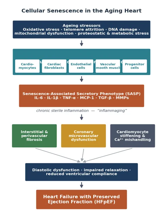
Figure 1. Accumulation of senescent cells in the ageing heart and its progression to HFpEF. Chronic stressors drive senescence across multiple cardiac cell types; the resulting SASP propagates “inflammaging,” fibrosis, microvascular dysfunction and cardiomyocyte stiffening, culminating in diastolic dysfunction and heart failure with preserved ejection fraction.[32]
Specific aging phenotypes can be traced to distinct cardiac lineages. Here, we summarize some of these aging phenotypes. We focus first on endothelial cell senescence. Endothelial cell senescence limits bioavailability of nitric oxide, creates a thrombotic surface, and deteriorates the perfusion of the distal coronary microvascular system. In aging mice models, senescent endothelial cells have been shown to participate in heart failure with preserved ejection fraction (HFpEF).[25],[26] Senescent vascular smooth muscle cells lead the stiffening of smooth muscle, the breakdown of the extracellular matrix, calcification of the cortical wall, and instability of plaques. Once cardiac fibroblasts reach senescence, they become pro-fibrotic and enlarge the collagen secretory profile. Cardiac myocytes also enlarge with senescence and become SASP factor secretors. SASP factors create a paracrine wave of cellular senescence and activate local myofibroblasts.[8] Unlike the other cell types, senescent cardiac myocytes retain the ability to secrete factors. During aging, cells remain in a persistent state and obstruct the heart’s ability to heal and regenerate. The removal of these cells restores the heart's ability to heal in experimental models.[27] In summary, the aging heart consists of a senescent integrated system with dysfunctional cardiac compartments.
"Inflammaging" describes the persistent low-grade inflammation seen in aging tissue and is believed to have its origins in the senescence-associated secretory phenotype (SASP).[28],[29] In cardiac tissue, the combination of sustained IL-6, IL-1β, and TNF-α exposure leads to a loss of myofilament calcium sensitivity, increased stiffness of titin, and TGF-β-associated fibrosis, which in turn causes a loss of compliance in the ventricles. Furthermore, the SASP associated with endothelial senescence progressively damages the coronary microcirculation and causes the microvascular ischemia and endothelial-cardiomyocyte signaling perturbations that are increasingly being associated with HFpEF.[30],[31]
Senescent cells have a strong resistance to apoptosis causing SASP-associated fibrosis and losing compliance to be increasingly pronounced in the aging heart. This pathophysiological process, which is characterized by the accumulation of senescent cells and SASP-related senescence, provides a theoretical basis for senomorphic therapies. The goal of senomorphic therapies is to halt the secretory phenotype within the SASP and alleviate the associated pathophysiological processes (Figure 1) even in the presence of senescent cells.[32]
This axis has a high clinically relevant association with senescence and its relationship to type 2 diabetes and the many metabolic stressors (e.g. hyperglycemia, dyslipidemia, and obesity) given that many older adults with heart failure and preserved ejection fraction (HFpEF) have a high degree of cardiometabolic comorbidities.[31],[33]
Cardiac ageing also describes phenomena involving the resident progenitor compartment.[18] It describe how senescence in cardiac progenitor and stem-like cells diminishes their mobility and the ability to proliferate or differentiate, further eroding the already limited potential of the heart to regenerate. Senescence in cardiac progenitor cells also spurs a paracrine SASP and induces senescence in the surrounding cells. Hence, a scarce population of cells can affect repair in a wider region. The removal of aged and senescent cells in an experiment increased the proliferation of cardiomyocytes and resident progenitors, demonstrating that the regenerative deficits in the old heart can be reversed.[27] The selective removal of the SASP of senescent progenitor cells can provide a means to minimize inflammation and improve the reparative ability of aging myocardium.
Table 1. Representative SASP factors and their cardiac consequences.
|
SASP factor |
Cardiac consequence |
|
IL-6, IL-1β, TNF-α |
Chronic inflammation; impaired myofilament Ca²⁺ sensitivity |
|
TGF-β |
Fibroblast activation; interstitial and perivascular fibrosis |
|
MMPs |
Extracellular-matrix remodelling; wall destabilisation |
|
MCP-1 / chemokines |
Immune-cell recruitment; amplified myocardial inflammation |
|
Reduced NO (endothelial) |
Impaired vasodilation; coronary microvascular dysfunction |
IL, interleukin; TNF-α, tumour necrosis factor-α; TGF-β, transforming growth factor-β; MMP, matrix metalloproteinase; MCP-1, monocyte chemoattractant protein-1; NO, nitric oxide.
SENOMORPHICS AS A THERAPEUTIC PARADIGM
Strategies related to the removal or management of senescent cells can be divided into three main types. First, senolytics aim to remove senescent cells by disrupting their anti-apoptotic networks. Senolytic drugs act particularly upon the BCL-2/BCL-XL, PI3K/AKT, p53/FOXO4 and closely related networks. As such, these drugs can be used in an intermittent manner[34],[35], due to the time delay in the re- accumulation of senescent cells. Prominent examples include the combination of dasatinib and quercetin and the flavonol fisetin, both of which show translational promise in the aging and diseased heart.[36],[37],[38]
Unlike senolytics, which aim to remove senescent cells, senomorphics either inhibit the SASP or obstruct the transition into senescence, thereby inhibiting the SASP while maintaining the arrested and non-proliferative (therefore tumor suppressive) senescent cell.[11] Earliest examples of senomorphics include rapamycin (mTOR), metformin (IKK/NF- kappa B), and JAK inhibitors,[23],[39],[40] while the gerostatics, a related class of senomorphics, aim to inhibit cellular senescence. For many natural products, there is a dose.
The early-stage clinical translation of senotherapeutics has progressed rapidly. Senotherapeutics clinical trials have evaluated the combination of dasatinib and quercetin for the reduction of the burden of senescent cells, first, in the study of pulmonary fibrosis, and later in the study of diabetic kidney disease.[41],[42] These trials, in addition to the studies of the safety and pharmacology of senolytic regimens, have described the basis for what has been termed the “hit-and-run” senolytics dosing.[43],[44] It is possible, however, that for a chronic and insidious condition such as heart aging the exact opposite strategy could be used: a continuous regimen of low-toxicity that places a more or less constant restraint on the SASP. This is where the use of well-tolerated senomorphics could play an important role.[45]
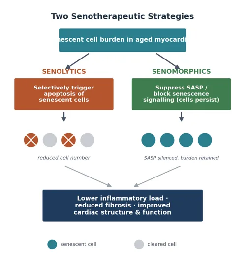
Figure 2. Two senotherapeutic strategies. Senolytics trigger apoptosis and reduce senescent-cell burden, whereas senomorphics silence the SASP while the arrested cells persist. Both converge on a lower inflammatory and fibrotic load and improved cardiac structure and function.
Master regulators constitute the limited set of nodes for orchestrating the SASP. NF-κB is a major node for the transcription of proinflammatory SASP genes, and the SASP secretome is significantly decreased with its inhibition.[20],[21] Additionally, the mTOR pathway, which promotes the translation of the SASP components IL-1α and MAPKAPK2, links the SASP with nutrient sensing and secretory output.[23],[46] The activation of SASP can occur through a DNA-damage-independent pathway as well via p38 MAPK.[22] The JAK/STAT signaling pathway creates a more extensive, systemic enhancement of cytokine signaling.[40]
SASP regulation relies similarly on counter-adaptive pathways. The SIRT1–AMPK pathway dampens NF-κB, promotes autophagy, and enhances mitochondrial quality control, thereby countering senescence.[47] The Nrf2/antioxidant response element pathway is a senescence counteracting pathway, as its activity reduces the burden of ROS, a hallmark of senescence and its aging. Its repression is a hallmark of premature aging.[48] The activation of NLRP3 inflammasome forms a link between cellular stress and the maturation of the cytokines IL-1β and IL-18, and its inhibition prevents cardiac aging in mice. The great interest in phytochemical senomorphics is attributable to the frequent engagement of many nodes in cellular aging by individual compounds.[49]
Three properties recommend plant-derived compounds for chronic cardiac senomorphic therapy. The first property is multi-target pharmacology, where a single polyphenol can inhibit NF-kB, activate SIRT1/AMPK, and induce Nrf2, thereby circumventing the redundancy problem of single-target drugs.[12] The second property relates to senomorphics’ long history of use in traditional and contemporary diets, which has promoted the development of strategies that incorporate senomorphics for use in polymedicine systems. The third property, exposure through further dietary intervention, is strongly supported by the availability and integration of senomorphics in the conventional diet of the population. These benefits may be negatively affected by real and significant marginal shortcomings of low potency, inadequate absorption (bioavailability), and variability of extract composition, as covered in the following section (Section 6).
HERBAL SENOMORPHICS: COMPOUND-BY-COMPOUND EVIDENCE
The subsequent subsections evaluate the main cardiac-senescence- and heart-failure-related phytochemicals, senomorphics. Table 2 compiles their targets and sources along with select findings.
First on the list is Resveratrol, which is a stilbene polyphenol primarily derived from grapes, and a SIRT1 natural activator. Resveratrol enhances autophagy and mitochondrial biogenesis, and mitigates the SASP via the inhibition and subsequent deacetylation of the NF-κB p65 Sunit. In human endothelial cells, it protected against HP-induced oxidative senescence in a SIRT1 dependent manner and reduced the senescence of endothelial progenitor cells by promoting telomerase.[50],[51] In aged myocardium, Resveratrol inhibits cardiomyocyte hypertrophy; SIRT1 also inhibits p16/p21. Ventricular dysfunction in aged subjects is mitigated by Resveratrol, which also inhibits the inflammation response and oxidative stress.[4] Recent small scale studies on patients that have recently experienced a myocardial infarction report that resveratrol improves diastolic function and results in endothelial vasodilation, however, due to the small sample size, the studies cannot be considered conclusive.[12]
Curcumin, the main curcuminoid in turmeric, acts as an NF- κB inhibitor, possessing both antioxidant and anti-fibrotic properties. In endothelial cells and vascular smooth muscle, it promotes NF- κB-dependent adhesion, increases monocyte adhesion, and increases smooth muscle proliferation and migration, which helps restore endothelial function.[12] In the cardiac ageing process, the curcumin/sirtuin 1/AMP-activated protein kinase/mammalian target of rapamycin (mTOR) signalling pathway assists curcumin in the restoration of autophagic flux and the suppression of markers of senescence, leading to a reduction in senescenceassociated β-galactosidase activity, in addition to a decrease in p16, p53, and levels of reactive oxygen species, in D-galactose stressed cardiomyocytes.[12],[14] The major obstacle to its therapeutic applications remains its poor solubility in water and rapid metabolic degradation, which limits its bioavailability and promotes an intensive effort in nanoformulations. [52],[53]
Quercetin, a widely used dietary flavonol, is one of the rare examples of a senotherapeutic agent that spans both therapeutic classes. It is a notable senolytic when used in combination with dasatinib, leading to a considerable restoration of cardiac and vascular function in aged and atherosclerotic animal models while decreasing markers of senescent cells and the senescence-associated secretory phenotype in early human studies (Zhu et al., 2015; Roos et al., 2016; Hickson et al., 2019; Nieto et al., 2024).[34],[36] Surprisingly, at lower concentrations quercetin acts as a senomorphic agent, decreasing NF- κB activation and the secretion of pro-inflammatory cytokines.[54] Quercetin’s unique, concentration-dependent, and dual activity makes it a highly flexible scaffold.[38],[42]
Fisetin is a flavonol that occurs in strawberries, some other fruits, and vegetables. Unbiased screens have led to the identification of Fisetin as one of the most powerful natural senotherapeutics. Fisetin reduces the burden of senescent cells and increases the healthspan and lifespan of aged mice.[55],[56] Because of its senescence-reducing effects, intermittent supplementation of Fisetin has been shown to improve arterial function in aged mice.[57] In the context of ischaemic cardiac injury, Fisetin inhibits RAGE/NF-κB signalling, decreases the levels of cytokines IL-6 and TNF-α, improves the contractility and the relaxation of the left ventricle, and facilitates the IGF-1R–PI3K/AKT survival pathway. Fisetin, as with many other flavonoids, has poor long-term oral bioavailability due to rapid metabolism and low absorption, which has been a major obstacle to its forwarding in clinical practice.[58]
Apigenin and Kaempferol are also flavones/flavonols that have significant senomorphic activity. Unlike Fisetin, which primarily targets senescence, the senomorphic action of Apigenin is primarily due to its ability to inhibit the NF-κB signalling pathway. Apigenin suppresses the SASP by blocking IL-1α signal transduction that is mediated by IRAK1/IRAK4, p38 MAPK and NF-κB cascades, and by reducing peroxiredoxin-6 thereby decreasing the senescent cell-induced pro-inflammatory paracrine signaling. Kaempferol inhibits the SASP by reducing NF-κB p65 activity and IκBζ expression by the IRAK1/IκBα pathway thereby reducing IL-1α.[59]
Senomorphic effects of EGCG that derive from sirtuin- and Nrf2-related pathways are also apparent in greeen tea. Among the various bioactives tested, EGCG most prominently decreased both IL-6 secretion and the p21 expression in senescence-induced preadipocytes through SIRT3/Nrf2 signalling.[60] In a study of vascular model systems, EGCG was shown to promote autophagy, alleviate ferroptosis through SIRT1, and reduce the senescence of vascular endothelial cells and aortic stiffness.[12] However, extremely low oral bioavailability and chemical instability limit the concentration of EGCG in tissues. [60]
Ginsenosides are the triterpenoid saponins of Panax ginseng, and show a variety of cardioprotective and anti-senescent effects. Ginsenoside Rg1 is the most prominent example and acts through PI3K/AKT and SIRT1 signalling to limit the expression of senescence-associated p16 and p21, reduce DNA damage and inflammation and restore telomere length in stress-induced senescence. In Rg1′s cardioprotective effects, Rg1 appears to improve bioenergetics and mitochondrial function during Ischemia-Reperfusion injury, and relieves endoplasmic reticulum stress through SIRT1 and reduces inflammation. These effects spanning multiple pharmacological concepts such as antioxidant, mitochondrial, and anti-inflammatory make ginsenosides a senomorphic delight for the ageing heart. [62]
Astragaloside IV is extracted from the plant Astragalus membranaceus and is a cardioprotective agent and anti-senescence compound. Integrative transcriptomic proteomic analysis showed astragaloside IV protects the heart from pulmonary fibrosis by regulating senescence.[61] In vascular smooth muscle, astragaloside IV protects against senescence by activating Parkin-mediated mitophagy, improving the integrity of mitochondria in bleomycin- and D-galactose-induced senescence.[12] In heart failure, astragaloside IV offers cells antioxidative protection which is mediated through the Nrf2 pathway. By increasing the integrity of mitochondria in cardiac cells as well as improving the antioxidant defenses and decreasing fibrosis, astragaloside IV brings solutions to several causes of cardiac senescence.[62]
Salidroside is a phenylpropanoid glycoside derived from Rhodiola rosea, known for its cardioprotective properties, and for extending the mechanisms of ginsenosides. With ginsenoside Rg1, salidroside helps in the protection of the heart from myocardial ischemia-reperfusion injury by improving mitophagy and the antioxidant response through the SIRT1/3–PGC-1α–Nrf2 pathway, in addition to supporting mitochondrial membrane potential and diminishing ROS.[12] Procyanidin C1, a new flavonoid compound, is a senomorphic compound, exhibited senotherapeutic activity, and increased the lifespan of mice. Rutin is a highly senomorphic compound which addresses the SASP and improves therapeutic outcomes.[63]
Apart from main senomorphic agents identified, varieties of phytochemicals are emerging including phytochemicals with components relevant to the cardiovascular system.[64] It show that sulforaphane (isothiocyanate from cruciferous vegetables) is a strong activator of Nrf2 and upregulates the expression of Antioxidant Response Element genes. Sulforaphane also prevents diabetic cardiomyopathy through an Nrf2 pathway while countering the oxidative drive to senescence. Berberine (isoquinoline from Coptis and Berberis species) engages AMPK and inhibits the signaling pathway of NF-κB which exerts broad anti-inflammatory, lipid-lowering, and cardioprotective effects that relate to the senomorphics.[65] Further, alkaloids like oxymatrine improve AMPK activation and oxidative stress-induced senescence. In addition to flavones like luteolin and isoflavones like genistein which offer NF-κB and Nrf2 anti-senescent activities. Together, they offer a broad spectrum of senomorphics beyond classical polyphenols, and they maintain a repeat theme of converging on a common regulatory network while being distinct. [66]
Table 2. Leading herbal senomorphics with cardiovascular relevance.
|
Compound |
Botanical source |
Principal molecular targets |
Cardiovascular / senescence evidence |
Model / system |
|
Resveratrol |
Grapes; Polygonum spp. |
SIRT1 ↑; NF-κB p65 ↓; telomerase ↑ |
Attenuates endothelial and cardiomyocyte senescence; lowers p16/p21; improves diastolic function |
HUVEC; aged mice; small human trials |
|
Curcumin |
Curcuma longa (turmeric) |
NF-κB ↓; SIRT1/AMPK/mTOR; autophagy ↑ |
Reduces SA-β-gal, p16, p53, ROS; suppresses adhesion molecules; anti-fibrotic |
D-gal cardiomyocytes; endothelium; VSMC |
|
Quercetin |
Onion, apple, capers |
NF-κB ↓ (senomorphic); pro-apoptotic (senolytic, with dasatinib) |
Clears senescent cells with dasatinib; improves cardiac/vascular function; lowers SASP |
Aged & atherosclerotic mice; human pilots |
|
Fisetin |
Strawberry, apple |
RAGE/NF-κB ↓; PI3K/AKT ↑; BCL-2 family |
Improves arterial function; preserves LV contractility in ischaemia; extends healthspan |
Old mice; isoproterenol injury; H9c2 |
|
Apigenin |
Parsley, celery, chamomile |
IL-1α/IRAK–p38–NF-κB ↓; PRDX6 ↓ |
Suppresses SASP and paracrine inflammation |
Senescent human fibroblasts / stroma |
|
Kaempferol |
Broccoli, kale, tea |
NF-κB/IκBζ ↓; Nrf2/HO-1 ↑ |
Lowers IL-1α/β, TNF-α, IL-6; attenuates vascular injury and atherosclerosis |
Senescent BJ cells; aortic tissue; rats |
|
EGCG |
Camellia sinensis (green tea) |
SIRT3/SIRT1; Nrf2 ↑; autophagy ↑; ferroptosis ↓ |
Reduces IL-6 and p21; lessens endothelial senescence and aortic stiffening |
Preadipocytes; endothelium; aorta |
|
Ginsenoside Rg1 |
Panax ginseng |
PI3K/AKT; SIRT1 ↑; ER-stress ↓; mitochondrial function ↑ |
Lowers p16/p21, restores telomere length; limits I/R injury and inflammation |
Stem cells; H9c2; rat I/R |
|
Astragaloside IV |
Astragalus membranaceus |
Parkin-mediated mitophagy ↑; Nrf2 ↑; senescence ↓ |
Protects against myocardial fibrosis via senescence regulation; antioxidative in HF |
VSMC; d-gal mice; HF models |
|
Salidroside |
Rhodiola rosea |
SIRT1/3–PGC-1α–Nrf2; mitophagy ↑ |
Enhances mitochondrial protection and antioxidant defence (with Rg1) in I/R |
Rat MIRI; H9c2 H/R |
|
Procyanidin C1 |
Grape seed |
Senomorphic (low dose) / senolytic (high dose); SASP ↓ |
Senotherapeutic activity; increases lifespan in mice |
Aged mice; senescent fibroblasts |
|
Rutin |
Buckwheat, citrus |
SASP ↓; NF-κB-linked signalling |
Potent senomorphic; improves therapeutic efficacy |
Senescent cell models; mice |
SA-β-gal, senescence-associated β-galactosidase; HUVEC, human umbilical-vein endothelial cells; VSMC, vascular smooth muscle cell; LV, left ventricular; I/R, ischaemia–reperfusion; MIRI, myocardial ischaemia–reperfusion injury; HF, heart failure; EGCG, epigallocatechin gallate. ↑ increase/activation; ↓ decrease/inhibition.
MECHANISTIC CONVERGENCE IN THE SENESCENT CARDIAC NICHE:
Although they have different chemical structures, herbal senomorphics point to the same signaling nodes that control the senescent phenotype (Figure 3; Table 3). The predominant shared action is the inhibition of NF-κB since it is the main transcriptional regulator of the pro-inflammatory SASP. Resveratrol, curcumin, quercetin, apigenin and kaempferol all decrease NF-κB with a corresponding decrease in the pro-inflammatory cytokines IL-6, IL-1β and TNF-α.[12],[20]
Since NF-κB is the convergence point of several pathways including p38 MAPK, the DNA-damage response, and inflammasome, its activity results in broad secretory outflow.
The activation of SIRT1/AMPK results in the restoration of autophagy and quality control of mitochondria. Resveratrol, curcumin, ginsenoside Rg1 and salidroside enhance this axis by the deacetylation of NF-κB, thus improving mitophagy and bioenergetics.[12],[47] The senomorphic action of many herbal compounds involves the activation of NRF2 and the consequent reduction of ROS, which also helps the antioxidant system.[48],[64] Many of the same compounds restrain the NLRP3 inflammasome and the mTOR pathway (resveratrol, EGCG and curcumin), which further limits the SASP.[23],[49] The induction of mitophagy by salidroside, astragaloside IV and ginsenosides removes stressed mitochondria that would worsen oxidative and inflammatory signalling.
Autophagy integrates the factors discussed. The age-associated decline in the mitophagic and autophagic flux permits the buildup of dysfunctional mitochondria and aggregates of proteins which, in turn, produce ROS and other inflammatory agents, which further aggravate senescence. The restoration of autophagy is cardioprotective in aged models. Some herbal senomorphics curcumin and resveratrol when taken via SIRT1/AMPK, astragaloside IV, and salidroside when taken via Parkin- and PINK1-mediated mitophagy, function at least in part by restoring autophagy, thus mediating their anti-inflammatory and anti-oxidative actions through a common mechanism.
The combination of effects described provides two distinct therapeutic implications. The self-limiting and redundant loops of SASP can be more effectively inhibited by one herbal compound than by multiple synthetic compounds targeting different pathways. Furthermore, since different herbal compounds may act on different pathways, the logical use of different herbal senomorphics may provide a more effective means of achieving the desired results of synectic or additive secretome suppression than the use of one compound. [67]
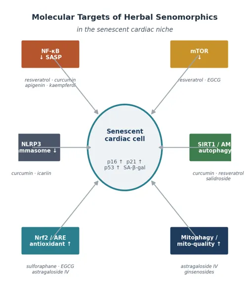
Figure 3. Convergent molecular targets of herbal senomorphics. Chemically diverse phytochemicals act on a compact set of nodes—NF-κB, mTOR, the NLRP3 inflammasome, SIRT1/AMPK, Nrf2 and mitophagy—that govern the senescent cardiac secretome.
TRANSLATIONAL CHALLENGES AND OPPORTUNITIES:
The biggest barrier to clinical application is pharmacokinetic. Curcumin, resveratrol, fisetin, EGCG and quercetin all demonstrate poor water solubility, high first-pass metabolism and high clearance, and thus, plasma levels after either dietary or conventional oral routes are likely to be subtherapeutic with regards to their senomorphic effects in vitro. Due to its advanced technology, nanotechnology has been the most successful at lowering the potency-bioavailability gap. These include liposomes, nanoparticles (including polymeric and lipid), phospholipid complexes, and nanomicelles. In several preclinical cardiac applications, all of these have improved drug exposure and delivery.
Internal tissue targeting is feasible with nanocarriers due to their surface modifiability. Further, to minimize systemic delivery them senomorphic effects, nanocarriers can be designed to deliver their load in the presence of SASP-associated proteases. In the case of curcumin, drug-loaded nanomicelles have improved cardiac function and also have shown positive effects on myocardial and mitochondrial integrity and inflammation compared to curcumin alone in models of ischaemic and age-related injury. Taken together with delivery methods of co-crystals and prodrugs, these are the most promising methods for achieving clinically relevant senomorphic effects with these phytochemicals. [52],[53]
Natural products may act as senomorphics at low concentrations and senolytics at higher concentrations. This flexibility disrupts dosing, as a single compound may either inhibit the SASP or eliminate the cells, depending on concentration, timing, and the tissue of interest. Additionally, the hormetic dose–response relationships—where low doses are often beneficial to cells and higher doses have the opposite effect—also require that cautious dosing is employed. Establishing concentration ranges for senomorphic and senolytic activity in cardiac tissue is critical for this research.[68]
Herbal medicine variants of a given formulation show considerable chemical formulation, and thus, considerable variability in the concentration of active constituents. Standardization of phytochemical extraction must be implemented to address such issues along with extensive safety monitoring of herb-drug interactions. The likely effects of coadministered phytomedicines and cardiovascular medications on Cytochrome-P450 metabolism need to be studied. While the historical safety data of these products as nutriceuticals is comforting, the required doses for senomorphic action may be greater than constituents used in the normal diet. [52]
The development of validated, minimally invasive biomarkers for assessing cardiac burden of cellular senescence and SASP and for measuring their targeting to address the unmet need for patient selection is essential to facilitate future clinical studies. Circulating SASP factors, senescence-associated microRNAs, and imaging correlates are adjunctive biomarkers that are currently under investigation.[69] A novel approach that is preferred is called the "one-two punch". Intermittent dosing of senolytics (e.g., D+Q, Fisetin) to remove the burden of senescent cells, is followed by continuous dosing of a well-tolerated plant-derived senomorphic agent to inhibit the remaining SASP and protect against its ongoing production. This may provide the synergistic benefit of burden-reducing senolysis, and chronic, low-harm secretome control, which is the likely role of dietary senomorphics.[70],[71]
With regard to anatomical focus, a large proportion of the age-associated failure of the heart involves the vasculature; senescence of the endothelium, increased stiffness of the large arteries, and dysfunction of the coronary microvasculature, as well as that of the myocardium. Several of the herbal senomorphics, such as Resveratrol, EGCG, and Astragaloside IV, are likely to be initially useful for the treatment of HFpEF through their ability to enhance the function of the microvasculature and endothelium. This is likely to be through improvement of microvascular and endothelial function before improving direct function of cardiomyocytes. [72],[73]
FUTURE DIRECTIONS:
Many factors will determine the incorporation of Senomorphic herbs into cardiac practice. First, single-cell transcriptomics with activity-based proteomics can assist cell-type specific target discovery by defining the cardiac senescent cell subtype and the target protein of each phytocompound. This can pave the way for personalized senotherapeutics, with less reliance on empirical approaches.[12] Second, innovative methodologies such as network pharmacology and machine-learning based screens of natural product libraries are advancing the discovery of new senomorphic frameworks and rational multi-component formulations. Third, targeted, senescence-responsive nanocarriers are likely to solve the bioavailability problem while minimizing off-target effects. Fourth, well designed randomized controlled trials with functional and structural endpoint and senescence biomarker incorporation are needed to study older adults with, or at risk of, HFpEF, as the preclinical evidence has been substantial, but the same can’t be said for clinical studies. Finally, the state of the botanical senomorphics and of standardization and regulatory frameworks should develop simultaneously to create the opportunity to evaluate new senomorphics in clinical practice as needed.[73]
CONCLUSION
Particularly with respect to HFpEF, the aging process and advancing heart failure have been characterized by the interplay of cellular senescence, fibrosis, and chronic inflammation. Theoretically, the development of agents that silence the senescence-associated secretory phenotype of senescent cells, rather than remove them, may offer an attractive mechanism to halt the progression of cardiovascular aging and the subsequent heart failure. Senomorphic herbs acting as agents (polyphenols) such as resveratrol, curcumin, quercetin, fisetin, apigenin, kaempferol, EGCG, ginsenosides, astragaloside IV, and salidroside, act upon a small network of druggable nodes, such as NF-κB, mTOR, NLRP3, SIRT1/AMPK, Nrf2, and Micro- and Mitochondrial Quality Control and Integrative Pharmacology. There is strong support for the development of agents with low phytochemical toxicity for senomorphic herbs. The pharmacological effect of herbal senomorphics and reduction of the senescent phenotype of cardiac fibroblasts and cardiomyocytes and reduction of the SASP has been demonstrated. However, its clinical use is limited by low bioavailability. Herbal senomorphics have great potential as an easily accessible and valuable adjunct therapy to prevent and treat age-related heart failure, if the hurdles of the development (i.e., defining senomorphic dose windows, standardizing preparations, developing senescence biomarkers, and performing rigorous randomised trials)
Table 3. Convergent molecular nodes and representative herbal senomorphics.
|
Molecular node |
Representative phytochemicals |
|
NF-κB (SASP driver ↓) |
Resveratrol, curcumin, quercetin, apigenin, kaempferol |
|
mTOR (↓) |
Resveratrol, EGCG |
|
NLRP3 inflammasome (↓) |
Curcumin |
|
SIRT1 / AMPK; autophagy (↑) |
Resveratrol, curcumin, ginsenoside Rg1, salidroside |
|
Nrf2 / ARE antioxidant (↑) |
EGCG, kaempferol, astragaloside IV |
|
Mitophagy / mito-quality (↑) |
Astragaloside IV, ginsenosides, salidroside |
|
p38 MAPK / IRAK (↓) |
Apigenin, kaempferol |
RE, antioxidant-response element. ↑ activation; ↓ inhibition.
REFERENCES

Sonali Sansare, Herbal Senomorphics: Novel Interventions for Age-Induced Heart Failure, Int. J. of Pharm. Sci., 2026, Vol 4, Issue 9, 2821-2837. https://doi.org/10.5281/zenodo.22912949
 10.5281/zenodo.22912949