View Article

  • Exploring The Anti-Inflammatory Potential Of Salvia Plebeia Through In-Vitro Studies

  • 1M.Pharm Scholar, Department of Pharmacology, Dr. C.V. Raman University, Bilaspur (C.G.), India
    2Assistant Professor, Department of Pharmacology, Dr. C.V. Raman University, Bilaspur (C.G.), India.
    3Principal, Dr. C.V. Raman University, Bilaspur (C.G.), India.
     

Abstract

The present study was undertaken to investigate the phytochemical profile and in-vitro anti-inflammatory activity of the ethanolic extract of Salvia plebeia R. Br. Leaves were collected, authenticated, dried, powdered, defatted with petroleum ether, and subsequently extracted with ethanol using Soxhlet extraction. From 300 g of powdered plant material, 36.5 g of dark greenish-brown extract was obtained, corresponding to a yield of 12.17% w/w. Preliminary phytochemical screening revealed the presence of alkaloids, flavonoids, tannins, phenolic compounds, saponins, and glycosides. The anti-inflammatory activity was evaluated using protein denaturation inhibition and protease inhibition assays at concentrations of 100, 200, 400, and 800 µg/mL, with diclofenac sodium as the standard drug. The extract demonstrated concentration-dependent inhibition of protein denaturation, increasing from 32.58% at 100 µg/mL to 69.56% at 800 µg/mL. Similarly, protease inhibition increased from 28.13% to 63.64% over the same concentration range. Diclofenac sodium exhibited higher inhibition in both assays, reaching 82.45% protein denaturation inhibition and 78.91% protease inhibition at 800 µg/mL. The findings demonstrate that S. plebeia ethanolic extract possesses appreciable in-vitro anti-inflammatory activity, which may be associated with its diverse phytoconstituents and warrants further pharmacological investigation.

Keywords

Salvia plebeia; Ethanolic Extract; Phytochemical Screening; Anti-inflammatory Activity; Protein Denaturation; Protease Inhibition

Introduction

× Popup Image
  1. Inflammation

Inflammation is a coordinated protective response of the body that occurs following infection, tissue injury, or exposure to harmful chemical or physical stimuli. It is an essential component of innate immunity, helping to recognize and eliminate potentially damaging agents while initiating tissue repair and restoration of normal physiological function. Although inflammation is generally protective, an excessive or unresolved inflammatory response can cause tissue injury and contribute to the progression of several chronic disorders.[1,2]

The inflammatory process involves a complex interaction between immune cells, blood vessels, and molecular mediators. Recognition of tissue damage or invading microorganisms by pattern-recognition receptors activates intracellular signaling pathways and promotes the production of cytokines, chemokines, prostaglandins, and other inflammatory mediators.[2,3] These mediators regulate vascular changes, leukocyte recruitment, and communication between immune and damaged cells. Depending on its duration and underlying cause, inflammation is generally classified as acute or chronic. Acute inflammation develops rapidly and is usually self-limiting, whereas chronic inflammation persists for prolonged periods and may result in continuous tissue remodeling and damage.[3,4]

Persistent inflammatory activity has been associated with the pathogenesis of several diseases, including rheumatoid arthritis, inflammatory bowel disorders, metabolic diseases, cardiovascular disorders, neurodegenerative conditions, and cancer.[4] Current pharmacological approaches include non-steroidal anti-inflammatory drugs, corticosteroids, and targeted biological therapies. Despite their therapeutic benefits, prolonged use of some anti-inflammatory agents may produce adverse effects, encouraging continued investigation of safer and potentially effective alternatives, particularly those derived from medicinal plants and other natural sources.[1,4] Consequently, evaluation of natural compounds with anti-inflammatory potential remains an important area of pharmaceutical and biomedical research.

2. MATERIALS AND METHOD

Materials

  • Collection of Plant Material

Salvia plebeia R. Br. leaves was purchased from a local market and subsequently authenticated by a qualified botanist or pharmacognosist to ensure their identity and purity before use in the experimental work.

Method

  • Preparation of Plant Extracts

Air-dried, coarsely powdered Salvia plebeia (300 g) was first defatted with petroleum ether (800 mL) using a Soxhlet apparatus for approximately 6 h at about 80°C [25]. The marc was separated and dried at room temperature to remove residual solvent [25,31]. It was subsequently extracted with analytical-grade ethanol (1000 mL) for approximately 8 h at the solvent boiling temperature (about 78°C). The ethanolic extract was filtered and concentrated under reduced pressure using a rotary evaporator below 50°C. The resulting extract was further dried, weighed to determine the extraction yield, and stored in airtight containers at 4°C until further investigation [34,39].

The percentage yield was calculated as:

Percentage yield (%) = (Weight of dried extract / Weight of initial plant material) × 100

  • Preliminary Phytochemical Screening [35,39]

Table 1: Procedure for Preliminary Phytochemical Screening of Ethanolic Extract of Salvia plebeian

Phytoconstituent

Test

Procedure

Alkaloids

Dragendorff’s test

2 mL of extract solution was treated with about 0.5 mL of Dragendorff’s reagent.

Mayer’s test

2 mL of extract solution was mixed with about 0.5 mL of Mayer’s reagent.

Wagner’s test

2 mL of extract solution was treated with about 0.5 mL of Wagner’s reagent.

Flavonoids

Shinoda test

2 mL of extract solution was mixed with magnesium turnings (about 20 mg), followed by 2 drops of concentrated HCl.

Tannins and phenolics

Ferric chloride test

2 mL of extract solution was treated with 2 drops of 5% ferric chloride solution.

Phenolic compounds

Lead acetate test

2 mL of extract solution was mixed with 1 mL of 10% lead acetate solution.

Saponins

Foam test

2 mL of extract was diluted with 5 mL distilled water and vigorously shaken for about 15 min.

Glycosides

Molisch’s test

2 mL of extract was treated with 1 mL Molisch’s reagent, followed by careful addition of 2 mL concentrated H₂SO₄ along the tube wall.

Benedict’s test

2 mL of extract was mixed with 2 mL Benedict’s reagent and heated in a water bath for 5 min.

  • In-Vitro Anti-inflammatory Activity

1. Protein Denaturation Inhibition Assay

The anti-inflammatory potential of the ethanolic extract of Salvia plebeia was assessed using the protein denaturation inhibition assay. Briefly, 0.2 mL of egg albumin was mixed with 2.8 mL of phosphate buffer (pH 6.4) and 2 mL of extract at concentrations of 100, 200, 400, and 800 µg/mL. The mixtures were incubated at 37°C for 20 min and subsequently heated at 70°C for 5 min to induce protein denaturation. After cooling to room temperature, absorbance was measured at 660 nm using a UV-Visible spectrophotometer. A reaction mixture without extract served as the control, while diclofenac sodium was used as the standard drug. All experiments were performed in triplicate. The percentage inhibition of protein denaturation was calculated using the absorbance values of the control and treated samples [26,36].

Inhibition (%) = [(Acontrol − Asample) / Acontrol] × 100

2. Protease Inhibition Assay

The anti-inflammatory activity of the ethanolic extract of  Salvia plebeia was evaluated using a protease inhibition assay. Briefly, 1 mL of trypsin solution (0.06 mg/mL), 1 mL of Tris-HCl buffer (pH 7.4), and 1 mL of extract at concentrations of 100, 200, 400, and 800 µg/mL were mixed and incubated at 37°C for 15 min. Subsequently, 1 mL of casein solution (0.8% w/v) was added, followed by incubation at 37°C for 20 min. The reaction was terminated by adding 2 mL of 10% trichloroacetic acid, and the mixture was centrifuged at 3000 rpm for 10 min. The absorbance of the resulting supernatant was measured at 280 nm using a UV-Visible spectrophotometer. A control without extract and diclofenac sodium as the standard were used for comparison [35,39].

The percentage inhibition was calculate by-

Inhibition (%) = [(Acontrol − Asample) / Acontrol] × 100

  • Comparative Analysis with Standard Drug

The anti-inflammatory activity of Salvia plebeia extracts was compared with diclofenac sodium as the standard drug using protein denaturation and protease inhibition assays. Extract concentrations of 100, 200, 400, and 800 µg/mL were evaluated under identical experimental conditions. The percentage inhibition was calculated and expressed as mean ± SD based on triplicate experiments [26, 31]. The inhibitory effects of the extracts were compared with those of diclofenac sodium at corresponding concentrations. Where applicable, IC₅₀ values were determined, with lower values indicating greater inhibitory potency. This comparison was used to assess the relative anti-inflammatory potential of the plant extract [35, 39].

3. RESULST

  • Preparation of Plant Extracts

Soxhlet extraction of Salvia plebeia was successfully performed after petroleum ether defatting. The ethanolic extraction produced a dark greenish-brown, semisolid extract weighing 36.5 g. From 300 g of powdered plant material, an extraction yield of 12.17% w/w was obtained, indicating satisfactory recovery of extractable constituents.                   

Table 2: Preparation of Salvia plebeia Extract

Parameter

Result

Plant material

Salvia plebeia

Powdered material

300 g

Defatting solvent

Petroleum ether

Defatting time

6 h

Extraction solvent

Ethanol

Extraction time

8 h

Dried extract

36.5 g

Percentage yield

12.17% w/w

Appearance

Dark greenish-brown

Storage

4°C

  • Preliminary Phytochemical Screening

Table  3 - Preliminary Phytochemical Screening

S. No.

Phytoconstituent

Test Performed

Observation

Result

1

Alkaloids

Dragendorff’s test

Reddish-brown precipitate

+ Present

Mayer’s test

Creamy white precipitate

+ Present

Wagner’s test

Brown precipitate

+ Present

2

Flavonoids

Shinoda test

Pink-crimson color

+ Present

3

Tannins

Ferric chloride test

Greenish-black color

+ Present

4

Phenolic compounds

Lead acetate test

White precipitate

+ Present

5

Saponins

Foam test

Stable froth (2 cm)

+ Present

6

Glycosides

Molisch’s test

Violet ring formation

+ Present

Benedict’s test

Brick-red precipitate

+ Present

  • In-Vitro Anti-inflammatory Activity

1. Protein Denaturation Inhibition Assay

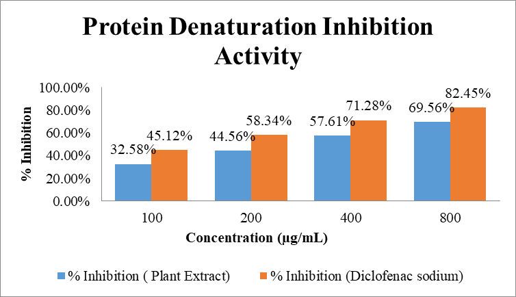
The ethanolic extract showed concentration-dependent inhibition of protein denaturation, increasing from 32.58% to 69.56% at 100–800 µg/mL, while diclofenac showed higher inhibition.

Table 4: Protein Denaturation Inhibition Activity of Ethanolic Extract of Salvia plebiea

S. No.

Concentration (µg/mL)

Absorbance (Extract)

% Inhibition (Extract)

% Inhibition (Diclofenac sodium)

1

100

0.62 ± 0.02

32.58%

45.12%

2

200

0.51 ± 0.01

44.56%

58.34%

3

400

0.39 ± 0.02

57.61%

71.28%

4

800

0.28 ± 0.01

69.56%

82.45%

Control Absorbance: 0.92 ± 0.02

Figure 1: Protein Denaturation Inhibition Activity Graph for Plant Extract and Standard Drug

2. Protease Inhibition Assay

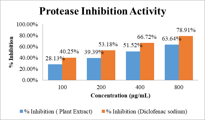
The ethanolic extract showed concentration-dependent protease inhibition, increasing from 28.13% to 63.64% at 100-800 µg/mL, while diclofenac sodium exhibited higher inhibition across all tested concentrations.

Table 5: Protease Inhibition Activity of Ethanolic Extract of Salvia plebiea

S. No.

Concentration (µg/mL)

Absorbance (Extract)

% Inhibition

( Plant Extract)

% Inhibition (Diclofenac sodium)

1

100

0.71 ± 0.02

28.13%

40.25%

2

200

0.60 ± 0.01

39.39%

53.18%

3

400

0.48 ± 0.02

51.52%

66.72%

4

800

0.36 ± 0.01

63.64%

78.91%

Control Absorbance: 0.99 ± 0.02

Figure 2: Protease Inhibition Activity for Plant Extract and Standard Drug

  • Comparative Analysis with Standard Drug

The ethanolic extract of Salvia plebeia showed concentration-dependent anti-inflammatory activity in both assays. Protein denaturation inhibition increased from 32.58% to 69.56%, while protease inhibition increased from 28.13% to 63.64%. Diclofenac sodium showed higher inhibition throughout, confirming greater potency, while the plant extract demonstrated promising anti-inflammatory activity at higher concentrations.

Table 6: Comparative Anti-inflammatory Activity of Salvia plebeia Extract and Diclofenac Sodium

S. No.

Concentration (µg/mL)

Protein Denaturation Inhibition (%)

Protein Denaturation Inhibition (%)

Protease Inhibition (%)

Protease Inhibition (%)


Plant Extract

Diclofenac Sodium

Plant Extract

Diclofenac Sodium

1

100

32.58%

45.12%

28.13%

40.25%

2

200

44.56%

58.34%

39.39%

53.18%

3

400

57.61%

71.28%

51.52%

66.72%

4

800

69.56%

82.45%

63.64%

78.91%

4. CONCLUSION

The present study was carried out to investigate the phytochemical composition and in-vitro anti-inflammatory activity of the ethanolic extract of Salvia plebeia The plant material was successfully collected, authenticated, and processed, followed by extraction using the Soxhlet method. The ethanolic extract yielded a satisfactory percentage recovery, indicating efficient extraction of bioactive constituents. Preliminary phytochemical screening confirmed the presence of important secondary metabolites such as alkaloids, flavonoids, tannins, phenolic compounds, saponins, and glycosides, which are known to possess various pharmacological properties. The anti-inflammatory activity of the extract was evaluated using protein denaturation inhibition and protease inhibition assays. The results demonstrated a clear concentration-dependent increase in inhibitory activity in both models. The extract showed significant inhibition of protein denaturation and protease activity, indicating its ability to stabilize proteins and inhibit inflammatory enzymes. Although the activity of the extract was slightly lower than that of the standard drug diclofenac sodium, it exhibited comparable effects at higher concentrations, suggesting promising therapeutic potential.

5. CONFLICT OF INTEREST

The authors declare that there is no conflict of interest associated with this research work.

REFERENCES

  1. Schmid-Schönbein, G. W. (2006). Analysis of inflammation. Annu. Rev. Biomed. Eng., 8(1), 93-151.
  2. Nathan, C., & Ding, A. (2010). Nonresolving inflammation. Cell, 140(6), 871-882.
  3. Ahmed, A. U. (2011). An overview of inflammation: mechanism and consequences. Frontiers in Biology, 6(4), 274-281.
  4. Punchard, N. A., Whelan, C. J., & Adcock, I. (2004). The journal of inflammation. Journal of inflammation, 1(1), 1.
  5. Soliman, A. M., & Barreda, D. R. (2022). Acute inflammation in tissue healing. International journal of molecular sciences, 24(1), 641.
  6. Pérez, S., & Rius-Pérez, S. (2022). Macrophage polarization and reprogramming in acute inflammation: a redox perspective. Antioxidants, 11(7), 1394.
  7. Hannoodee, S., & Nasuruddin, D. N. (2024). Acute inflammatory response. In StatPearls [Internet]. StatPearls Publishing.
  8. Liu, X., Ou, X., Zhang, T., Li, X., Qiao, Q., Jia, L., ... & Zhang, Z. (2024). In situ neutrophil apoptosis and macrophage efferocytosis mediated by Glycyrrhiza protein nanoparticles for acute inflammation therapy. Journal of Controlled Release, 369, 215-230.
  9. Serhan, C. N., Ward, P. A., & Gilroy, D. W. (Eds.). (2010). Fundamentals of inflammation. Cambridge University Press.
  10. Herrero-Cervera, A., Soehnlein, O., & Kenne, E. (2022). Neutrophils in chronic inflammatory diseases. Cellular & molecular immunology, 19(2), 177-191.
  11. Stenvinkel, P., Chertow, G. M., Devarajan, P., Levin, A., Andreoli, S. P., Bangalore, S., & Warady, B. A. (2021). Chronic inflammation in chronic kidney disease progression: role of Nrf2. Kidney international reports, 6(7), 1775-1787.
  12. Saito, S., Shahbaz, S., Osman, M., Redmond, D., Bozorgmehr, N., Rosychuk, R. J., ... & Elahi, S. (2024). Diverse immunological dysregulation, chronic inflammation, and impaired erythropoiesis in long COVID patients with chronic fatigue syndrome. Journal of autoimmunity, 147, 103267.
  13. Nathan, C. (2002). Points of control in inflammation. Nature, 420(6917), 846-852.
  14. Ospelt, C., & Gay, S. (2010). TLRs and chronic inflammation. The international journal of biochemistry & cell biology, 42(4), 495-505.
  15. Ryan, G. B., & Majno, G. (1977). Acute inflammation. A review. The American journal of pathology, 86(1), 183.
  16. Varela, M. L., Mogildea, M., Moreno, I., & Lopes, A. (2018). Acute inflammation and metabolism. Inflammation, 41(4), 1115-1127.
  17. Levy, B. D., & Serhan, C. N. (2014). Resolution of acute inflammation in the lung. Annual review of physiology, 76(1), 467-492.
  18. Kaur, T., Bhat, H. A., Bhat, R., Kumar, A., Bindu, K., Koul, S., & Vyas, D. (2015). Physio-chemical and antioxidant profiling of Salvia sclarea L. at different climates in north-western Himalayas. Acta Physiologiae Plantarum, 37(7), 132.
  19. Kumar, R., Sharma, S., & Pathania, V. (2013). Effect of shading and plant density on growth, yield and oil composition of clary sage (Salvia sclarea L.) in north western Himalaya. Journal of essential oil research, 25(1), 23-32.
  20. Kačániová, M., Vukovic, N. L., Čmiková, N., Galovičová, L., Schwarzová, M., Šimora, V., ... & Vukic, M. D. (2023). Salvia sclarea essential oil chemical composition and biological activities. International journal of molecular sciences, 24(6), 5179.
  21. Yaseen, M., Singh, M., Ram, D., & Singh, K. (2014). Production potential, nitrogen use efficiency and economics of clarysage (Salvia sclarea L.) varieties as influenced by nitrogen levels under different locations. Industrial Crops and Products, 54, 86-91.
  22. Singh, V., Sood, R., Ramesh, K., & Singh, B. (2008). Effects of growth regulator application on growth, flower, oil yield, and quality of clary sage (Salvia sclarea L.). Journal of herbs, spices & medicinal plants, 14(1-2), 29-36.
  23. Chand, S., Yaseen, M., Rajkumari, & Patra, D. D. (2015). Application of heavy metal rich tannery sludge on sustainable growth, yield and metal accumulation by clarysage (Salvia sclarea L.). International journal of phytoremediation, 17(12), 1171-1176.
  24. Aćimović, M., Kiprovski, B., Rat, M., Sikora, V., Popović, V., Koren, A., & Brdar-Jokanović, M. (2018). Salvia sclarea: Chemical composition and biological activity. Journal of Agronomy, Technology and Engineering Management (JATEM), 1(1), 18-28.
  25. Randjelovic, M., Miladinović, B., Milutinović, M., Jovanović, M., Branković, S., Kitić, N., ... & Kitić, D. (2025). Antioxidant, antimicrobial, and spasmolytic effects of the clary sage (Salvia sclarea L.) hydroethanolic extracts prepared by digestion. Acta medica Medianae, 64(2).
  26. Ajaz, N., & Rehman, F. (2025). Fabrication & evaluation of anti-inflammatory effect of clary sage oil (Salvia sclarea L.) loaded hydrogel films. RADS Journal of Pharmacy and Allied Health Sciences, 3(1), 13-24.
  27. Hans, M., Nayik, G. A., & Salaria, A. (2023). Clary sage essential oil. In Essential Oils (pp. 459-478). Academic Press.
  28. Sienkiewicz, M., Głowacka, A., Poznańska-Kurowska, K., Kaszuba, A., Urbaniak, A., & Kowalczyk, E. (2015). The effect of clary sage oil on staphylococci responsible for wound infections. Advances in Dermatology and Allergology/Postępy Dermatologii i Alergologii, 32(1), 21-26.
  29. Tulukcu, E., Sagdic, O., Albayrak, S. E. V. İ. L., Ekici, L. Ü. T. F. İ. Y. E., & Yetim, H. (2009). Effect of collection time on biological activity of clary sage (Salvia sclarea). Journal of applied Botany and food quality, 83(1), 44-49.
  30. Singh, S., Chishti, S. A., Singh, S., Bhatt, U. H., & Khan, M. M. A. (2023). Bioactive Compounds and Pharmacological Values of Salvia Sclarea L. In Bioactives and Pharmacology of Lamiaceae (pp. 289-296). Apple Academic Press.
  31. Alomar, H. A., Elkady, W. M., Abdel-Aziz, M. M., Ibrahim, T. A., & Fathallah, N. (2023). Anti-heliobacter pylori and anti-inflammatory potential of Salvia officinalis metabolites: in vitro and in silico studies. Metabolites, 13(1), 136.
  32. Chehade, S., Kobeissy, M., Kanaan, H., & Haddad, M. (2022). Comparison between the chemical compositions and the in-vitro antidiabetic and anti-inflammatory activities of Salvia Libanotica’and Salvia officinalis’ leaves essential oils. Eur. J. Pharm. Med. Res, 93, 34-43.
  33. Randjelović, M., Branković, S., Jovanović, M., Kitić, N., Živanović, S., Mihajilov-Krstev, T., ... & Kitić, D. (2023). An In Vitro and In Silico Characterization of Salvia sclarea L. Methanolic Extracts as Spasmolytic Agents. Pharmaceutics, 15(5), 1376.
  34. Quradha, M. M., Duru, M. E., Kucukaydin, S., Tamfu, A. N., Iqbal, M., Bibi, H., ... & Ceylan, O. (2024). Comparative assessment of phenolic composition profile and biological activities of green extract and conventional extracts of Salvia sclarea. Scientific Reports, 14(1), 1885.
  35. Margetts, G., Kleidonas, S., Zaibi, N. S., Zaibi, M. S., & Edwards, K. D. (2022). Evidence for anti-inflammatory effects and modulation of neurotransmitter metabolism by Salvia officinalis L. BMC Complementary Medicine and Therapies, 22(1), 131.
  36. Kačániová, M., Vukovic, N. L., Čmiková, N., Galovičová, L., Schwarzová, M., Šimora, V., ... & Vukic, M. D. (2023). Salvia sclarea essential oil chemical composition and biological activities. International journal of molecular sciences, 24(6), 5179.
  37. Bendaas, R., Messaadia, L., Bekkar, Y., Bourougaa, L., & Messaoudi, A. (2025). Comprehensive in vitro and molecular docking analysis of antioxidant and antimicrobial properties of salvia officinalis L. extracts and essential oils. Journal of Molecular Structure, 1331, 141621.
  38. Kubatka, P., Mazurakova, A., Koklesova, L., Kuruc, T., Samec, M., Kajo, K., ... & Kello, M. (2024). Salvia officinalis L. exerts oncostatic effects in rodent and in vitro models of breast carcinoma. Frontiers in Pharmacology, 15, 1216199.
  39. Boufadi, M. Y., Keddari, S., MOULAI-HACENE, F., & Chaa, S. (2021). Chemical composition, antioxidant and anti-inflammatory properties of Salvia officinalis extract from Algeria. Pharmacognosy Journal, 13(2).

Reference

  1. Schmid-Schönbein, G. W. (2006). Analysis of inflammation. Annu. Rev. Biomed. Eng., 8(1), 93-151.
  2. Nathan, C., & Ding, A. (2010). Nonresolving inflammation. Cell, 140(6), 871-882.
  3. Ahmed, A. U. (2011). An overview of inflammation: mechanism and consequences. Frontiers in Biology, 6(4), 274-281.
  4. Punchard, N. A., Whelan, C. J., & Adcock, I. (2004). The journal of inflammation. Journal of inflammation, 1(1), 1.
  5. Soliman, A. M., & Barreda, D. R. (2022). Acute inflammation in tissue healing. International journal of molecular sciences, 24(1), 641.
  6. Pérez, S., & Rius-Pérez, S. (2022). Macrophage polarization and reprogramming in acute inflammation: a redox perspective. Antioxidants, 11(7), 1394.
  7. Hannoodee, S., & Nasuruddin, D. N. (2024). Acute inflammatory response. In StatPearls [Internet]. StatPearls Publishing.
  8. Liu, X., Ou, X., Zhang, T., Li, X., Qiao, Q., Jia, L., ... & Zhang, Z. (2024). In situ neutrophil apoptosis and macrophage efferocytosis mediated by Glycyrrhiza protein nanoparticles for acute inflammation therapy. Journal of Controlled Release, 369, 215-230.
  9. Serhan, C. N., Ward, P. A., & Gilroy, D. W. (Eds.). (2010). Fundamentals of inflammation. Cambridge University Press.
  10. Herrero-Cervera, A., Soehnlein, O., & Kenne, E. (2022). Neutrophils in chronic inflammatory diseases. Cellular & molecular immunology, 19(2), 177-191.
  11. Stenvinkel, P., Chertow, G. M., Devarajan, P., Levin, A., Andreoli, S. P., Bangalore, S., & Warady, B. A. (2021). Chronic inflammation in chronic kidney disease progression: role of Nrf2. Kidney international reports, 6(7), 1775-1787.
  12. Saito, S., Shahbaz, S., Osman, M., Redmond, D., Bozorgmehr, N., Rosychuk, R. J., ... & Elahi, S. (2024). Diverse immunological dysregulation, chronic inflammation, and impaired erythropoiesis in long COVID patients with chronic fatigue syndrome. Journal of autoimmunity, 147, 103267.
  13. Nathan, C. (2002). Points of control in inflammation. Nature, 420(6917), 846-852.
  14. Ospelt, C., & Gay, S. (2010). TLRs and chronic inflammation. The international journal of biochemistry & cell biology, 42(4), 495-505.
  15. Ryan, G. B., & Majno, G. (1977). Acute inflammation. A review. The American journal of pathology, 86(1), 183.
  16. Varela, M. L., Mogildea, M., Moreno, I., & Lopes, A. (2018). Acute inflammation and metabolism. Inflammation, 41(4), 1115-1127.
  17. Levy, B. D., & Serhan, C. N. (2014). Resolution of acute inflammation in the lung. Annual review of physiology, 76(1), 467-492.
  18. Kaur, T., Bhat, H. A., Bhat, R., Kumar, A., Bindu, K., Koul, S., & Vyas, D. (2015). Physio-chemical and antioxidant profiling of Salvia sclarea L. at different climates in north-western Himalayas. Acta Physiologiae Plantarum, 37(7), 132.
  19. Kumar, R., Sharma, S., & Pathania, V. (2013). Effect of shading and plant density on growth, yield and oil composition of clary sage (Salvia sclarea L.) in north western Himalaya. Journal of essential oil research, 25(1), 23-32.
  20. Ka?ániová, M., Vukovic, N. L., ?miková, N., Galovi?ová, L., Schwarzová, M., Šimora, V., ... & Vukic, M. D. (2023). Salvia sclarea essential oil chemical composition and biological activities. International journal of molecular sciences, 24(6), 5179.
  21. Yaseen, M., Singh, M., Ram, D., & Singh, K. (2014). Production potential, nitrogen use efficiency and economics of clarysage (Salvia sclarea L.) varieties as influenced by nitrogen levels under different locations. Industrial Crops and Products, 54, 86-91.
  22. Singh, V., Sood, R., Ramesh, K., & Singh, B. (2008). Effects of growth regulator application on growth, flower, oil yield, and quality of clary sage (Salvia sclarea L.). Journal of herbs, spices & medicinal plants, 14(1-2), 29-36.
  23. Chand, S., Yaseen, M., Rajkumari, & Patra, D. D. (2015). Application of heavy metal rich tannery sludge on sustainable growth, yield and metal accumulation by clarysage (Salvia sclarea L.). International journal of phytoremediation, 17(12), 1171-1176.
  24. A?imovi?, M., Kiprovski, B., Rat, M., Sikora, V., Popovi?, V., Koren, A., & Brdar-Jokanovi?, M. (2018). Salvia sclarea: Chemical composition and biological activity. Journal of Agronomy, Technology and Engineering Management (JATEM), 1(1), 18-28.
  25. Randjelovic, M., Miladinovi?, B., Milutinovi?, M., Jovanovi?, M., Brankovi?, S., Kiti?, N., ... & Kiti?, D. (2025). Antioxidant, antimicrobial, and spasmolytic effects of the clary sage (Salvia sclarea L.) hydroethanolic extracts prepared by digestion. Acta medica Medianae, 64(2).
  26. Ajaz, N., & Rehman, F. (2025). Fabrication & evaluation of anti-inflammatory effect of clary sage oil (Salvia sclarea L.) loaded hydrogel films. RADS Journal of Pharmacy and Allied Health Sciences, 3(1), 13-24.
  27. Hans, M., Nayik, G. A., & Salaria, A. (2023). Clary sage essential oil. In Essential Oils (pp. 459-478). Academic Press.
  28. Sienkiewicz, M., G?owacka, A., Pozna?ska-Kurowska, K., Kaszuba, A., Urbaniak, A., & Kowalczyk, E. (2015). The effect of clary sage oil on staphylococci responsible for wound infections. Advances in Dermatology and Allergology/Post?py Dermatologii i Alergologii, 32(1), 21-26.
  29. Tulukcu, E., Sagdic, O., Albayrak, S. E. V. ?. L., Ekici, L. Ü. T. F. ?. Y. E., & Yetim, H. (2009). Effect of collection time on biological activity of clary sage (Salvia sclarea). Journal of applied Botany and food quality, 83(1), 44-49.
  30. Singh, S., Chishti, S. A., Singh, S., Bhatt, U. H., & Khan, M. M. A. (2023). Bioactive Compounds and Pharmacological Values of Salvia Sclarea L. In Bioactives and Pharmacology of Lamiaceae (pp. 289-296). Apple Academic Press.
  31. Alomar, H. A., Elkady, W. M., Abdel-Aziz, M. M., Ibrahim, T. A., & Fathallah, N. (2023). Anti-heliobacter pylori and anti-inflammatory potential of Salvia officinalis metabolites: in vitro and in silico studies. Metabolites, 13(1), 136.
  32. Chehade, S., Kobeissy, M., Kanaan, H., & Haddad, M. (2022). Comparison between the chemical compositions and the in-vitro antidiabetic and anti-inflammatory activities of Salvia Libanotica’and Salvia officinalis’ leaves essential oils. Eur. J. Pharm. Med. Res, 93, 34-43.
  33. Randjelovi?, M., Brankovi?, S., Jovanovi?, M., Kiti?, N., Živanovi?, S., Mihajilov-Krstev, T., ... & Kiti?, D. (2023). An In Vitro and In Silico Characterization of Salvia sclarea L. Methanolic Extracts as Spasmolytic Agents. Pharmaceutics, 15(5), 1376.
  34. Quradha, M. M., Duru, M. E., Kucukaydin, S., Tamfu, A. N., Iqbal, M., Bibi, H., ... & Ceylan, O. (2024). Comparative assessment of phenolic composition profile and biological activities of green extract and conventional extracts of Salvia sclarea. Scientific Reports, 14(1), 1885.
  35. Margetts, G., Kleidonas, S., Zaibi, N. S., Zaibi, M. S., & Edwards, K. D. (2022). Evidence for anti-inflammatory effects and modulation of neurotransmitter metabolism by Salvia officinalis L. BMC Complementary Medicine and Therapies, 22(1), 131.
  36. Ka?ániová, M., Vukovic, N. L., ?miková, N., Galovi?ová, L., Schwarzová, M., Šimora, V., ... & Vukic, M. D. (2023). Salvia sclarea essential oil chemical composition and biological activities. International journal of molecular sciences, 24(6), 5179.
  37. Bendaas, R., Messaadia, L., Bekkar, Y., Bourougaa, L., & Messaoudi, A. (2025). Comprehensive in vitro and molecular docking analysis of antioxidant and antimicrobial properties of salvia officinalis L. extracts and essential oils. Journal of Molecular Structure, 1331, 141621.
  38. Kubatka, P., Mazurakova, A., Koklesova, L., Kuruc, T., Samec, M., Kajo, K., ... & Kello, M. (2024). Salvia officinalis L. exerts oncostatic effects in rodent and in vitro models of breast carcinoma. Frontiers in Pharmacology, 15, 1216199.
  39. Boufadi, M. Y., Keddari, S., MOULAI-HACENE, F., & Chaa, S. (2021). Chemical composition, antioxidant and anti-inflammatory properties of Salvia officinalis extract from Algeria. Pharmacognosy Journal, 13(2).

Photo
Pooja Kumbhkar
Corresponding author

M.Pharm Scholar, Department of Pharmacology, Dr. C.V. Raman University, Bilaspur (C.G.), India

Photo
Sarika Tiwari
Co-author

Assistant Professor, Department of Pharmacology, Dr. C.V. Raman University, Bilaspur (C.G.), India.

Photo
Dr.Saket Singh Chandel
Co-author

Principal, Dr. C.V. Raman University, Bilaspur (C.G.), India.

Pooja Kumbhkar*, Sarika Tiwari, Dr.Saket Singh Chandel, Exploring The Anti-Inflammatory Potential Of Salvia Plebeia Through In-Vitro Studies, Int. J. of Pharm. Sci., 2026, Vol 4, Issue 10, 1844-1852. https://doi.org/10.5281/zenodo.23338028

More related articles
Dual- And Multiphasic-Release Drug Formulations In...
Dr. C. Sankar, Dhanapriya S., Naresh Kumar S., Sneha R., Yokeshwa...
Nanostructured Lipid Carrier-Based Thermoresponsiv...
Gowri Prasad G .S. , G. Neha, Ajitha Cherian...
A Review on Multifaceted Therapeutic Potential of ...
Pranay Pawar, Shikha Shukla, Ruchita Dhangar, Sunita Ogale...
An Overview of Analyticalmethods on Content Analysis of Empagliflozin in Pharmac...
R. Revathi, P. Vishalakshi , D. Sivabalan, S. Pearline, P. Mohammed Rizwan, R. Jeya Prakash, C. S....
Related Articles
A Comprehensive Review on Sunscreen: Guidelines and Natural Alternatives...
Tanvi Kothawale, Radhesham Yadav, Kshitij Kamble, Vaishnavi Kalingan, Manasi Kalsekar, Samita Kadam...
Dual- And Multiphasic-Release Drug Formulations In Sports Medicine: Pharmacokine...
Dr. C. Sankar, Dhanapriya S., Naresh Kumar S., Sneha R., Yokeshwaran S....
More related articles
Dual- And Multiphasic-Release Drug Formulations In Sports Medicine: Pharmacokine...
Dr. C. Sankar, Dhanapriya S., Naresh Kumar S., Sneha R., Yokeshwaran S....
A Review on Multifaceted Therapeutic Potential of Ipriflavone...
Pranay Pawar, Shikha Shukla, Ruchita Dhangar, Sunita Ogale...
Dual- And Multiphasic-Release Drug Formulations In Sports Medicine: Pharmacokine...
Dr. C. Sankar, Dhanapriya S., Naresh Kumar S., Sneha R., Yokeshwaran S....
A Review on Multifaceted Therapeutic Potential of Ipriflavone...
Pranay Pawar, Shikha Shukla, Ruchita Dhangar, Sunita Ogale...