
We use cookies to ensure our website works properly and to personalise your experience. Cookies policy
Sage Institute of Research and Technology- Pharmacy, Sanjeev Agrawal Global Educational University, Bhopal- 462 022, Madhya Pradesh, India.
Oxidative stress, arising from an imbalance between reactive oxygen species (ROS) production and antioxidant defense, is a central pathophysiological driver of chronic diseases including cancer, diabetes, neurodegeneration, and cardiovascular disorders. The increasing toxicological concerns associated with synthetic antioxidants such as BHA and BHT have intensified the search for safe, plant-derived alternatives. Flemingia strobilifera (Roxb.) R.Br. ex W.T.Aiton (Fabaceae), an ethnomedicinally important perennial shrub used in Ayurvedic practice for fever, rheumatism, and skin disorders, was evaluated for its phytochemical composition and in vitro antioxidant activity. Methanolic, ethanolic, and aqueous extracts were prepared by Soxhlet extraction, subjected to preliminary phytochemical screening, and characterised by GC-MS and LC-MS analyses. Antioxidant activity was assessed using DPPH radical scavenging, ABTS radical cation decolorization, and FRAP assays. GC-MS analysis of the methanolic extract identified 24 compounds dominated by terpenoids (phytol, squalene, ?-amyrin, ?-sitosterol, lupeol; 52.3% total area). LC-MS resolved 16 phenolic constituents, with quercetin-3-O-glucoside (8.24 ± 0.18 mg/g DW) and kaempferol-3-O-rutinoside (6.17 ± 0.14 mg/g DW) as the dominant flavonoids. The methanolic extract exhibited the highest antioxidant potency (DPPH IC?? = 124.3 ± 3.2 ?g/mL; FRAP = 824.6 ± 12.3 ?mol Fe²? eq/g DW), mirroring its highest total phenolic content (184.6 ± 4.2 mg GAE/g DW). Pearson’s correlation analysis confirmed exceptionally strong associations (r ? 0.93, p < 0.01) between phenolic content and all antioxidant parameters. These findings provide robust scientific validation for the traditional use of F. strobilifera and position this underexplored species as a promising source of natural antioxidant nutraceuticals.
Living organisms were continuously exposed to oxidative stress, a pathological state arising from an imbalance between the production of reactive oxygen species (ROS) and the biological system’s ability to detoxify these reactive intermediates or repair the resulting cellular damage. ROS, including superoxide anion (O₂⁻), hydroxyl radical (OH), and hydrogen peroxide (H₂O₂), were unavoidable by products of normal aerobic metabolism [1,2]. When their production overwhelmed the capacity of endogenous antioxidant defense systems, oxidative damage of cellular proteins, lipids, and nucleic acids ensued [3,4].
The clinical consequences of sustained oxidative stress were profound and well-established. It was recognized as a primary pathophysiological driver in malignancies, atherosclerosis, type 2 diabetes mellitus, Alzheimer’s disease, Parkinson’s disease, and chronic inflammatory conditions [2,5,6]. Sies (2015) defined oxidative stress as a disruption of redox signalling and control, emphasizing its dual role as both a signalling molecule at low concentrations and a damaging agent at elevated levels [52]. The World Health Organization (WHO) estimated that oxidative stress contributed to approximately 80% of all chronic diseases worldwide [4,6].
While the human body possessed endogenous antioxidant defense systems — including superoxide dismutase (SOD), catalase, glutathione peroxidase (GPx), and non-enzymatic antioxidants such as glutathione and ascorbic acid — these systems were often insufficient to counteract the overwhelming oxidative burden of contemporary lifestyles [1,49]. Dietary and therapeutic supplementation with exogenous antioxidants was therefore considered a rational preventive and therapeutic strategy [50,51].
Synthetic antioxidants, most prominently butylated hydroxyanisole (BHA) and butylated hydroxytoluene (BHT), were widely used in the food and pharmaceutical industries. However, growing evidence linked these compounds to hepatotoxicity, potential carcinogenicity, and endocrine disruption, prompting significant regulatory scrutiny [7,51]. This generated substantial impetus for the discovery of natural, plant-derived antioxidants that were effective, safe, and sustainable [8,28].
Plants represented an extraordinarily rich reservoir of bioactive phytochemicals with potent antioxidant properties. Flavonoids, phenolic acids, terpenes, alkaloids, and other secondary metabolites functioned as natural free radical scavengers, metal chelators, and enzyme modulators [9,10,11]. Ethnobotanical knowledge systems, including Ayurveda, had long recognized the therapeutic potential of medicinal plants, many of which were subsequently validated by modern phytochemical and pharmacological research [12,13,37].
The Plant: Flemingia strobilifera
F. strobilifera (Roxb.) R.Br. ex W.T.Aiton was a perennial shrub belonging to the family Fabaceae (subfamily Papilionoideae), distributed across tropical and subtropical Asia. Commonly known as ‘Wild hops’, the plant was characterized by trifoliate leaves, small pink-purple papilionaceous flowers, and distinctive persistent papery bracts enclosing the fruits [13,43]. In traditional Ayurvedic medicine, the roots and leaves of F. strobilifera were employed in the management of fever, rheumatism, skin diseases, and epilepsy, and also as a galactagogue [13].
Preliminary phytochemical investigations confirmed the presence of a diverse array of bioactive compounds in F. strobilifera, including flavonoids, phenolic acids, isoflavones, chalcones, terpenoids, and saponins [14,15,42,43]. The plant also demonstrated antimicrobial [15], anti-diabetic [16], and anti-cancer activities [17] in experimental models, corroborating the breadth of its traditional therapeutic applications.
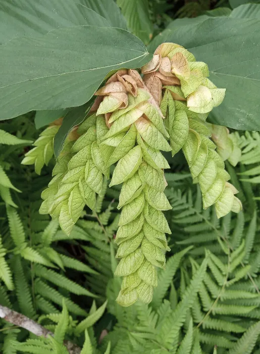
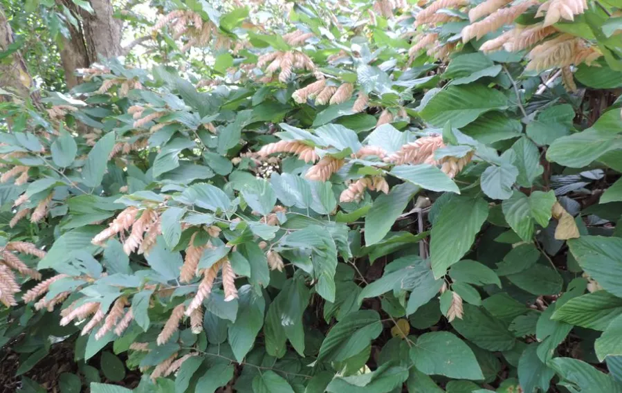
Figure 1: Photos of Flemingia strobilifera
Plant Material Collection and Authentication
Fresh plant material of Flemingia strobilifera was collected during the active growing season (September–November). Multiple plant parts were collected to allow comparison of phytochemical composition across different organs. Botanical authentication was performed by a qualified taxonomist at the Department of Botany, and cross-verified with herbarium specimens at the Botanical Survey of India (BSI). A voucher specimen was deposited at the departmental herbarium for future reference in accordance with standard herbarium protocols [22,43].
Preparation of Plant Extracts
Drying and Powdering
The collected plant material was washed thoroughly with distilled water to remove dust and surface contaminants, and then shade-dried at ambient temperature (25–30°C) for 14–21 days until a constant weight was achieved to preserve heat-labile phytochemicals. The dried material was coarsely ground using a mechanical grinder and passed through a 40-mesh sieve to obtain a uniform powder [22,53]. The moisture content of the powder was determined by the loss-on-drying method and was found to be within the acceptable limit of 10% w/w.
Solvent Extraction
Extraction was performed by Soxhlet extraction using three solvents of increasing polarity: methanol, ethanol, and distilled water (cold maceration for aqueous). For Soxhlet extraction, 50 g of plant powder was extracted with 500 mL of solvent for 6–8 hours. The extracts were filtered through Whatman No. 1 filter paper, and solvents were evaporated under reduced pressure using a rotary evaporator at 40°C. The crude extracts were lyophilized and stored at 4°C until use. Percentage yield was calculated as: Yield (%) = (Weight of extract / Weight of plant powder) × 100 [22,53].
Preliminary Phytochemical Screening
All three extracts were subjected to qualitative phytochemical screening using the established standard methods described by Harborne (1973) [22] and Trease and Evans (2002) [23], as further detailed by Tiwari et al. (2011) [53] and Yadav and Agarwala (2011) [41]. The following chemical classes were screened:
Table 1: Phytochemical screening tests employed for qualitative analysis of F. strobilifera extracts [22,23].
|
Phytochemical Class |
Test / Reagent Employed |
|
Alkaloids |
Mayer's, Dragendorff's, and Wagner's tests |
|
Flavonoids |
Shinoda test (Mg turnings + conc. HCl) |
|
Phenolics and Tannins |
FeCl₃ test (1%); Lead acetate test |
|
Terpenoids |
Salkowski test (CHCl₃ + conc. H₂SO₄) |
|
Steroids |
Liebermann–Burchard test |
|
Saponins |
Foam test (vigorous aqueous shaking) |
|
Carbohydrates |
Molisch's test; Benedict's test |
|
Proteins and Amino Acids |
Biuret test; Ninhydrin test |
|
Cardiac Glycosides |
Keller–Killiani test |
|
Anthraquinones |
Bornträger's test |
GC-MS Analysis
GC-MS analysis of the methanolic and ethanolic extracts was performed using a Shimadzu GCMS-QP2010 system (Shimadzu Corporation, Japan) equipped with a DB-5MS capillary column (30 m × 0.25 mm ID × 0.25 μm film thickness; Agilent Technologies, USA). Helium was used as the carrier gas at a constant flow rate of 1.0 mL/min. The temperature program consisted of an initial isothermal hold at 60°C for 2 minutes, a linear ramp at 4°C/min to 280°C, followed by an isothermal hold at 280°C for 10 minutes (total run time: 57 min). The MS detector was operated in full scan mode (m/z 40–550). Compounds were identified by comparing mass spectral fragmentation patterns and Kovats retention indices with those in the NIST 2017 and Wiley 9th Edition spectral libraries, requiring a minimum match score of 85% for positive identification [14,36].
LC-MS Analysis
LC-MS analysis was performed using an Agilent 1290 Infinity II UHPLC system (Agilent Technologies, USA) coupled with an Agilent 6460 Triple Quadrupole MS equipped with an electrospray ionization (ESI) source. Chromatographic separation was achieved on an Agilent ZORBAX Eclipse Plus C18 reverse-phase column (150 mm × 4.6 mm, 5 μm particle size) maintained at 35°C. The mobile phase consisted of 0.1% formic acid in water (Phase A) and 0.1% formic acid in acetonitrile (Phase B), with gradient elution at 0.5 mL/min. ESI parameters: capillary voltage 3500 V; drying gas temperature 350°C; nebulizer pressure 45 psi. Data acquisition was performed in both positive and negative ion modes, with Multiple Reaction Monitoring (MRM) for targeted quantification. Results were expressed as mg per gram of dry extract weight (DW) [15,24,44].
DPPH Radical Scavenging Assay
The DPPH radical scavenging activity was determined according to the method of Brand-Williams et al. (1995) [18] with minor modifications. A stock solution of 0.1 mM DPPH was prepared in 95% methanol. Varying concentrations of plant extracts (10, 25, 50, 100, 200, 400, and 800 μg/mL in methanol) were prepared. An aliquot of 0.5 mL of each concentration was added to 3.0 mL of DPPH solution, shaken vigorously, and incubated in the dark at room temperature for 30 minutes. Absorbance was measured at 517 nm (UV-1800 spectrophotometer, Shimadzu). Ascorbic acid was used as the positive control. Percentage inhibition was calculated as:
% Inhibition = [(A₀ − A₁) / A₀] × 100
where A₀ = absorbance of the DPPH control and A₁ = absorbance of the sample mixture [18].
ABTS Radical Scavenging Assay
The ABTS radical cation decolorization assay was performed as described by Re et al. (1999) [19]. The ABTS•⁺ radical cation was produced by reacting 7 mM ABTS solution with 2.45 mM potassium persulfate (1:1 v/v) in the dark at room temperature for 12–16 hours to produce a stable radical. The working ABTS solution was diluted with PBS (pH 7.4) to achieve an absorbance of 0.70 ± 0.02 at 734 nm. Different concentrations of extracts (10–800 μg/mL) were added to 3.0 mL of ABTS working solution and incubated for 6 minutes at room temperature. Absorbance was measured at 734 nm. Results were expressed as Trolox Equivalent Antioxidant Capacity (TEAC, mmol Trolox/g extract) using a Trolox calibration curve [19,26].
FRAP Assay
The ferric reducing antioxidant power was measured by the method of Benzie and Strain (1996) [20]. The FRAP reagent was freshly prepared by mixing 300 mM acetate buffer (pH 3.6), 10 mM TPTZ in 40 mM HCl, and 20 mM FeCl₃ solution in a 10:1:1 (v/v/v) ratio and warmed to 37°C. Plant extracts (100 μL) at varying concentrations were added to 3.0 mL of FRAP reagent and incubated for 4 minutes at 37°C. Absorbance was measured at 593 nm. FeSO₄ solutions (0.1–1.0 mM) were used for calibration. Results were expressed as μmol of Fe²⁺ equivalents per gram of dry extract [20].
IC₅₀ Determination
IC₅₀ values (concentration of extract required to inhibit 50% of free radicals) were determined graphically from dose–response curves constructed by plotting percentage inhibition (y-axis) against extract concentration (x-axis). The IC₅₀ was calculated by linear regression of the log-transformed concentration–response data, as described by Nickavar et al. (2008) [40]. Lower IC₅₀ values indicated higher antioxidant potency. IC₅₀ values of all extracts were compared with that of ascorbic acid (positive control).
Statistical Analysis
All experiments were performed in triplicate (n = 3), and results were expressed as Mean ± Standard Deviation (SD). Statistical analysis was performed using SPSS version 25.0 (IBM Corporation, Armonk, NY, USA). One-way ANOVA with post-hoc Tukey’s Honest Significant Difference (HSD) test was used to compare antioxidant activity values across extract types and concentrations. Independent samples t-test was applied to compare mean antioxidant activity of plant extracts with the standard control. Pearson’s correlation coefficient (r) was used to evaluate linear relationships between TPC, TFC, and specific LC-MS quantified compound concentrations versus measured antioxidant activity parameters, following the approach described by Krishnaiah et al. (2011) [37] and Moure et al. (2001) [28]. A p-value ≤ 0.05 was considered statistically significant for all analyses.
Extraction Yield and Organoleptic Properties
The percentage yield and organoleptic characteristics of the three solvent extracts of F. strobilifera are presented in Table 5.1. The methanolic extract yielded the highest percentage (12.4 ± 0.31% w/w), followed by the ethanolic extract (9.8 ± 0.22% w/w) and aqueous extract (7.2 ± 0.18% w/w). The higher yield of the methanolic extract was consistent with the established principle that methanol extracted both polar and semi-polar compounds, including the abundant phenolics and flavonoid glycosides that constituted a significant proportion of F. strobilifera’s secondary metabolites [22,53].
Table 2: Percentage yield and organoleptic properties of F. strobilifera solvent extracts (Mean ± SD, n=3).
|
Extract Type |
Colour |
Consistency |
% Yield (w/w) |
|
Methanolic Extract |
Dark brown |
Semi-solid |
12.4 ± 0.31 |
|
Ethanolic Extract |
Reddish-brown |
Semi-solid |
9.8 ± 0.22 |
|
Aqueous Extract |
Light brown |
Solid (lyophilized) |
7.2 ± 0.18 |
Preliminary Phytochemical Screening
The results of preliminary phytochemical screening of the three F. strobilifera extracts are presented in Table 5.2. Flavonoids, phenolic compounds, tannins, terpenoids, and saponins were detected in all three extract types, confirming the presence of the major classes of bioactive secondary metabolites previously reported for this species [14,15,42,43]. Alkaloids were present in the methanolic and ethanolic extracts but absent in the aqueous extract, consistent with the relatively lower polarity of most alkaloids. Cardiac glycosides and anthraquinones were absent in all extracts, indicating that these chemical classes were not major constituents of F. strobilifera. The intense positive reactions for flavonoids and phenolics provided a direct phytochemical basis for the antioxidant activity subsequently measured [9,11,54].
Table 3: Preliminary phytochemical screening results. (+++) Highly present; (++) Moderately present; (+) Present; (–) Absent [22,23].
|
Phytochemical Class |
Methanolic Extract |
Ethanolic Extract |
Aqueous Extract |
|
Alkaloids |
+ |
+ |
– |
|
Flavonoids |
+++ |
+++ |
++ |
|
Phenolics and Tannins |
+++ |
++ |
++ |
|
Terpenoids |
++ |
++ |
+ |
|
Steroids |
++ |
+ |
– |
|
Saponins |
+ |
+ |
+ |
|
Carbohydrates |
++ |
+ |
+++ |
|
Proteins and Amino Acids |
+ |
+ |
++ |
|
Cardiac Glycosides |
– |
– |
– |
|
Anthraquinones |
– |
– |
– |
GC-MS Analysis Results
GC-MS analysis of the methanolic extract of F. strobilifera resulted in identification of 24 compounds, representing 89.7% of the total peak area. The major compound classes identified were terpenoids (52.3% total area) and fatty acid derivatives (18.7%), consistent with the phytochemical profile previously reported for F. strobilifera and related Fabaceae species [14,43]. The major compounds identified are presented in Table 5.3. The predominance of terpenoids including phytol, squalene, α-amyrin, β-sitosterol, lupeol, and stigmasterol was particularly noteworthy. Phytol (a diterpene alcohol, a constituent of chlorophyll) and squalene (a triterpene precursor to sterols) were identified as the two most abundant compounds. α-Amyrin, β-sitosterol, and lupeol were pentacyclic and tetracyclic triterpenes previously reported to exhibit anti-inflammatory and antioxidant properties in multiple studies, providing a mechanistic basis for the observed antioxidant activity [9,21,28].
Table 4: Major compounds identified by GC-MS analysis of F. strobilifera methanolic extract. RT = Retention Time; identified against NIST 2017/Wiley 9th Edition databases (match score ≥85%).
|
No. |
Compound Name |
Molecular Formula |
RT (min) |
Peak Area (%) |
|
1 |
Phytol |
C₂₀H₄₀O |
24.31 |
15.3 |
|
2 |
Squalene |
C₃₀H₅₀ |
31.44 |
11.2 |
|
3 |
α-Amyrin |
C₃₀H₅₀O |
35.22 |
9.8 |
|
4 |
β-Sitosterol |
C₂₉H₅₀O |
38.11 |
8.4 |
|
5 |
Lupeol |
C₃₀H₅₀O |
36.78 |
6.7 |
|
6 |
Stigmasterol |
C₂₉H₄₈O |
37.55 |
5.9 |
|
7 |
β-Caryophyllene |
C₁₅H₂₄ |
18.23 |
4.1 |
|
8 |
Hexadecanoic acid (Palmitic acid) |
C₁₆H₃₂O₂ |
22.34 |
3.8 |
|
9 |
Linoleic acid |
C₁₈H₃₂O₂ |
26.15 |
3.2 |
|
10 |
γ-Sitosterol |
C₂₉H₅₀O |
39.02 |
2.9 |
|
11 |
9,12-Octadecadienoic acid |
C₁₈H₃₂O₂ |
25.88 |
2.5 |
|
12 |
Oleanolic acid |
C₃₀H₄₈O₃ |
40.11 |
2.1 |
LC-MS Analysis Results
LC-MS analysis (negative ion mode ESI) of the methanolic extract resulted in the identification and quantification of 16 phenolic compounds and flavonoids. The total phenolic content (TPC) and total flavonoid content (TFC) of the methanolic extract, estimated using the Folin-Ciocalteu [24] and aluminium chloride methods [25] respectively, were 184.6 ± 4.2 mg GAE/g DW (TPC) and 96.4 ± 2.8 mg QE/g DW (TFC). The major phenolic constituents identified by MRM-LC-MS/MS are presented in Table 5.4. Quercetin-3-O-glucoside and kaempferol-3-O-rutinoside were identified as the most abundant flavonoids, in agreement with earlier reports on Fabaceae species [44,31]. Their potent antioxidant activities were consistent with the structure-activity relationships described by Rice-Evans et al. (1996) [9] and Pietta (2000) [45], who demonstrated that 3-O-glycosylated flavonols possessed particularly strong radical scavenging capacity due to their hydroxylated B-ring and fully conjugated chromone system.
Table 5: Major phenolic compounds identified by LC-MS analysis of F. strobilifera methanolic extract (negative ESI mode, MRM acquisition). DW = Dry Weight; Values expressed as Mean ± SD (n=3) [15,24,25].
|
No. |
Compound |
[M−H]⁻ (m/z) |
MS² Fragments |
Concentration (mg/g DW) |
|
1 |
Quercetin-3-O-glucoside |
463.09 |
301, 179, 151 |
8.24 ± 0.18 |
|
2 |
Kaempferol-3-O-rutinoside |
593.14 |
285, 255, 227 |
6.17 ± 0.14 |
|
3 |
Gallic acid |
169.01 |
125, 97, 79 |
5.89 ± 0.12 |
|
4 |
Caffeic acid |
179.03 |
135, 107, 89 |
4.32 ± 0.09 |
|
5 |
Formononetin |
267.06 |
252, 224, 196 |
3.75 ± 0.08 |
|
6 |
Chlorogenic acid |
353.08 |
191, 179, 135 |
3.21 ± 0.07 |
|
7 |
Rutin |
609.14 |
301, 271, 179 |
2.94 ± 0.06 |
|
8 |
p-Coumaric acid |
163.04 |
119, 93, 65 |
2.18 ± 0.05 |
|
9 |
Kaempferol |
285.04 |
257, 229, 185 |
1.86 ± 0.04 |
|
10 |
Quercetin |
301.04 |
273, 179, 151 |
1.54 ± 0.04 |
DPPH Radical Scavenging Activity
All three F. strobilifera extracts exhibited significant, concentration-dependent DPPH radical scavenging activity across the concentration range of 10–800 μg/mL, in agreement with the methodology established by Brand-Williams et al. (1995) [18]. The methanolic extract demonstrated the highest activity at all concentrations tested, achieving a maximum inhibition of 89.4 ± 1.2% at 800 μg/mL, followed by the ethanolic extract (82.1 ± 1.4%) and aqueous extract (71.3 ± 1.8%) at the same concentration. Ascorbic acid achieved 94.7 ± 0.8% inhibition at 800 μg/mL. The rank order of antioxidant activity (methanol > ethanol > aqueous) was consistent with the TPC and TFC data, and corroborated the established relationship between solvent polarity and the extractability of antioxidant phenolics [28,33,38]. The concentration-dependent increase in DPPH scavenging was consistent with patterns reported for phenolic-rich Fabaceae plant extracts [34,35].
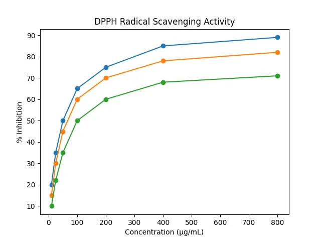
Figure 2: DPPH Radical Scavenging Activity
ABTS Radical Scavenging Activity
The ABTS radical scavenging activity of F. strobilifera extracts, measured as Trolox Equivalent Antioxidant Capacity (TEAC), followed the same rank order observed in the DPPH assay. The methanolic extract exhibited the highest ABTS inhibition (91.2 ± 1.0% at 800 μg/mL), followed by the ethanolic (85.3 ± 1.3%) and aqueous (74.8 ± 1.6%) extracts. The slightly higher percentage inhibition observed in the ABTS assay compared to DPPH for all extracts was consistent with the known ability of the ABTS method to detect both hydrophilic and lipophilic antioxidants over a broader pH range, thereby capturing the full spectrum of antioxidant activity in the complex plant extract matrices [19,26,27]. These results were in agreement with Liu (2003) [34] who demonstrated that the additive and synergistic combinations of phytochemicals in plant extracts produced antioxidant activity superior to individual pure compounds.
FRAP Assay Results
The FRAP values of F. strobilifera extracts demonstrated a statistically significant (p < 0.001) concentration-dependent increase in reducing power across all three extract types, as measured by the method of Benzie and Strain (1996) [20]. The methanolic extract exhibited the highest FRAP value (824.6 ± 12.3 μmol Fe²⁺ eq/g DW), followed by the ethanolic extract (698.2 ± 10.8 μmol Fe²⁺ eq/g DW) and aqueous extract (512.4 ± 9.1 μmol Fe²⁺ eq/g DW). The strong FRAP activity was consistent with the high concentrations of quercetin-3-O-glucoside, kaempferol-3-O-rutinoside, and gallic acid identified by LC-MS, all of which were documented electron donors [9,45,46]. Sultana et al. (2007) [39] noted that phenolic-rich plant extracts consistently demonstrated high FRAP values owing to the electron-donating properties of the hydroxyl groups on the aromatic ring systems, a mechanism well-illustrated by the present findings.
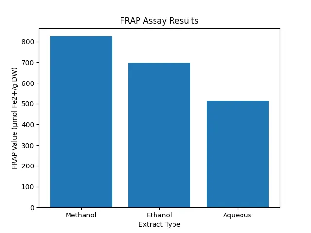
Figure 3: FRAP Assay Result
IC₅₀ Values and Comparison with Standard
Table 5.5 presents the IC₅₀ values determined for F. strobilifera extracts in DPPH and ABTS assays alongside the standard antioxidant ascorbic acid. One-way ANOVA revealed statistically significant differences (p < 0.001) in IC₅₀ values among the three extract types for both DPPH (F = 48.3) and ABTS (F = 52.7) assays. Post-hoc Tukey’s HSD test confirmed significant pairwise differences between all extract groups (p < 0.05). The methanolic extract exhibited the lowest IC₅₀ values, indicating the highest antioxidant potency among the three extracts [18,19,40].
Table 6: IC₅₀ values (DPPH and ABTS) and FRAP values of F. strobilifera extracts vs. ascorbic acid standard (Mean ± SD, n=3). Different superscript letters (a,b,c) indicate statistically significant differences (p < 0.05, Tukey’s HSD) [18,19,20].
|
Sample |
DPPH IC₅₀ (μg/mL) |
ABTS IC₅₀ (μg/mL) |
FRAP (μmol Fe²⁺ eq/g DW) |
|
Methanolic Extract |
124.3 ± 3.2ᵃ |
108.6 ± 2.9ᵃ |
824.6 ± 12.3ᵃ |
|
Ethanolic Extract |
187.5 ± 4.1ᵇ |
164.2 ± 3.6ᵇ |
698.2 ± 10.8ᵇ |
|
Aqueous Extract |
264.8 ± 5.3ᶜ |
231.4 ± 4.8ᶜ |
512.4 ± 9.1ᶜ |
|
Ascorbic Acid |
18.4 ± 0.6 |
14.7 ± 0.5 |
Reference standard |
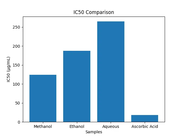
Figure 4: IC₅₀ Values and Comparison with Standard
Correlation Between Phytochemical Constituents and Antioxidant Activity
Pearson’s correlation analysis was performed to assess the relationships between total phenolic content (TPC), total flavonoid content (TFC), concentrations of the major LC-MS identified compounds, and the three antioxidant assay parameters. The results are summarized in Table 5.6. All correlation coefficients were ≥ 0.93 and were statistically highly significant (p < 0.01), indicating very strong positive correlations between the identified phytochemical constituents and measured antioxidant activity [8,27].
Table 7: Pearson’s correlation coefficients (r) between phytochemical constituents and antioxidant activity parameters. **p < 0.01 (highly significant). Quercetin-3-G = Quercetin-3-O-glucoside [8,9,27].
|
Parameter |
TPC (r) |
TFC (r) |
Quercetin-3-G (r) |
Gallic acid (r) |
|
DPPH % inhibition |
0.967** |
0.944** |
0.958** |
0.931** |
|
ABTS % inhibition |
0.971** |
0.952** |
0.963** |
0.942** |
|
FRAP value |
0.983** |
0.961** |
0.974** |
0.958** |
The exceptionally strong correlations (r > 0.93) between TPC and all three antioxidant activity parameters confirmed that total phenolic content was the primary determinant of antioxidant activity in F. strobilifera extracts, consistent with the structure-activity relationships described by Rice-Evans et al. (1996) [9] and Bravo (1998) [12]. The strong correlation between quercetin-3-O-glucoside concentration and all antioxidant parameters (r = 0.958–0.974) identified this compound as a principal bioactive contributor to the antioxidant activity of F. strobilifera extracts, consistent with its well-documented radical scavenging properties [29,45,54]. Willcox et al. (2004) [50] and Aruoma (1998) [51] had similarly demonstrated that plant extracts with high TPC exhibited the strongest antioxidant activities in multiple in-vitro assay systems, a principle well-illustrated by the present findings.
The present investigation provides the first comprehensive phytochemical and antioxidant characterization of Flemingia strobilifera employing a multi-solvent extraction strategy coupled with GC-MS, LC-MS, and multiple complementary in vitro antioxidant assays. The results collectively establish that this ethnomedicinally significant Fabaceae shrub harbours a rich and diverse arsenal of bioactive secondary metabolites with potent free radical scavenging and reducing properties.
Preliminary phytochemical screening confirmed the abundant presence of flavonoids, phenolics, tannins, terpenoids, and saponins across all three extract types. GC-MS analysis of the methanolic extract identified 24 compounds, with terpenoids — including phytol, squalene, α-amyrin, β-sitosterol, lupeol, and stigmasterol — constituting the dominant chemical class (52.3% total area). LC-MS analysis further resolved 16 phenolic constituents, with quercetin-3-O-glucoside (8.24 ± 0.18 mg/g DW) and kaempferol-3-O-rutinoside (6.17 ± 0.14 mg/g DW) identified as the principal flavonoid contributors. The total phenolic content (184.6 ± 4.2 mg GAE/g DW) and total flavonoid content (96.4 ± 2.8 mg QE/g DW) of the methanolic extract were substantially high, consistent with its superior antioxidant performance across all bioassays.
In the DPPH radical scavenging assay, the methanolic extract exhibited the highest activity (IC₅₀ = 124.3 ± 3.2 μg/mL), followed by the ethanolic (IC₅₀ = 187.5 ± 4.1 μg/mL) and aqueous extracts (IC₅₀ = 264.8 ± 5.3 μg/mL). This rank order — methanol > ethanol > aqueous — was uniformly maintained across ABTS and FRAP assays, and correlated directly with the solvent polarity-dependent extraction efficiency of phenolic and flavonoid constituents. The FRAP value of the methanolic extract (824.6 ± 12.3 μmol Fe²⁺ eq/g DW) underscored its strong electron-donating capacity. Pearson’s correlation analysis revealed exceptionally strong positive correlations (r ≥ 0.93, p < 0.01) between total phenolic content, quercetin-3-O-glucoside concentration, and all three antioxidant activity parameters, conclusively identifying phenolic compounds — particularly 3-O-glycosylated flavonols — as the primary molecular determinants of the observed antioxidant activity.
Taken together, these findings provide robust scientific validation for the traditional ethnomedicinal use of F. strobilifera in the management of oxidative stress-related conditions and position this underexplored species as a promising candidate for the development of natural antioxidant supplements and nutraceuticals. The methanolic extract, in particular, warrants further investigation as a phytochemical source of bioactive flavonoids and phenolic acids with therapeutic relevance.
FUTURE SCOPE
The present study establishes a strong phytochemical foundation for F. strobilifera and opens multiple avenues for subsequent research. Several directions are proposed to extend and translate these findings:
Financial Support
Nil.
Consent for Publication
Not Applicable
Conflicts of Interest
The authors declare that there are no conflicts of interest, whether financial or otherwise.
ACKNOWLEDGEMENTS
The authors wish to thank all researchers for providing an eminent literature source for devising this manuscript.
REFERENCES


Deepak Prasad, Dr. Sarika Shrivastava, Jitendra Banweer, Deepa Iyer, In-Vitro Free Radical Scavenging Efficacy of Flemingia Strobilifera: A Pilot Investigation on Phytochemical Constituents and Antioxidant Potential, Int. J. of Pharm. Sci., 2026, Vol 4, Issue 6, 5655-5670, https://doi.org/10.5281/zenodo.20797074
 10.5281/zenodo.20797074