

We use cookies to ensure our website works properly and to personalise your experience. Cookies policy
1,2,3 Sagar College of Pharmacy, Barabanki, Uttar Pradesh, India
4,5,6 J.S. Singh Institute of Pharmacy, Sitapur, Uttar Pradesh, India 261207
The present study investigated the in vivo anti-inflammatory and wound-healing potential of a hydroalcoholic extract of Matricaria chamomilla L. flower heads. The extract was subjected to preliminary phytochemical screening and evaluated using carrageenan-induced paw edema, excision wound and incision wound models in Wistar rats. The extractive yield was 15.84% w/w, with positive reactions for flavonoids, phenolics, terpenoids, tannins and other phytochemical constituents. In the acute inflammation model, the extract reduced paw edema in a dose-dependent manner, with the 400 mg/kg dose showing greater activity than the 200 mg/kg dose. In wound-healing models, topical M. chamomilla extract improved wound contraction, shortened the epithelialization period, and increased tensile strength and hydroxyproline content. Biochemical evaluation showed reductions in TNF-?, IL-6, MPO and MDA levels following treatment. Histopathological evaluation further demonstrated improved epithelial continuity, fibroblast proliferation and collagen deposition, particularly in the 10% MCE-treated group. Overall, the findings demonstrate the promising anti-inflammatory and wound-healing potential of hydroalcoholic extract of M. chamomilla L. and support its further investigation as a potential herbal therapeutic candidate.
Inflammation is a protective biological response to tissue injury, infection, or irritation. Although the inflammatory response is essential for tissue defense and repair, excessive or prolonged inflammation may cause tissue damage and delay wound healing.[9,15] Wound healing is a complex and highly regulated process involving overlapping phases of haemostasis, inflammation, proliferation, collagen deposition, epithelialization, and tissue remodeling.
Matricaria chamomilla L., commonly known as German chamomile, belongs to the family Asteraceae. The dried flower heads are the principal medicinal part and contain several bioactive constituents, including flavonoids, phenolic acids, terpenoids, coumarins, and volatile-oil components such as α-bisabolol and chamazulene. These constituents have been associated with anti-inflammatory, antioxidant, and tissue-repair-related activities.[9]
Previous experimental studies have reported the potential of M. chamomilla in inflammatory conditions and wound repair. However, the anti-inflammatory and wound-healing effects may vary depending on the plant material, extraction method, dose, experimental model, and outcome parameters.[23] The thesis specifically identifies the need for an integrated evaluation combining inflammatory and wound-healing endpoints.
Therefore, the present study was designed to evaluate the in vivo anti-inflammatory and wound-healing activities of a hydroalcoholic extract of M. chamomilla flower heads. Anti-inflammatory activity was assessed using the carrageenan-induced paw edema model, while wound-healing activity was evaluated using excision and incision wound models in Wistar rats. Wound contraction, epithelialization period, tensile strength, hydroxyproline content, inflammatory and oxidative stress markers, and histopathological changes were considered to obtain an integrated assessment of the pharmacological effects of the extract.[24]
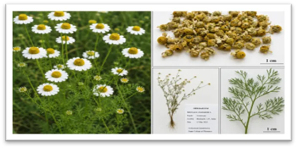
Figure 1. Matricaria chamomilla flower heads / authenticated plant material
Matricaria chamomilla flower heads and authenticated plant material. The characteristic white ray florets and yellow central disc florets of M. chamomilla are shown along with the collected plant material used for extraction.
2. MATERIALS AND METHODS
2.1 Plant Material and Extraction
Dried flower heads of Matricaria chamomilla L. were procured from an authenticated herbal source. The plant material was cleaned to remove foreign matter and stored in an airtight container protected from direct sunlight and moisture. The dried flower heads were coarsely powdered and passed through sieve No. 40. A total of 250 g of powdered material was extracted with 2.5 L of 70% ethanol by maceration for 72 h with intermittent shaking. The extract was filtered through muslin cloth followed by Whatman filter paper.[19] The marc was re-macerated once with fresh solvent, and the combined filtrate was concentrated under reduced pressure below 45°C and finally dried in a vacuum desiccator. The percentage yield was calculated based on the weight of the dried extract relative to the starting crude drug.[19,12]
2.2 Preliminary Phytochemical Screening
The hydroalcoholic extract was subjected to preliminary qualitative phytochemical screening for alkaloids, flavonoids, phenolics, tannins, saponins, glycosides, steroids and terpenoids using standard phytochemical tests.
2.3 Experimental Animals and Ethical Considerations
Wistar rats were used for the in vivo pharmacological studies. The animals were maintained under standard laboratory conditions and experimental procedures were performed in accordance with applicable CPCSEA guidelines and after approval from the Institutional Animal Ethics Committee (IAEC). [PBRI/IAEC/27-11-24/019]
2.4 Carrageenan-Induced Paw Edema
The extract and standard drug were administered orally using 0.5% sodium carboxymethyl cellulose (CMC) as the vehicle. One hour after treatment, acute inflammation was induced by sub-plantar injection of 0.1 mL of 1% w/v carrageenan suspension in normal saline into the right hind paw. Paw volume was measured using a plethysmometer at 0, 1, 2, 3, 4 and 5 h after carrageenan administration. Percentage inhibition of edema was calculated in comparison with the disease-control group.[24-29]
2.5 Excision Wound Model
Animals were anesthetized and the dorsal skin was shaved and disinfected. A circular full-thickness excision wound of approximately 500 mm² was created on the dorsal thoracic region using sterile surgical instruments.[9] The animals were assigned to control ointment, standard povidone-iodine, 5% w/w MCE ointment and 10% w/w MCE ointment groups. Treatments were applied topically once daily. Wound areas were recorded on days 0, 3, 7, 10 and 14, and percentage wound contraction was calculated relative to the initial wound area.[25]
2.6 Incision Wound Model
A 3 cm paravertebral full-thickness incision was made under anesthesia. The wound was sutured at 1 cm intervals and treated topically with the assigned formulations. Sutures were removed on day 8, and tensile strength was measured on day 10 using a tensiometer or continuous water-flow method. Increased tensile strength was considered indicative of improved collagen maturation and tissue repair.[13,25]
2.7 Biochemical and Histopathological Evaluation
TNF-α and IL-6 were evaluated as inflammatory markers, while MPO activity was considered an indicator of neutrophil infiltration and MDA as an indicator of lipid peroxidation. Hydroxyproline content was assessed as an indirect marker of collagen deposition. For histopathological evaluation, skin samples were fixed in 10% neutral buffered formalin, processed, sectioned and stained with hematoxylin and eosin. Histological parameters included inflammatory cell infiltration, fibroblast proliferation, collagen deposition and epithelialization.[17-18]
2.8 Statistical Analysis
Data were expressed as mean ± SD for six animals per group. The exact statistical test, post-hoc test and significance values should be reported according to the original statistical output. The thesis methodology mentions one-way or two-way ANOVA followed by Tukey's post-hoc test, with p < 0.05 considered statistically significant.
3. RESULTS AND DISCUSSION
3.1 Extract Yield and Phytochemical Profile
The hydroalcoholic extraction of Matricaria chamomilla flower heads was carried out using 70% ethanol. From 250 g of crude drug, 39.6 g of dried extract was obtained, corresponding to an extractive yield of 15.84% w/w. The prepared extract was dark brown in colour and possessed a characteristic aromatic odour, with a semisolid/sticky dried consistency.
Preliminary phytochemical screening indicated the presence of several major classes of secondary metabolites, including flavonoids, phenolics, terpenoids, tannins, alkaloids, saponins, glycosides and steroids. Flavonoids and phenolics showed strong positive reactions, whereas terpenoids and tannins showed moderate reactions. These phytochemical constituents may contribute to the observed anti-inflammatory and wound-healing effects of the extract.
Table 1. Extractive yield and basic properties of M. chamomilla extract
|
Parameter |
Observation |
|
Weight of crude drug |
250 g |
|
Solvent system |
70% ethanol |
|
Weight of dried extract |
39.6 g |
|
Percentage yield |
15.84% w/w |
|
Colour |
Dark brown |
|
Odour |
Characteristic aromatic |
|
Consistency |
Semisolid/sticky dried mass |
Table 2. Preliminary phytochemical screening of M. chamomilla extract
|
Phytochemical |
Test/Observation |
Result |
|
Alkaloids |
Mayer/Wagner: faint turbidity |
+ |
|
Flavonoids |
Shinoda: pink-red colour |
+++ |
|
Phenolics |
Ferric chloride: bluish-green |
+++ |
|
Terpenoids |
Salkowski: reddish-brown interface |
++ |
|
Tannins |
Ferric chloride: greenish-black |
++ |
|
Saponins |
Froth test: persistent foam |
+ |
|
Glycosides |
Keller-Killiani: brown ring |
+ |
|
Steroids |
Liebermann-Burchard: green colour |
+ |
Preliminary phytochemical screening of the hydroalcoholic extract of M. chamomilla. The extract showed strong positive reactions for flavonoids and phenolics, moderate reactions for terpenoids and tannins, and mild reactions for alkaloids, saponins, glycosides and steroids.
Note: + = mild positive reaction; ++ = moderate positive reaction; +++ = strong positive reaction.
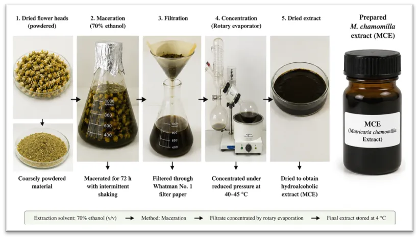
Figure 2. Extraction and prepared M. chamomilla extract
Preparation of hydroalcoholic extract of M. chamomilla. The extraction process involved powdered flower material, maceration with 70% ethanol, filtration, concentration and drying to obtain the hydroalcoholic M. chamomilla extract (MCE).
3.2 Anti-Inflammatory Activity
Carrageenan administration produced a progressive increase in paw edema in the disease-control group. Treatment with diclofenac sodium and M. chamomilla extract reduced paw edema compared with the disease-control group. The inhibitory effect of the extract increased with dose, with the 400 mg/kg MCE group showing greater activity than the 200 mg/kg group.
At 3 h, paw volume was 1.04 ± 0.06 mL in the disease-control group, whereas the values were 0.50 ± 0.06 mL for diclofenac, 0.68 ± 0.06 mL for MCE 200 mg/kg and 0.56 ± 0.06 mL for MCE 400 mg/kg.
Table 3. Effect of M. chamomilla extract on carrageenan-induced paw edema volume
|
Time (h) |
Normal control |
Disease control |
Diclofenac 10 mg/kg |
MCE 200 mg/kg |
MCE 400 mg/kg |
|
0 |
0.18 ± 0.02 |
0.19 ± 0.02 |
0.18 ± 0.02 |
0.18 ± 0.02 |
0.19 ± 0.02 |
|
1 |
0.19 ± 0.04 |
0.56 ± 0.04 |
0.38 ± 0.04 |
0.45 ± 0.04 |
0.41 ± 0.04 |
|
2 |
0.20 ± 0.05 |
0.82 ± 0.05 |
0.48 ± 0.05 |
0.60 ± 0.05 |
0.52 ± 0.05 |
|
3 |
0.20 ± 0.06 |
1.04 ± 0.06 |
0.50 ± 0.06 |
0.68 ± 0.06 |
0.56 ± 0.06 |
|
4 |
0.19 ± 0.05 |
0.92 ± 0.05 |
0.42 ± 0.05 |
0.58 ± 0.05 |
0.47 ± 0.05 |
|
5 |
0.18 ± 0.04 |
0.79 ± 0.04 |
0.34 ± 0.04 |
0.49 ± 0.04 |
0.39 ± 0.04 |
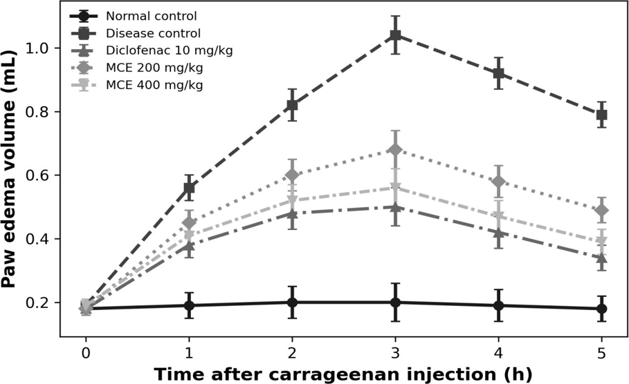
Figure 3. Effect of M. chamomilla extract on carrageenan-induced paw edema
Effect of M. chamomilla extract on carrageenan-induced paw edema in rats. Paw edema volume was measured at different time intervals following carrageenan administration. MCE treatment reduced paw edema compared with the disease-control group, with the 400 mg/kg dose showing greater activity than the 200 mg/kg dose.
3.3 Wound-Healing Activity
3.3.1 Excision Wound Model
Topical treatment with M. chamomilla extract enhanced wound contraction compared with the control ointment group. Wound contraction increased progressively with treatment duration in all groups. On day 14, the percentage wound contraction was 78.4 ± 2.6% in the control group, 96.5 ± 2.6% in the povidone iodine group, 90.7 ± 2.6% in the MCE 5% group and 95.2 ± 2.6% in the MCE 10% group. The 10% MCE formulation produced wound contraction close to that of the standard treatment, indicating a pronounced wound-healing response.
Table 4. Effect of M. chamomilla extract on percentage wound contraction
|
Day |
Control ointment |
Povidone iodine |
MCE 5% |
MCE 10% |
|
0 |
0.0 ± 0.0 |
0.0 ± 0.0 |
0.0 ± 0.0 |
0.0 ± 0.0 |
|
3 |
18.6 ± 2.1 |
31.4 ± 2.1 |
25.8 ± 2.1 |
29.6 ± 2.1 |
|
7 |
43.2 ± 3.4 |
67.9 ± 3.4 |
58.6 ± 3.4 |
64.3 ± 3.4 |
|
10 |
61.7 ± 3.1 |
84.8 ± 3.1 |
77.3 ± 3.1 |
83.6 ± 3.1 |
|
14 |
78.4 ± 2.6 |
96.5 ± 2.6 |
90.7 ± 2.6 |
95.2 ± 2.6 |
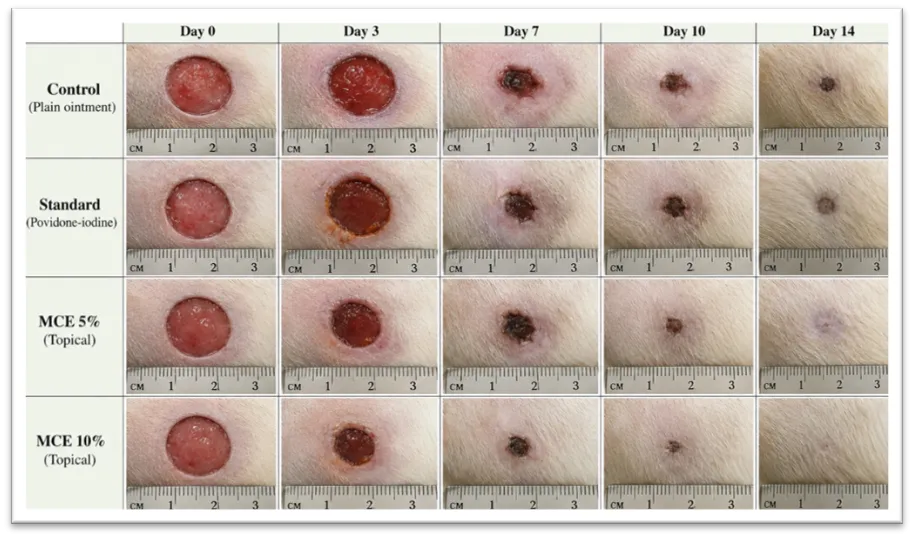
Figure 4. Representative photographs of excision wounds: Day 0, 3, 7, 10 and 14 (Control / Standard / MCE 5% / MCE 10%)
Representative photographs of excision wounds showing progressive wound healing on days 0, 3, 7, 10 and 14 in control, standard, MCE 5% and MCE 10% treated groups.
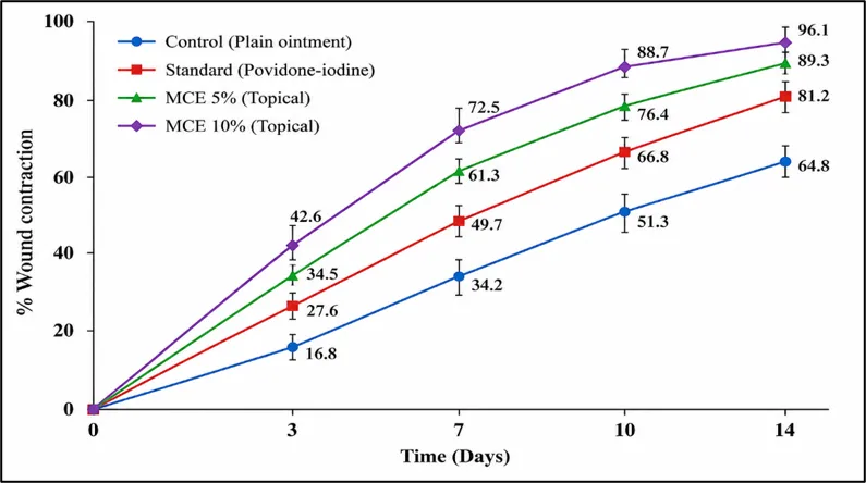
Figure 5. Percentage wound contraction versus time
Effect of M. chamomilla extract on percentage wound contraction in the excision wound model. Wound contraction increased progressively with treatment duration, with MCE 10% showing a response close to that of the standard treatment.
3.3.2 Epithelialization Period, Tensile Strength and Hydroxyproline Content
The wound-healing effect of M. chamomilla extract was further evaluated by determining the epithelialization period, tensile strength and hydroxyproline content. The epithelialization period was reduced in the extract-treated groups compared with the control ointment group. The MCE 10% formulation showed a shorter epithelialization period than MCE 5%.
Tensile strength was increased in the MCE-treated groups, indicating improved tissue strength and collagen maturation. Similarly, hydroxyproline content was higher in the extract-treated groups compared with the control group, suggesting enhanced collagen deposition during wound repair. The 10% MCE formulation showed better performance than the 5% formulation.
Table 5. Effect of M. chamomilla extract on epithelialization period, tensile strength and hydroxyproline content
|
Group |
Epithelialization period (days) |
Tensile strength (g) |
Hydroxyproline (µg/g tissue) |
|
Control ointment |
18.5 ± 1.1 |
318.4 ± 24.5 |
34.8 ± 3.6 |
|
Povidone iodine |
12.2 ± 0.8 |
522.7 ± 31.6 |
62.5 ± 4.1 |
|
MCE ointment 5% |
14.6 ± 0.9 |
451.8 ± 27.9 |
52.7 ± 3.8 |
|
MCE ointment 10% |
12.8 ± 0.7 |
506.5 ± 29.4 |
59.4 ± 4.0 |
Effect of M. chamomilla extract on epithelialization period, tensile strength and hydroxyproline content. Values are reported as mean ± SD.
The reduction in epithelialization period together with increased tensile strength and hydroxyproline content suggests enhanced collagen formation, maturation and overall tissue repair following topical MCE treatment. The comparatively better response of MCE 10% indicates a concentration-related wound-healing effect.
3.4 Biochemical Markers
The effect of M. chamomilla extract on inflammatory and oxidative stress markers was evaluated by determining TNF-α, IL-6, MPO and MDA levels. The disease/wound-control group showed higher levels of all four markers, indicating an enhanced inflammatory response, neutrophil accumulation and oxidative stress. Treatment with MCE reduced TNF-α, IL-6, MPO and MDA levels compared with the disease/wound-control group. The high-dose MCE group showed a greater reduction than the low-dose group, indicating a dose-related response.
Table 6. Effect of M. chamomilla extract on inflammatory and oxidative stress markers
|
Group |
TNF-α (pg/mL) |
IL-6 (pg/mL) |
MPO (U/g tissue) |
MDA (nmol/mg protein) |
|
Normal control |
38.4 |
42.6 |
1.22 |
2.18 |
|
Disease/ wound control |
104.8 |
118.5 |
4.86 |
6.74 |
|
Diclofenac/ standard |
52.7 |
59.4 |
2.05 |
3.02 |
|
MCE low dose |
68.5 |
76.8 |
2.84 |
4.21 |
|
MCE high dose |
56.2 |
63.5 |
2.22 |
3.36 |
Values are presented as reported in the source thesis. TNF-α and IL-6 are expressed in pg/mL, MPO in U/g tissue, and MDA in nmol/mg protein.
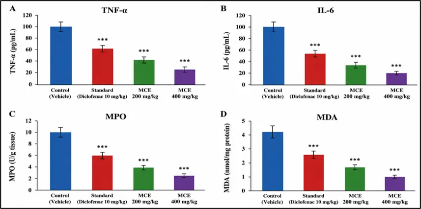
Figure 6. Effect of treatment on TNF-α, IL-6, MPO and MDA
Effect of treatment on TNF-α, IL-6, MPO and MDA. The disease/wound-control group showed elevated levels of inflammatory and oxidative stress markers, whereas MCE treatment reduced these markers, with a greater response observed at the higher dose.
3.5 Histopathological Findings
Histopathological examination was performed to assess tissue organization and the extent of inflammatory cell infiltration, fibroblast proliferation, collagen deposition and epithelialization in the healed skin. The control ointment group showed comparatively higher inflammatory cell infiltration with lower fibroblast proliferation, collagen deposition and epithelialization scores. In contrast, the povidone iodine and MCE-treated groups showed improved histological features. The MCE 10% group showed reduced inflammatory cell infiltration along with increased fibroblast proliferation, collagen deposition and epithelialization, indicating improved tissue repair.
Table 7. Semi-quantitative histopathological scoring of healed skin
|
Group |
Inflammatory cells (0–3) |
Fibroblast proliferation (0–3) |
Collagen deposition (0–3) |
Epithelialization (0–3) |
|
Control ointment |
3 |
1 |
1 |
1 |
|
Povidone iodine |
1 |
3 |
3 |
3 |
|
MCE ointment 5% |
2 |
2 |
2 |
2 |
|
MCE ointment 10% |
1 |
3 |
3 |
3 |
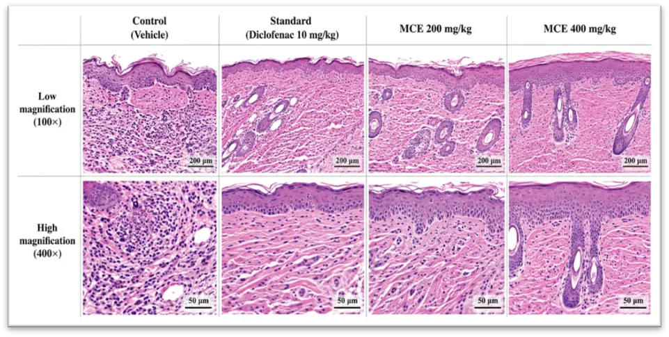
Figure 7. Representative H&E histopathological images of healed skin (4 groups)
Representative H&E histopathological images of healed skin from the experimental groups. MCE-treated groups showed improved epithelialization, fibroblast proliferation and collagen deposition with reduced inflammatory cell infiltration compared with the control group.
4. DISCUSSION
The present study demonstrated that the hydroalcoholic extract of Matricaria chamomilla L. possesses promising anti-inflammatory and wound-healing properties in experimental animal models. The extract showed a dose-related reduction in carrageenan-induced paw edema, indicating its potential to suppress the acute inflammatory response. The higher activity observed with MCE 400 mg/kg compared with MCE 200 mg/kg suggests a concentration-dependent pharmacological response.
The wound-healing results further supported the activity of the extract. Topical administration of MCE increased the rate of wound contraction and improved the overall healing process. The 10% MCE formulation produced a response close to that of povidone-iodine by day 14.
The reduction in epithelialization period together with increased tensile strength and hydroxyproline content indicates improved tissue repair and collagen maturation. The 10% MCE formulation showed better performance than the 5% formulation, supporting a dose-related wound-healing response.
Biochemical findings provided additional evidence for the anti-inflammatory and antioxidant effects of the extract. The disease/wound-control group showed elevated TNF-α, IL-6, MPO and MDA levels, whereas MCE treatment reduced these markers. This suggests that the extract may attenuate inflammatory mediator release, neutrophil-associated activity and oxidative stress during tissue repair.
Histopathological findings were consistent with the biochemical and macroscopic observations. MCE-treated groups showed reduced inflammatory cell infiltration along with improved fibroblast proliferation, collagen deposition and epithelialization. The 10% MCE group showed a particularly favorable histological profile.
The observed pharmacological effects may be associated with the phytochemical constituents of M. chamomilla, particularly flavonoids, phenolic compounds and terpenoid constituents. These phytochemical classes have been discussed in the thesis as possible contributors to modulation of inflammatory and oxidative processes and tissue repair.
Overall, the combined findings from the acute inflammation, wound-healing, biochemical and histopathological assessments provide preliminary evidence supporting the potential of hydroalcoholic M. chamomilla extract for further investigation in inflammatory and wound-related conditions.
5. CONCLUSION
The present study demonstrated that the hydroalcoholic extract of Matricaria chamomilla L. possesses promising anti-inflammatory and wound-healing activities in experimental animal models. The extract produced a dose-dependent reduction in carrageenan-induced paw edema, with the 400 mg/kg dose showing greater activity than the 200 mg/kg dose. Topical application of MCE improved wound contraction and was associated with a shorter epithelialization period, increased tensile strength and higher hydroxyproline content. The biochemical findings further indicated reductions in TNF-α, IL-6, MPO and MDA levels, while histopathological evaluation demonstrated improved epithelialization, fibroblast proliferation and collagen deposition, particularly with the 10% MCE formulation.
Overall, these findings support the pharmacological potential of M. chamomilla as a promising herbal candidate for further investigation in inflammatory and wound-healing conditions. Further studies involving extract standardization, detailed phytochemical characterization, mechanistic investigations and controlled experimental studies are warranted to establish its therapeutic potential.
6. DECLARATIONS
Ethics approval: The animal experiments were conducted in accordance with CPCSEA guidelines and were approved by the Institutional Animal Ethics Committee (IAEC). [PBRI/IAEC/27-11-24/019]
Funding: This research work did not receive any specific grant from funding agencies in the public, commercial, or not-for-profit sectors.
Conflict of interest: The authors declare that they have no conflict of interest.
Data availability: The data supporting the findings of this study are available from the corresponding author upon reasonable request.
Author contributions: Arvind Kumar Verma¹: Conceptualization, methodology, Investigation, Data curation, Writing – original draft. Santosh Kumar Mishra²: Supervision, Validation, Writing – review & editing. Amrita Gupta³: Co-supervision, Methodological guidance, Writing—review & editing. Shivam Jaiswal⁴, Ram Sevak Verma⁵, and Abhishek Mishra⁶: Investigation, Data collection, experimental support, and manuscript review.
REFERENCES





Arvind Kumar Verma, Santosh Kumar Mishra, Amrita Gupta, Shivam Jaiswal, Ram Sevak Verma, Abhishek Mishra, In Vivo Evaluation of Anti-Inflammatory and Wound-Healing Activities of Hydroalcoholic Extract of Matricaria chamomilla L., Int. J. of Pharm. Sci., 2026, Vol 4, Issue 8, 3416-3428. https://doi.org/10.5281/zenodo.22046547
 10.5281/zenodo.22046547