

We use cookies to ensure our website works properly and to personalise your experience. Cookies policy
Nalanda Institute of Pharmaceutical Sciences, Siddharth Nagar, Kantepudi, Sattenapalle, Guntur, Andhra Pradesh, India 522438
Chronic inflammation is widely recognized as a critical driver of tumor genesis, linking immune system dysregulation directly to carcinogenesis. The inflammatory tumor microenvironment promotes genomic instability, enhances cell proliferation, resists apoptosis, and stimulates angiogenesis and tissue metastasis. Key molecular pathways, including NF-KB, STAT3, COX-2, and various pro- inflammatory cytokines, serve as crucial links bridging chronic inflammatory states to malignant transformation. Target identification and validation within these interconnected signaling networks have opened new avenues for targeted therapeutic interventions. Small molecule inhibitors designed to selectively disrupt these molecular targets offer promising strategies to halt or reverse inflammation driven progression. This review discusses the complex molecular mechanisms underlying the inflammation-carcinogenesis axis, identifies key targetable pathways, and evaluates current small molecule intervention strategies aimed at cancer prevention and treatment.
The intricate relationship between chronic inflammation and the development of malignant tumors has been a subject of intensive scientific investigation for over a century, tracing back to Rudolf Virchow's pioneering observations in the 19th century. In the last few decades, it has become increasingly evident that oncologic diseases cannot be fully understood through a cancer-cell-centric lens. Inflammation is an evolutionary process involving the recruitment, activation and action innate and adaptive immune cells . In addition to its role in host defense against pathogens, inflammation plays a critical role in tissue repair, regeneration, and remodeling, and mild inflammation is necessary to maintain tissue homeostasis . The canonical inflammatory process is characterized by a series of vascular changes, the release of inflammatory mediators, and recruitment of inflammatory immune cells in inflammatory sites . Normally, inflammation can broadly be categorized as acute or chronic based on its duration and underlying mechanisms. Under physiological conditions, acute inflammation is the primary response to any invasion, and it is tightly controlled and self-limiting. In solid tumors, including gastrointestinal (GI) cancers, molecular features of cancer cell. Inflammation, regardless of diseases from which it originates, has an important effect on the formation of the cellular composition of the TME.
Carcinogenesis is a multi-step process during which the cells undergo many severe changes. It is char- acterized by excessive proliferation followed by escape surveillance by the immune system and metastases. Malignant tumors are not lonely ‘creatures’, and a height-ended understanding of tumor-host crosstalk is evidently warranted.1 A malignant tumor exerts effects on the or-ganism just as the organism’s physiology exert effects on the tumor. In fact, the body’s responses to the tumor may be beneficial (e.g., immune defenses) and/or detrimental (e.g., inflammation).Hallmarks of cancer are common traits that govern the transformation of normal cells to cancer cells .Hence, inflammation is recognized as a hallmark feature of cancer development and progression .During cancer progression, cancer cells develop strategies to evade immune surveillance, such as down regulation of antigen presentation mechanism and induction of immune checkpoint molecules (5, 6, 8, 10, 11, 17). Additionally, cancer-extrinsic inflammation induced by environmental factors, such as obesity, smoking .In this review article, we focus on the molecular and cellular mechanisms involved in the pathogenesis of cancer-associated inflammation, as well as the dynamic and complex interactions between cancer-associated inflammation, cancer cells, and immune system. Understanding all aspects of this crosstalk will pave the way for the way for the development of more effective molecular targeted therapies for cancer treatment.
MECHANISM LINKAGE INFLAMMATION AND CARCINOGENESIS
Inflammation is a protective biological response that helps eliminate harmful stimuli and promotes tissue repair. While acute inflammation is beneficial and self-limiting, chronic inflammation can contribute to the initiation and progression of cancer. Persistent inflammatory conditions caused by chronic infections, autoimmune diseases, obesity, smoking, or prolonged exposure to environmental toxins create a tissue microenvironment that favors carcinogenesis.
During chronic inflammation, immune cells such as macrophages, neutrophils, dendritic cells, and T lymphocytes infiltrate the affected tissue and release various inflammatory mediators, including cytokines (tumor necrosis factor-α [TNF-α], interleukin-1β [IL-1β], and interleukin-6 [IL-6]), chemokines, growth factors, and prostaglandins. These mediators activate intracellular signaling pathways that regulate cell survival, proliferation, angiogenesis, and immune responses. Continuous activation of these pathways promotes the transformation of normal cells into malignant cells. Several molecular signaling pathways play key roles in linking inflammation
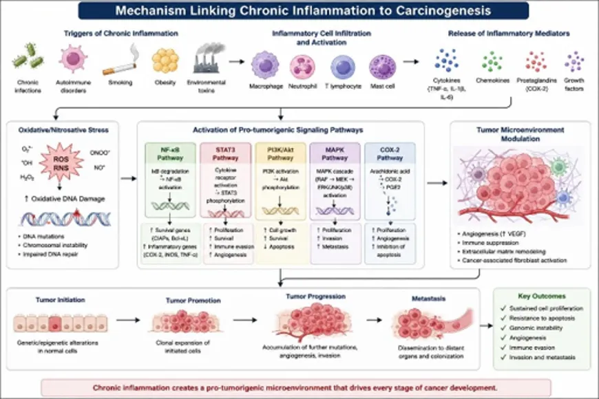
Fig.1 Molecular Mechanisms Linking Chronic Inflammation to Carcinogenesis
to cancer. NF-κB is one of the most important transcription factors activated during inflammation. It induces the expression of genes involved in cell proliferation, inflammation, angiogenesis, and inhibition of apoptosis. Similarly, the STAT3 signaling pathway promotes tumor cell growth, survival, immune evasion, and metastasis. Other pathways, including PI3K/Akt, MAPK, and COX-2, further enhance tumor progression by stimulating cell proliferation, inhibiting programmed cell death, and promoting the formation of new blood vessels required for tumor growth. Stimulating cell proliferation, inhibiting programmed cell death, and promoting the formation of new blood vessels required for tumor growth.
Chronic inflammation also modifies the tumor microenvironment by increasing angiogenesis through vascular endothelial growth factor (VEGF), suppressing anti-tumor immune responses, and remodeling the extracellular matrix. These changes facilitate tumor invasion into surrounding tissues and promote metastasis to distant organs .Overall, chronic inflammation contributes to every stage of carcinogenesis, including tumor initiation, promotion, progression, and metastasis. Understanding the molecular mechanisms that connect inflammation and cancer has led to the identification of several therapeutic targets and the development of small-molecule drugs aimed at interrupting these pathways, thereby reducing cancer risk and improving treatment outcomes.
MAJOR MOLECULAR TARGETS:
1. JAK-STAT.
The Janus kinase (JAK) signal transducer and activator of transcription (JAK-STAT) pathway is an evolutionary conserved signaling pathway that functions in several crucial physiological processes, including hematopoiesis, differentiation, metabolism, and inflammation. The JAK protein family contains four members: JAK1, JAK2, JAK3, and TYK2. The STAT family involves seven members: STAT1, STAT2, STAT3, STAT4, STAT5A, STAT5B, and STAT6.
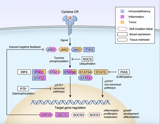
Figure 2. JAK–STAT Signaling Pathway in Inflammation and Carcinogenesis
Among the seven STAT family members, STAT3 and STAT5 have attracted the most oncological attention.The JAK–STAT signaling pathway is a key intracellular signaling mechanism involved in regulating immune responses, inflammation, cell proliferation, differentiation, and cell survival. The pathway begins when cytokines bind to cytokine receptors (CR), activating Janus kinases (JAK1, JAK2, JAK3, and TYK2). Activated JAKs phosphorylate the receptor, allowing STAT proteins to bind and become phosphorylated. The activated STAT proteins form dimers and translocate into the nucleus, where they regulate the expression of genes involved in inflammation, metabolism, cell growth, and apoptosis. The pathway is negatively regulated by suppressor of cytokine signaling (SOCS) proteins, protein inhibitors of activated STAT (PIAS), protein tyrosine phosphatases (PTPs), USP18, and ISG15, which prevent excessive signaling. Persistent activation of the JAK–STAT pathway, particularly STAT3, contributes to chronic inflammation, angiogenesis, immune evasion, and cancer progression, making it an important therapeutic target in inflammatory diseases and carcinogensis.
2. STAT3 SIGNALING PATHWAY
Signal Transducer and Activator of Transcription (STAT3) is activated primarily by interleukin-6 (IL-6) receptor binding, which induces janus kinase (JAK) phosphorylation. Once phosphorylated, STAT3 homodimerizes and translocates to the nucleus to induce gene transcription. In many epithelial and hematological malignancies, STAT3 remains constitutively active due to continuous exposure to inflammatory cytokines within the tumor microenvironment. Constitutive STAT3 activation actively represses anti-tumor immune responses while promoting crucial hallmarks of cancer, including angiogenesis via VEGF up-regulation and resistance to apoptosis .Notably, STAT3, in particular, plays a central role in promoting immune evasion by upregulating immune checkpoint molecules such as PD-L1 and inhibiting the dendritic cell maturation. These immune alterations bypass the antitumor surveillance and therefore facilitate malignant progression. Consequently, aberrant JAK/STAT signaling serves as a molecular bridge that links chronic inflammation to oncogenesis and has become a compelling therapeutic target. This pathway does not operate in isolation as NF-κB-driven IL-6 secretion feeds directly into STAT3 activation, while oxidative stress amplifies signaling through both pathways. STAT3 has been implicated in the regulation of oxidative phosphorylation and metabolic flexibility which enables the oncogenic cells to adapt to fluctuating energy demands. In parallel, JAK mediates histone modifications and interacts with chromatin remodeling complexes which help to link inflammatory inputs to durable.
3. NF-κB Signaling.
Network The Nuclear Factor-kappa B (NF-κB) pathway acts as a central gatekeeper linking chronic inflammation to cancer development. In response to inflammatory stimuli such as TNF-α, IL-1β, or lipopolysaccharides (LPS), the IκB kinase (IKK) complex is activated, leading to the phosphorylation and subsequent proteasomal degradation of IκB proteins. This allows the NF-κB dimers (typically p50/p65) to translocate into the nucleus, where they bind to specific promoter elements. This transcription factor regulates a vast array of genes involved in cell survival (BCL-2, BCL-XL, c-IAP), cell cycle progression (Cyclin D1), and the sustained production of pro-inflammatory cytokines, thereby establishing a self-amplifying feedback loop that drives malignant cell proliferation .Inside the nucleus, activated NF-κB binds to specific DNA sequences called κB response elements, initiating the transcription of numerous genes associated with inflammation and cancer. These target genes include Cyclin D1, which promotes cell-cycle progression; BCL-2, BCL-XL, and c-IAP, which inhibit apoptosis; vascular endothelial growth factor (VEGF), which stimulates angiogenesis; matrix metalloproteinases (MMP-2 and MMP-9).
4. ARACHIDONIC ACID PATHWAY (eicosanoid biology).
The arachidonic acid pathway is a metabolic process of converting phospholipids into bioactive lipid mediators, which control fever, pain, and inflammation. It mainly comprises of cyclooxygenase-2 (COX-2), prostaglandins, and their downstream metabolite prostaglandin E2 (PGE2) and lipoxygenases (LOX). Cyclooxygenases are the rate-limiting enzymes in the arachidonic acid cascade, which catalyse the biosynthesis of prostanoids, among which PGE2 is the most abundant and biological active. Beyond classical prostaglandin synthesis, COX enzymes can also metabolise endocannabinoids to generate prostaglandin glycerol esters and prostaglandin ethanolamides, which are collectively termed as prostamides. Under physiological conditions, COX-2 expression is momentarily high during inflammatory responses. However, sustained COX-2 overexpression and excessive PGE2 production are frequently observed across a wide spectrum of human malignancies and are closely associated with the establishment of a tumour-promoting inflammatory microenvironment.
5. ROS/RNS-mediated oxidative stress.
ROS were historically viewed as harmful byproducts of cellular metabolism because of their capacity to damage nucleic acids, proteins, and lipids. However, literature precedents have reported that ROS, along with RNS (reactive nitrogen species), are bona fide signalling mediators that participate in the redox-sensitive pathways. These pathways govern proliferation, differentiation, immune activation, and metabolic flexibility. Then the question arises is that What keeps this signalling functional rather than destructive? The simple answer is the presence of sophisticated antioxidant and redox-buffering infra-structure that maintains a dynamic equilibrium between ROS generation and detoxification. However, when that equilibrium is lost due to any reason, the resulting oxidative stress becomes a contributor to cancer pathogenesis and a range of other chronic disorders by causing genetic imbalance and activation of other pathways. At the immune level, oxidative stress impairs effector cell function and tilts the TME towards immunosuppression, further facilitating disease progression and therapeutic resistance. Therefore, ROS serve as critical regulators of cancer biology, functioning as indispensable signalling molecules under physiological conditions but acting as potent drivers of genomic damage, inflammation, metabolic dys-regulation, and immune evasion when present in excess. Understanding the context-dependent nature of ROS signalling is therefore essential for the development of therapeutic strategies that effectively target redox imbalances in cancer.
6. CYTOKINSE AND CHEMOKINASE.
Cytokines are low-molecular-weight signalling proteins that regulate cellular proliferation, differentiation, survival, and apoptosis through receptor-mediated activation of intracellular transcriptional programs. Major cytokine families, including interferons, interleukins, TNF superfamily members, chemokines, and growth factors, play essential roles in physiological homeostasis and disease pathogenesis. Dysregulated cytokine networks contribute to cancer initiation and progression by sustaining chronic inflammation and facilitating immune evasion within the TME. Several cytokines, such as
IFN-α, IFN-γ, IL-2, IL-12, IL-15, and GM-CSF, demonstrate antitumour activity by directly suppressing tumour growth or enhancing immune-mediated clearance (Figure 3).
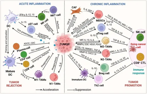
Fig 3. Divergent roles of Immune cells, cytokines, and chemokines in the acute and chronic inflammation in tumor progression (figure was drawn by the authors using BioRender software).
7. INTERLEUKINS (ILS)
ILs represent a diverse class of cytokines that are known to critically regulate immune homeostasis, inflammation, and antitumour immunity. IL-2, originally identified as a T-cell growth factor, plays a central role in adaptive immune responses by promoting the proliferation, differentiation, and cytotoxic activity of T lymphocytes and natural killer cells. Generally, it comprises three receptor subunits, viz. IL-2Rα, IL-2Rβ, and IL-2Rγ-whose assembly determines binding affinity and downstream signalling through JAK-STAT, MAPK, and PI3K pathways. Noteworthy to mention that, IL-2 maintains the immune balance by enhancing the effector T-cell responses and immune surveillance, and it also supports regulatory T-cell expansion. However, impaired IL-2 signalling is associated with poor outcomes in several malignancies, whereas therapeutic administration can stimulate antitumour immunity and improve immunotherapeutic responses. Similarly, IL-10 exerts complex and context-dependent functions in cancer .As a consequence, induction of anti-inflammatory mediators helps to suppress excessive immune activation. This immunosuppressive capacity can facilitate tumour progression by dampening antigen presentation and effector T-cell responses. Paradoxically, IL-10 can also enhance antitumour activity by stimulating tumour-resident CD8+ T cells by increasing interferon-γ production and cytotoxic potential, and thereby restraining tumour growth in specific set-tings. Additionally, IL-12 and IL-15 serve as key immunostimulatory cytokines that reinforce antitumour immunity. IL-12, primarily produced by antigen-presenting cells, and signals through IL-12Rβ1/β2 to activate JAK2-TYK2-STAT4 pathways, which promote T helper 1 differentiation and interferon-γ production while suppressing Th2 polarisation.
Anti-inflammatory therapeutics for cancer
Targeting inflammation has emerged as a rational strategy in cancer therapy, given its central role in tumour initiation, progression, and resistance. From a medicinal chemistry perspective, diverse classes of anti-inflammatory therapeutics have been explored to modulate key signalling pathways and the tumour microenvironment. These include anti-infective agents that indirectly suppress inflammation-driven carcinogenesis, NSAIDs, and small molecules or biologics that modulate inflammatory cytokines and chemokine networks. In addition, natural anti-inflammatory compounds and their derivatives have gained attention as structurally diverse scaffolds with multitarget potential. Collectively, these approaches highlight the chemical and pharmacological diversity available for intervention along the inflammation-cancer axis. In the following subsections, each of these therapeutic classes will be discussed in detail with emphasis on their molecular targets and translational relevance.
Anti-infective therapeutics
As mentioned above, various infectious diseases are the primary cause of inflammation-induced Therefore, anti-infective therapeutics, viz. antiviral, antibacterial, and antifungal therapies, represent a genuinely preventive tier of cancer medicine, Viral infections such as the hepatitis B and C are leading causes of hepatocellular carcinoma, where continuous immune activation and inflammatory signalling contribute to liver damage and tumorigenesis. persistent infection with high-risk HPV is responsible for the majority of cervical cancers. Prophylactic HPV vaccination has demonstrated remarkable success in reducing infection rates and precancerous lesions, and it underlines that the infection can be intervened at the early stage before inflammatory damage accumulates. Some oncogenic viruses, such as Epstein-Barr virus, by contrast, remain therapeutically unaddressed despite their association with multiple malignancies and they display a conspicuous gap in the field. Similarly, among bacterial pathogens, H. pylori carries the strongest evidence base for the inflammation-induced gastric cancer. Its eradication with antibiotic regimens reduces both the incidence and recurrence of gastric cancer, and this relationship is very well-established to inform clinical guidelines. Fusobacterium nucleatum has more recently been implicated in colorectal cancer progression through inflammatory pathway modulation. However, due to a lack of specific antibiotics, broad-spectrum antibiotic use remains limited due to potential disruption of beneficial microbiota and lack of target specificity. Moreover, fungal infection contributions to cancer-associated inflammation are less characterised but not negligible. For instance, associations of Candida species with oesophageal squamous cell carcinoma suggest antifungal strategies may carry preventive value in specific contexts, though clinical translation remains at an early stage. Therefore, these findings position anti-infective therapy as a legitimate and underutilised instrument in cancer prevention, particularly when deployed with appropriate specificity.
Small-molecule intervention strategies
Considering the critical involvement of key inflammatory pathways, including NF-κB, ROS-mediated signalling, JAK/STAT, inflammasomes, and cytokine networks, extensive efforts have been directed towards the development of small-molecule therapeutics targeting these axes. Over the past 5 years, a diverse range of compounds has been reported, encompassing single-target inhibitors, dual inhibitors, multitarget-directed ligands, and prodrug strategies designed to modulate these interconnected pathways. These approaches reflect a shift towards rational design aimed at overcoming pathway redundancy and therapeutic resistance. In the following section, recent advances in small-molecule development are summarised, with particular emphasis on design strategies, structure-activity relationships (SAR), and their in vitro and in vivo pharmacological evaluations.
Small molecule therapeutic intervention for RNS modulation
Reported a novel series of nonquinone-based substrates as NAD(P)H: quinone oxidoreductase 1 (NQO1) modulators to induce intracellular ROS generation for the treatment of drug-resistant NSCLC. For this study, they utilised their screening hit molecule 1, which contains a tetracyclic nonquinone scaffold, and conducted extensive structure-activity relationship (SAR) studies. This led to the identification of 3 (tricyclic 2,3-dicyano indenopyrazinone), as the most potent analogue. During the initial in vitro screening, 3 has selectively inhibited the proliferation of NQO1-overexpressing A549 and drug-resistant A549/Taxol cells. Mechanistic investigations further revealed that 3 undergoes NQO1-catalyzed redox cycling, leading to a significant elevation of intracellular ROS levels, which subsequently triggered PARP-1-mediated apoptosis in an NQO1- and ROS-dependent manner. Furthermore, in vivo studies showed that 3 effectively suppressed tumour growth in A549/Taxol xenograft models without any observable systemic toxicity.
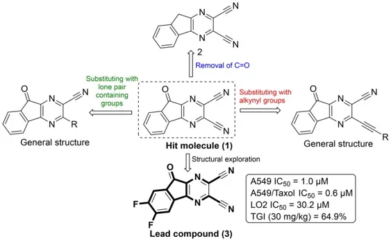
Fig4: Nonquinone substrates as ROS modulators by targeting NQO1 in NSCLC (figure was drawn by the authors using ChemDraw software).
Wang and the research group accomplished a series of 2-aminopyrimidine derivatives as potent and selective inhibitors of IKKβ. Structural optimisation of the aminopyrimidine scaffold was undertaken to improve kinase inhibitory potency and selectivity. SAR studies helped to reveal compound 17 as the most promising candidate among the synthesised analogues. It has displayed remarkable inhibitory potency against IKKβ with an IC50 value of 7.5 nM and excellent kinome selectivity. Initial in vitro evaluation demonstrated that 17 significantly suppressed cell viability and proliferation in human colorectal cancer cell lines RKO and HCT116, which is many-fold better than the reference inhibitor BMS-345541. Further, mechanistic investigations revealed that 17 attenuated NF-κB signalling, and simultaneously induced autophagy and G2/M phase cell cycle arrest.In vivo studies using RKO and MC38 xenograft mouse models, as well as an MC38-derived syngeneic model, confirmed that 17 effectively suppressed tumour growth without causing significant toxicity. Therefore, these findings identify LP46 as a potent and selective IKKβ inhibitor with strong therapeutic potential for the treatment of colorectal cancer.
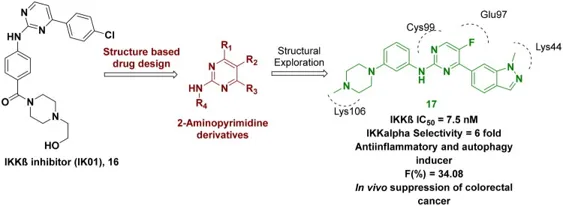
Fig6: 2-Aminopyrimidine derivatives as a selective IKKβ inhibitor in colorectal cancer (figure was drawn by the authors using ChemDraw software.
Applications of Small Molecule Intervention Strategies
CONCLUSION
Chronic inflammation plays a central role in the initiation, promotion, and progression of carcinogenesis by activating multiple signaling pathways, including NF-κB, JAK/STAT, COX-2, PI3K/Akt, and MAPK. These pathways regulate cell proliferation, apoptosis, angiogenesis, immune evasion, and metastasis, making them attractive therapeutic targets. Small-molecule intervention strategies have emerged as effective approaches to inhibit these molecular pathways, suppress inflammatory responses, and reduce tumor development. Several small-molecule inhibitors have demonstrated significant therapeutic potential in both preclinical and clinical studies, either as monotherapy or in combination with conventional anticancer treatments. Despite challenges such as drug resistance, off-target effects, and toxicity, continued advances in medicinal chemistry, precision medicine, and artificial intelligence–assisted drug discovery are accelerating the development of safer and more selective compounds.
REFERENCES

K. Malleswari, Dr. D. Rama Brahma Reddy, A. Lakshmi Prasanna Sahithi, Inflammation and Carcinogenesis: Molecular Targets and Small Molecule Intervention Strategies, Int. J. of Pharm. Sci., 2026, Vol 4, Issue 9, 3728-3738. https://doi.org/10.5281/zenodo.23022516
 10.5281/zenodo.23022516