

We use cookies to ensure our website works properly and to personalise your experience. Cookies policy
Dhanalakshmi Srinivasan College of Pharmacy, Thuraiyur Road, Perambalur 621212.
Oxidative stress and inflammation are major contributors to chronic diseases. This study evaluated the antioxidant and anti-inflammatory activities of methanolic leaf extract of Vigna stipulacea (VSMLE). Anti-inflammatory activity was assessed by protein denaturation assay, while antioxidant activity was evaluated in H?O?-induced H9c2 cardiomyocyte cells using MTT assay and antioxidant biomarkers. VSMLE showed concentration-dependent anti-inflammatory activity (IC??: 56.1 mg/mL) and significantly improved cell viability against oxidative damage. These findings suggest that Vigna stipulacea leaves possess promising antioxidant and anti-inflammatory potential.
In vitro screening refers to experiments conducted outside a living organism using isolated biological components such as molecules, enzymes, tissues, or cells under controlled laboratory conditions. The term in vitro means “in glass,” originating from the traditional use of test tubes and Petri dishes for biological experiments. These models allow researchers to study biological processes at cellular and molecular levels without the complexity of a whole living organism. Modern in vitro systems include cell lines, primary cell cultures, recombinant enzymes, and molecular assays designed to reproduce specific biochemical and cellular processes occurring in living organisms. They are particularly useful for evaluating the effects of herbal extracts on gene expression, receptor interactions, metabolism, cellular responses, and toxicity. Compared with animal experiments, in vitro methods are considered more ethical, economical, reproducible, reliable, and relevant to human health. They can also reduce the need for extensive animal testing and are useful for high-throughput screening of potential therapeutic compounds.[1]
Oxidative stress occurs when there is an imbalance between the production of reactive oxygen species (ROS) and the body's antioxidant defense system. According to Sies (1985), oxidative stress results from an imbalance between oxidants and antioxidants, which can cause cellular damage. ROS are naturally generated during normal metabolic processes and can also increase in response to various external and internal factors. When ROS accumulate beyond the capacity of antioxidants to neutralize them, they can damage important cellular components.[2]
Free radicals are highly reactive molecules containing unpaired electrons. Antioxidants help protect cells by neutralizing free radicals and interrupting oxidative chain reactions. During excessive oxidative stress, ROS can damage DNA, lipids, proteins, and cell membranes. DNA damage may cause mutations and strand breaks, while lipid peroxidation can disrupt membrane structure and permeability. Protein oxidation can alter protein structure and interfere with enzyme function.[3]
Oxidative stress has been associated with several diseases and pathological conditions, including cancer, aging, Alzheimer's disease, Parkinson's disease, cardiovascular disorders, diabetes, obesity, atherosclerosis, diabetic complications, inflammation, and autoimmune disorders. Both ROS and reactive nitrogen species (RNS) have important physiological roles in normal cellular functions and defense against infections. However, excessive production can contribute to disease development and progression.[4]
Oxidative stress may develop through increased auto-oxidation of endogenous or externally introduced substances, depletion of low-molecular-weight antioxidants, inactivation of antioxidant enzymes, reduced production of antioxidant molecules and enzymes, or a combination of these factors. Therefore, maintaining an appropriate balance between oxidants and antioxidants is essential for protecting cells and tissues from oxidative damage.[5]
Reactive oxygen species (ROS) are oxygen-derived molecules that include both free radicals and non-radicals, such as hydrogen peroxide, superoxide, hydroxyl radicals, and singlet oxygen. They are generated mainly in mitochondria, peroxisomes, the endoplasmic reticulum, and by NADPH oxidases. At low levels, ROS function as important signalling molecules involved in cell proliferation, differentiation, and immune responses. However, excessive ROS production causes oxidative stress, damaging lipids, proteins, and DNA, and contributing to cellular dysfunction and diseases such as cancer.[6]
Any molecular species with an unpaired electron in an atomic orbital that can exist independently is referred to as a free radical. The majority of radicals share certain characteristics due to the presence of an unpaired electron. A lot of radicals are extremely reactive and unstable. They behave as oxidants or reductants by having the ability to give or receive electrons from other molecules.[7]
Antioxidant activity refers to the ability of substances to neutralize free radicals and ROS, thereby preventing oxidative damage to lipids, proteins, and DNA. It maintains oxidative balance through mechanisms such as hydrogen atom transfer, electron transfer, and metal chelation, reducing oxidative stress. [8]
Antioxidants are substances that prevent or delay oxidation and protect cells from oxidative damage by scavenging free radicals, chelating metals, and quenching singlet oxygen. They are important for maintaining food quality and human health. Plants contain various antioxidants, including nutrients such as vitamins C and E, carotenoids, lipoic acid, and glutathione. Antioxidant enzymes such as superoxide dismutase, glutathione peroxidase, and glutathione reductase help neutralize free radicals. Metal-binding proteins, including ferritin, lactoferrin, albumin, and ceruloplasmin, also reduce oxidative stress. Additionally, many plants contain diverse antioxidant phytochemicals that provide further protective effects against oxidative damage. [9,10,11]
ENZYMATIC ANTIOXIDANT DEFENCE: SOD, CAT, GSH, AND GSH-PX
Reactive oxygen species (ROS), such as the superoxide radical and hydrogen peroxide, are produced during normal cell metabolism and increase under stress conditions, causing damage to cell membranes, proteins, and DNA. To protect against this damage, cells possess a first line of enzymatic antioxidant defence consisting of four key components: superoxide dismutase (SOD), catalase (CAT), glutathione (GSH), and glutathione peroxidase (GSH-Px). These work together to safely convert harmful ROS into water and oxygen, thereby protecting the cell from oxidative stress.
Brief Mechanism of Each Component
1. Superoxide Dismutase (SOD): A metalloenzyme (Cu/Zn-SOD, Mn-SOD, or Fe-SOD, depending on subcellular location and organism) that catalyses the dismutation of two superoxide radicals into hydrogen peroxide and molecular oxygen, forming the first and most powerful line of antioxidant defence.
2. Catalase (CAT): A haem-containing enzyme that does not require any external reductant. It decomposes H2O2 in a two-step reaction – H2O2 first oxidises the enzyme's Fe to form an intermediate (Compound I), and a second H2O2 molecule then reduces this intermediate, releasing water and oxygen.
3. Glutathione (GSH): A tripeptide (glutamate–cysteine–glycine) that serves as the essential reducing cofactor for GSH-Px. During the reaction, GSH is oxidised to glutathione disulfide (GSSG), which is then reduced back to GSH by glutathione reductase (GR) using NADPH, completing the redox cycle.
4. Glutathione Peroxidase (GSH-Px): A thiol-based enzyme that reduces H2O2 (and lipid hydroperoxides) to water/lipid alcohols at the expense of GSH, following a ping-pong catalytic mechanism involving oxidation and subsequent reduction of the active site. [12]
Classification of Antioxidants
Antioxidants are classified into five major types: primary antioxidants that terminate free-radical chain reactions, oxygen scavengers that remove molecular oxygen, secondary antioxidants that prevent oxidation, enzymatic antioxidants that detoxify ROS, and synergistic antioxidants that enhance antioxidant activity by chelating metal ions and inhibiting oxidation.[10]
INFLAMMATION
Inflammation is a protective biological response of the immune and vascular systems to harmful stimuli such as infections, tissue injury, irritants, or abnormal cell death. It plays an important role in eliminating pathogens, removing damaged cells, and promoting tissue repair and wound healing. The major signs of inflammation include redness, swelling, pain, warmth, stiffness, and loss of function. Inflammation can be classified into acute and chronic inflammation.
Acute inflammation is a short-term response that usually lasts from a few minutes to several days. It is characterized by increased vascular permeability, capillary changes, and migration of leukocytes to the site of injury. Examples include acute pancreatitis, acute kidney injury, and acute asthma.
Chronic inflammation persists for several months or years and occurs when the inflammatory response continues for a prolonged period. It is characterized by infiltration of mononuclear immune cells, macrophage and monocyte activity, fibroblast activation, angiogenesis, and fibrosis. Chronic inflammation is associated with conditions such as atherosclerosis, rheumatoid arthritis, inflammatory bowel disease, and psoriasis. Thus, controlled inflammation is essential for tissue protection and repair, whereas prolonged inflammation may contribute to tissue damage and chronic diseases. [13–15]
Triple Response of Inflammation
The Triple Response of Lewis is an immediate localized skin reaction associated with early acute inflammation, mainly caused by the release of histamine and other chemical mediators. It occurs in three successive phases. Red spot (5–15 seconds): localized redness caused by dilation of capillaries and arterioles. Flare (30–45 seconds): a surrounding reddish area produced by arteriolar dilation through axon reflexes and release of neuropeptides. Wheal (1–3 minutes): localized swelling caused by increased capillary and venular permeability, resulting in fluid accumulation and edema. Thus, the triple response demonstrates the vascular changes occurring during inflammation. (16)
Inflammatory mediators are signalling molecules that initiate and regulate inflammation. Major mediators include histamine, bradykinin, prostaglandins, leukotrienes, cytokines, nitric oxide, and platelet-activating factor. They regulate vasodilation, vascular permeability, pain, fever, immune-cell activity, and pathogen defense. Excessive mediator production may cause chronic inflammation and tissue damage. (17)
Anti-inflammatory activity refers to the ability of substances or mechanisms to reduce or control excessive inflammation while preserving normal immune functions. It supports tissue healing through the regulation of immune cells and inflammatory mediators such as cytokines, interleukins, IL-1, and IL-2, thereby controlling inflammation and preventing disease progression. (18,19)
Anti-Inflammatory Mechanisms:
Anti-inflammatory treatments target several stages of the inflammatory process:
Anti-inflammatory agents are substances or drugs that reduce inflammation by interfering with the biological pathways responsible for the inflammatory response. They help minimize tissue damage, relieve symptoms, and improve patient comfort. These agents act on specific inflammatory mediators and pathways while aiming to maintain normal immune functions.
Anti-inflammatory agents are broadly classified into steroidal and non-steroidal anti-inflammatory drugs (NSAIDs). NSAIDs, such as aspirin and selective COX-2 inhibitors (coxibs), reduce inflammation mainly by inhibiting cyclooxygenase enzymes and decreasing the production of inflammatory prostaglandins. However, prolonged use of NSAIDs may increase the risk of gastrointestinal and cardiovascular complications. Glucocorticoids are steroidal agents that suppress inflammation by regulating the expression of several inflammatory cytokines and genes. Although highly effective, their long-term use can cause significant adverse effects.
Another important group is biological agents, including monoclonal antibodies that specifically target inflammatory cytokines such as TNF-α, IL-6, IL-1, IL-12, and IL-17. They are commonly used for autoimmune and chronic inflammatory diseases such as rheumatoid arthritis, Crohn’s disease, and psoriasis [20,21]
Classification of Anti-Inflammatory Agents
Anti-inflammatory agents are mainly classified into steroidal and non-steroidal anti-inflammatory drugs (NSAIDs). Common NSAIDs include aspirin and coxibs. Prolonged NSAID use may cause cardiovascular and gastrointestinal complications. Therefore, natural-product-based preparations are being explored as potential alternatives to conventional anti-inflammatory agents with fewer adverse effects. [22]
PLANT PROFILE:
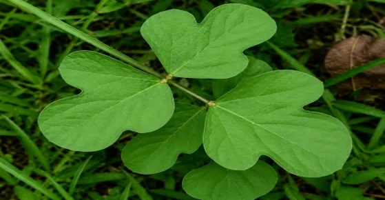
Fig 1: Vigna stipulacea
Synonyms:
Biological Source:
Dried leaf extract of Vigna stipulacea (Lam.) Kuntz [24]
Family:
It belongs to the family of Fabaceae [23]
Taxonomical Classification:
Vernacular Names:
Morphological Description
Vigna stipulacea is an uncommon twining or trailing plant with angular, sparsely hairy stems. Its stipules are large, ovate, pointed, and serve as an important identifying feature. The leaves are trifoliate, shiny, and possess a characteristically trilobed terminal leaflet. The flowers are bright yellow with purplish keel tips and occur in axillary pseudo-racemes on stems that may reach approximately 30 cm. The seeds are black, elliptical, and possess a small protruding aril and whitish oblong hilum. The pods are linear, 4.4–5.6 cm long, sparsely hairy, and contain about 12–14 seeds. At maturity, the pods become brownish-black. [23]
Geographical Source
Vigna stipulacea is distributed in several Indian states, including Andhra Pradesh, Tamil Nadu, Odisha, Madhya Pradesh, Goa, Kerala, and Chhattisgarh. It is also reported from Madagascar, Vietnam, Myanmar, Sri Lanka, Bangladesh, and Yemen. [25]
Ethnobotany
The plant is mainly utilized as animal feed and green manure. It is also used as a food ingredient in preparations such as sambar, condiments, and dosa. The species has also been reported to provide resistance against certain pests and diseases. [26]
Cultivation
Vigna stipulacea is primarily cultivated during the kharif season, with flowering generally occurring from September to October. Seeds are commonly sown in August by broadcasting. The crop may be cultivated before paddy farming for seed production, human food, animal feed, and green manure. It can grow naturally in moist clay soils both inside and outside paddy fields. [24]
Chemical Constituents:
Phytochemical Analysis:
Traditional Uses:
Particularly in southern India, it is used for animal feed, fertilizer production, and inclusion in classic Indian dishes like idly and vada. Despite its potential in traditional food systems being underutilized, this species is still prized for its nutritious grains.[28]
Plant Extraction:
Fresh Vigna stipulacea leaves
↓
Wash with distilled water
↓
Shade dry and powder
↓
Weigh 40 g powder
↓
Add to Soxhlet apparatus
↓
Add 200 mL methanol
↓
Extract at 60°C for 6–8 h
↓
Cool and filter the extract
↓
Concentrate at 40–45°C
↓
Collect VSMLE
↓
Store in airtight container at 4°C [29,30]
ANTIOXIDANT ASSAY:
Antioxidant Assay by H2O2 Induced Oxidative Stress in H9c2 Cardiomyocyte Cells:
The methodology for inducing oxidative stress in H9c2 cells using H2O2 involved several precise steps. Initially, H9c2 cells were cultured and adjusted to a concentration of 1 × 10⁴ cells/mL, then seeded into 96-well plates 200 μL per well and allowed to adhere and grow at 37°C for 24 hours under a 5% CO2 atmosphere. To establish the oxidative damage model, the cells were treated with 0.3 mmol/L hydrogen peroxide (H2O2) for 4 hours, which was added directly to the culture medium, inducing oxidative stress and cellular injury. After this exposure, the cells were subjected to varying concentrations of Malus doumeri leaf extract (MDLE) at 0, 40, 100, and 160 μg/mL, which were added to the wells to assess protective effects against H2O2-induced damage. The treatment with MDLE lasted for an additional 24 hours. Subsequently, cell viability was evaluated using the MTT assay: the medium was removed, and 200 μL of MTT solution (5 mg/mL) was added to each well, followed by incubation at 37°C for 4 hours, allowing metabolically active cells to convert MTT into formazan. After incubation, the medium was aspirated, and 200 μL of DMSO was added to dissolve the formed formazan crystals, which was then shaken for 30 minutes. The absorbance was measured at 490 nm using a spectrophotometer, providing quantitative data on cell viability.[32] In addition, apoptosis and cell death were analysed via flow cytometry by staining the cells with annexin V-FITC and propidium iodide after detachment, washing, and suspension in PBS, then incubating them in the dark at 37°C for 15 minutes. This detailed approach allowed the researchers to evaluate the extent of oxidative injury induced by H2O2 and the potential protective effects of MDLE on cell survival and apoptosis.[31]
ANTI-INFLAMMATORY ASSAY:
Inhibition of Protein Denaturation Assay Methodology:
This method evaluates the ability of herbal extracts to prevent protein denaturation, a process associated with inflammation-related disorders such as rheumatoid arthritis, diabetes, and cancer. (34) The assay involves preparing reaction mixtures with varying concentrations of the herbal extract (typically 100-500 μg/mL) and adding it to a solution containing egg albumin or bovine serum albumin (approximately 200-450 μL).(35-37) The mixture is initially incubated at 37°C for 15 minutes to allow interaction, then heated at 70°C for 5 minutes to induce denaturation of the proteins.(38) After cooling under tap water, the samples are measured spectrophotometrically at 660 nm. (39) The degree of protein denaturation is indicated by absorbance; lesser absorbance in the presence of the extract suggests protective effects against denaturation.
The percentage inhibition of protein denaturation is calculated using the formula:
% Inhibition = [(1 - D/C) x 100],
where D is the absorbance of the test sample and C is that of the negative control without extract. Standard drugs such as acetyl salicylic acid or diclofenac sodium are used as positive controls to validate the assay. (40)
RESULTS:
In Vitro Anti-Inflammatory Activity of Vigna stipulacea Methanolic Leaf Extract (VSMLE)
The anti-inflammatory activity of Vigna stipulacea methanolic leaf extract (VSMLE) was evaluated using the albumin denaturation inhibition assay, which measures the ability of a test substance to prevent heat-induced protein denaturation. VSMLE demonstrated a clear concentration-dependent inhibitory effect over the range of 20–100 mg/mL. The percentage inhibition increased from 35% at 20 mg/mL to 45%, 55%, 65%, and 75% at 40, 60, 80, and 100 mg/mL, respectively. This progressive increase indicates a dose-dependent anti-inflammatory response and significant protein-stabilizing activity. Aspirin, used as the standard drug, showed higher inhibition, ranging from 40% to 90% across the same concentrations. The calculated IC₅₀ value was 56.1 mg/mL for VSMLE and 34.45 mg/mL for aspirin. Since a lower IC₅₀ indicates greater potency, aspirin was approximately 1.6 times more potent than VSMLE. However, the consistent inhibition exhibited by VSMLE confirms its appreciable in vitro anti-inflammatory potential and supports its possible therapeutic value.
Table 1: Effect of VSMLE on Albumin Denaturation Inhibition (In Vitro Anti-Inflammatory Activity)
|
Sr. No |
Concentration of VSMLE (mg/mL) |
% Protein Denaturation (VSMLE) |
% Protein Denaturation (Aspirin) |
|
1. |
20 |
35 |
40 |
|
2. |
40 |
45 |
55 |
|
3. |
60 |
55 |
66 |
|
4. |
80 |
65 |
76 |
|
5. |
100 |
75 |
90 |
|
IC50 value |
56.1 mg/mL |
34.45 mg/mL |
|
Effect of VSMLE on Cell Viability in H₂O₂-Induced Oxidative Stress in H9c2 Cells
The cytoprotective effect of Vigna stipulacea methanolic leaf extract (VSMLE) was evaluated in H9c2 rat cardiomyoblast cells exposed to 0.3 mM H₂O₂ using the MTT assay. Normal control cells showed 100.00 ± 0.00% viability, whereas H₂O₂ exposure significantly reduced cell survival to 43.51 ± 1.82%, confirming successful induction of oxidative stress. Treatment with VSMLE significantly improved cell viability in a concentration-dependent manner. At 80 µg/mL, VSMLE increased cell survival to 65.84 ± 2.10%, while 160 µg/mL further increased viability to 84.35 ± 1.75%, approaching normal control levels. Statistical analysis showed significant differences among all treatment groups (p < 0.05). These findings demonstrate that VSMLE effectively protects H9c2 cells against H₂O₂-induced oxidative cytotoxicity, with greater protection observed at higher concentrations. Thus, VSMLE exhibits significant dose-dependent cytoprotective and antioxidant potential.
Table 2: Effect of VSMLE on Cell Viability (Survival Rate) in H₂O₂-Induced Oxidative Stress in H9c2 Cells
|
Group |
Normal Control |
Oxidative Stress Control (H2O2 0.3 mM) |
H2O2 + VSMLE (80µg/mL) |
H2O2 + VSMLE (160µg/mL) |
|
Survival Rate (%) |
100.00 ± 0.00a |
43.51± 1.82d |
65.84 ± 2.10c |
84.35 ± 1.75b |
Effect of VSMLE on Antioxidant Enzyme Activities and Lipid Peroxidation in H9c2 Cells
The effect of Vigna stipulacea methanolic leaf extract (VSMLE) on oxidative stress markers was evaluated in H9c2 cells by measuring SOD, GSH, GSH-Px, CAT, and MDA levels. H₂O₂ exposure significantly impaired the cellular antioxidant defense system, while VSMLE treatment produced a concentration-dependent protective effect.
SOD activity decreased from 8.45 ± 0.52 to 3.10 ± 0.28 U/mg protein after H₂O₂ exposure. VSMLE restored SOD activity to 5.35 ± 0.38 and 7.12 ± 0.41 U/mg protein at 80 and 160 µg/mL, respectively. Similarly, GSH content declined from 0.86 ± 0.06 to 0.30 ± 0.04 µmol/mg protein but increased to 0.53 ± 0.04 and 0.71 ± 0.05 µmol/mg protein following VSMLE treatment.
GSH-Px activity decreased from 5.60 ± 0.31 to 2.15 ± 0.22 U/mg protein after oxidative injury and was restored to 3.62 ± 0.29 and 4.68 ± 0.25 U/mg protein by VSMLE. CAT activity also decreased from 3.50 ± 0.26 to 0.85 ± 0.15 U/mg protein, while VSMLE increased it to 1.96 ± 0.22 and 2.81 ± 0.24 U/mg protein.
Conversely, MDA levels increased markedly from 0.15 ± 0.02 to 0.82 ± 0.07 nmol/mg protein following H₂O₂ exposure. VSMLE significantly reduced MDA to 0.51 ± 0.04 and 0.32 ± 0.03 nmol/mg protein at 80 and 160 µg/mL, respectively. All parameters showed significant differences (p < 0.05). Overall, VSMLE enhanced antioxidant defenses and reduced lipid peroxidation, demonstrating significant dose-dependent antioxidant and cytoprotective activity in H9c2 cells. [34]
Table 3: Effect of VSMLE on SOD, GSH, GSH-Px, CAT Activity and MDA Levels in H₂O₂-Induced Oxidative Stress in H9c2 Cells
|
Parameters |
Normal Control |
Oxidative Stress Control (H2O2 0.3 mM) |
H2O2 + VSMLE (80 µg/mL) |
H2O2 + VSMLE (160 µg/mL) |
|
SOD (U/mg protein) |
8.45 ± 0.52 a |
3.10 ± 0.28 d |
5.35 ± 0.38 c |
7.12 ± 0.41 b |
|
GSH (µmol/ mg protein) |
0.86 ± 0.06 a |
0.30 ± 0.05 d |
0.53 ± 0.04 c |
0.71 ± 0.05 b |
|
GSH-Px (U/mg protein) |
5.60 ± 0.31 a |
2.15 ± 0.22 d |
3.62 ± 0.29 c |
4.68 ± 0.25 b |
|
CAT (U/mg protein) |
3.50 ± 0.26 a |
0.85 ± 0.15 d |
1.96 ± 0.22 c |
2.81 ± 0.24 b |
|
MDA (nmol/mg protein) |
0.15 ± 0.02 d |
0.82 ± 0.07 a |
0.51 ± 0.04b |
0.32 ± 0.03 c |
DISCUSSION:
The present study investigated the in vitro antioxidant and anti-inflammatory activities of Vigna stipulacea methanolic leaf extract (VSMLE) using three complementary experimental approaches: albumin denaturation inhibition, H₂O₂-induced oxidative stress in H9c2 cardiomyoblast cells, and estimation of antioxidant defense markers including SOD, GSH, GSH-Px, CAT, and MDA. Overall, the results demonstrated that VSMLE possesses significant, concentration-dependent antioxidant, cytoprotective, and anti-inflammatory properties. [18]
The albumin denaturation assay showed that VSMLE produced a clear concentration-dependent anti-inflammatory effect. Inhibition increased from 35% at 20 mg/mL to 75% at 100 mg/mL, indicating progressive protein-stabilizing activity. Aspirin showed greater potency, with an IC₅₀ of 34.45 mg/mL, compared with 56.1 mg/mL for VSMLE, making aspirin approximately 1.6 times more potent. However, the appreciable activity of the crude plant extract is noteworthy because it contains a complex mixture of phytoconstituents. Phenolic and flavonoid compounds may contribute to this effect by stabilizing proteins and preventing heat-induced denaturation. These findings are consistent with previous reports on plant extracts showing concentration-dependent anti-inflammatory activity. [32,40]
The cytoprotective activity of VSMLE was assessed using H₂O₂-induced oxidative injury in H9c2 cells. H₂O₂ exposure reduced cell viability from 100.00 ± 0.00% to 43.51 ± 1.82%, confirming successful induction of oxidative stress. VSMLE significantly improved cell survival to 65.84 ± 2.10% at 80 µg/mL and 84.35 ± 1.75% at 160 µg/mL. The dose-dependent improvement indicates that VSMLE protects cardiomyoblast cells against oxidative damage and supports its antioxidant potential. Similar protective effects have been observed with other plant extracts in H₂O₂-induced H9c2 cell models. [31,33]
The biochemical findings further supported the protective mechanism of VSMLE. H₂O₂ treatment markedly reduced SOD, GSH, GSH-Px, and CAT levels while increasing MDA, indicating depletion of antioxidant defenses and enhanced lipid peroxidation. VSMLE treatment reversed these changes in a concentration-dependent manner. At 160 µg/mL, SOD, GSH, GSH-Px, and CAT recovered to approximately 84%, 83%, 84%, and 80% of normal control values, respectively. MDA was reduced from 0.82 ± 0.07 to 0.32 ± 0.03 nmol/mg protein, demonstrating suppression of membrane lipid peroxidation. Thus, VSMLE appears to protect cells by maintaining endogenous antioxidant defenses and limiting oxidative damage. [10,31]
The consistent concentration-dependent responses observed across the assays strengthen the reliability of the findings. Oxidative stress and inflammation are closely interconnected, as excessive ROS can activate inflammatory pathways, while inflammation can further increase ROS production. Therefore, the antioxidant and anti-inflammatory effects of VSMLE may act together to interrupt this harmful cycle. The activity may be associated with phenolic and flavonoid constituents reported in V. stipulacea and related Vigna species. [18,27,29]
However, the study has certain limitations. Anti-inflammatory activity was evaluated using only one in vitro assay, while the antioxidant study involved only two concentrations and one cell line. Further studies using complementary inflammatory assays, additional cell models, broader concentration ranges, and bioassay-guided fractionation are required to identify the active constituents. In vivo studies are also necessary to establish efficacy and safety.
Overall, the findings provide consistent evidence that VSMLE possesses meaningful concentration-dependent antioxidant, cytoprotective, and anti-inflammatory activity. Restoration of endogenous antioxidant enzymes and reduction of lipid peroxidation appear to be important mechanisms underlying its protective effect against oxidative stress.
CONCLUSION
The present study successfully evaluated the in vitro antioxidant, cytoprotective, and anti-inflammatory activities of Vigna stipulacea methanolic leaf extract (VSMLE) using complementary experimental models. In the albumin denaturation assay, VSMLE demonstrated a clear concentration-dependent anti-inflammatory effect, with inhibition increasing from 35% to 75% across 20–100 mg/mL. The IC₅₀ value of VSMLE was 56.1 mg/mL, compared with 34.45 mg/mL for aspirin, indicating lower potency than the standard drug but significant protein-stabilizing activity.
In the H₂O₂-induced oxidative stress model using H9c2 cardiomyoblast cells, VSMLE significantly improved cell viability from the oxidative stress control level of 43.51% to 65.84% and 84.35% at 80 and 160 µg/mL, respectively. Biochemical analysis further showed that VSMLE restored the activities of SOD, GSH, GSH-Px, and CAT, which were depleted following H₂O₂ exposure. It also significantly reduced the elevated MDA level, indicating suppression of lipid peroxidation.
Overall, the findings provide consistent evidence that VSMLE possesses dose-dependent antioxidant and anti-inflammatory properties, possibly associated with its phenolic and flavonoid constituents. Further phytochemical characterization, complementary in vitro studies, and in vivo investigations are recommended to establish its therapeutic potential.
REFERENCES

A. Gowtham Sashtha, S. Mohamed Halith, M. Janani, S. Janani, T. Javith Ahamed, J. Jayaraman, G. Jayasri, Invitro Screening of Antioxidant and Anti-Inflammatory Activities of Vigna stipulacea Methanolic Leaf Extract, Int. J. of Pharm. Sci., 2026, Vol 4, Issue 8, 4428-4441. https://doi.org/10.5281/zenodo.22120523
 10.5281/zenodo.22120523