

We use cookies to ensure our website works properly and to personalise your experience. Cookies policy
Department of pharmaceutics, KMCH College of Pharmacy, Coimbatore-641048
Despite being one of the world's biggest killers and causes of morbidity, the traditional chemotherapy approach, with its systemic toxicity, poor tumour specificity, multidrug resistance, and sub-optimal pharmacokinetics, still has a significant effect on therapeutic outcomes. Nanotechnology is seen to be one avenue that could address these problems by designing novel drug delivery systems that enhance drug stability, bioavailability and targeted delivery to the site of action. One of these is lipid–polymer hybrid nanoparticles (LPHNPs), which combine the features of polymeric nanoparticles (controlled drug release and structural stability) with the excellent biocompatibility and biomimetic properties of lipid-based carriers. The current review outlines the design, fabrication techniques, physicochemical characterization, and its therapeutic applications for targeted cancer therapy using LPHNPs. The manuscript covers some of the most important preparation methods, characterization methods and advances in stimuli-responsive nanocarriers that are able to release drugs in response to endogenous or exogenous tumor-specific stimuli. The current applications in breast, pancreatic, liver and lung cancers are critically summarized, where there are improvements in targeted delivery, therapeutic efficacy, pharmacokinetics and multidrug resistance reversal. The review also considers the key challenges to clinical translation, such as large-scale manufacturing, physicochemical stability, immunogenicity, regulatory challenges, and quality control. Overall, lipid–polymer hybrid nanoparticles are potentially exciting, versatile, and multifunctional drug delivery platforms that have great promise for enhancing the safety, efficacy, and clinical performance of anticancer therapeutics, but require additional multidisciplinary research and regulatory standardization to realize their successful clinical translation.
Global Cancer Burden
Cancer is also among the most common causes of death in the world, but it is estimated that the number of its causes is about 9.7- 10 million people die each year1–4. Approximately 20 million of new cases were diagnosed in 2022 alone, and it is estimated that the number will increase by 77% by the year 2050 3,5. It has been shown that one out of every six women and one out of five men will have a tumour in their lifetime, which means that the disease poses a major challenge to human wellbeing and survival in the 21 st century6,7.
Problems of Conventional Chemotherapy.
In spite of its universal application, traditional chemotherapy has fatal clinical drawbacks1,5,8.
• Systemic Toxicity: A majority of the chemotherapeutic agents are not selective and will kill both malignant and normal cells4,9. The effects resulting include severe, and debilitating side effects like anaemia, bone marrow suppression, cardiotoxicity, pulmotoxicity and loss of hair4,9,10.
• Drug Resistance: Multidrug resistance (MDR) is a significant agent of treatment failure9–11. To survive, cancer cells develop different strategies such as overexpression of efflux pumps (such as P-glycoprotein) that release drugs out of the cell, or increased repair pathways in DNA2,12,13.
• Ineffective Bioavailability: Several potent anticancer drugs including taxanes and camptothecin have low water solubility and low absorption. These medications are frequently quickly removed out of the blood or broken down by enzymes before getting to the intended destination, and more aggressive, more toxic amounts are required8,13,14.
Targeted Drug Delivery Requirement and Significance of Nanotechnology.
The immediate requirement is the therapeutic strategies that can specifically act upon the cancerous cells, and do not have significant off-target action on the normal tissues. Nanotechnology has become a platform of change to overcome these bottlenecks5,6,9. Being characterized as high surface-area-volume ratios between 1 and 100 nm in diameter, nanoparticles (NPs) have tunable properties and are capable of:
Hybrid Systems and Nanocarriers.
Nanocarriers have been designed in wide variety to utilize precision therapy and they include polymeric, inorganic, and lipid-based nanocarriers19.
Lipid Nanoparticles (LNPs): such as liposomes, solid lipid nanoparticles (SLNs) and others are of great interest due to their biocompatibility and low toxicity, as they mimic natural cell membranes. Nonetheless, they are prone to such issues as inadequate bodily stability and untimely drug spills18,20.
Hybrid Lipid-Polymer Nanoparticles (LPHNPs): To address the weaknesses of each system, LPHNPs combine the mechanical capabilities of the polymeric cores with the biomimetic benefits of the lipid shells. Known functions of the polymeric core are structural stability and regulated release profile, whereas the lipid layer acts as molecular barrier, which enhances drug loading and prevents drug release. This two-cell architecture allows the simultaneous delivery of two or more therapeutic agents, and can be functionalized to support active targeting to overexpressed tumour receptors13,21.
Aim
This review intends to deliver a critical discussion of significant improvements in intelligent nanocarrier design to targeted cancer therapy. It dwells on the mechanistic approaches to site-specific drug delivery, the generation of stimuli responsive systems based on the tumour microenvironment and the clinical opportunities of hybrid lipid-polymer platforms7,22. Finally, the review aims to inform the logical development of the next generation nanomedicines that provide enhanced levels of safety, precision of treatment and efficacy to cancer patients.
2.Nanotechnology in Drug kinetics.
Nanotechnology has transformed the medical sphere since it allows the creation of nanocarriers (1-100 nm) able to glide past the biological barriers, enhance the solubility of drugs, and deliver them to the diseased tissues precisely23,24. The systems boost the therapeutic index of drugs by increasing the circulation time and providing control of release that will reduce systemic toxicity and off-target effects22,25.
2.1 Types of Nanocarriers
Polymeric Nanoparticles: The polymeric nanoparticles are biodegradable systems that can be polymerized by using synthetic polymers such as PLGA, PLA, and PCL or natural polymers such as chitosan and albumin. They are divided into nanospheres (matrix systems) and nanocapsules (core-shell systems). Polymeric NPs are also valued due to their mechanical stability as well as ability to offer prolonged drug release due to polymer hydrolysis or erosion of the matrix5,18,23.
Liposomes: liposomes are made of amphiphilic phospholipid bilayers around an aqueous core and are one of the most highly successful drug delivery system because of their biocompatibility and capacity to resemble natural cell membranes. Its distinctive structure enables them to entrap both hydrophilic drugs in the aqueous core and lipophilic drugs in the lipid bi layer25,26.
Lipid Nanoparticles (LNPs): This division encompasses Solid Lipid Nanoparticles (SLNs) and Nanostructured Lipid Carriers (NLCs). SLNs are based on a firm lipid matrix to encapsulate drugs and NLCs (second-generation) are based on a blend of solid lipids and liquid lipids to form an imperfect matrix that enhances drug loading capacity and prevents drug expulsion during storage. LNPs are now the most popular types of vectors to use in delivering therapeutic oligonucleotide (RNA)1,27.
Nanogels: These are three-dimensional crosslinked hydrophilic networks of polymer commonly referred to as nanogels or nanohydrogels or hydrogel nanoparticles. They portray enhanced swelling capacity in liquid systems and this degree of swelling is considered to maximize biologic drug-loading capacity of proteins, DNA, and RNA. Nanogels may be programmed to be stimuli-responsive, and adapt their structure to release cargo in response to stimuli such as acidic pH in the tumor microenvironment28,29.
Hybrid Nanoparticles: These are known as Lipid-Polymer Hybrid Nanoparticles (LPHNPs) and are the next level of nanocarriers that combines both a polymeric core with a lipid shell. They build synergistic advantages of the mechanical strength and controlled release of the polymers and biomimetic benefits and the high loading efficiency of lipids. This hybrid structure has the effect of overcoming the shortcomings of individual systems, which include the drug leakage of liposomes and the low cell interaction of polymeric particles21–23.
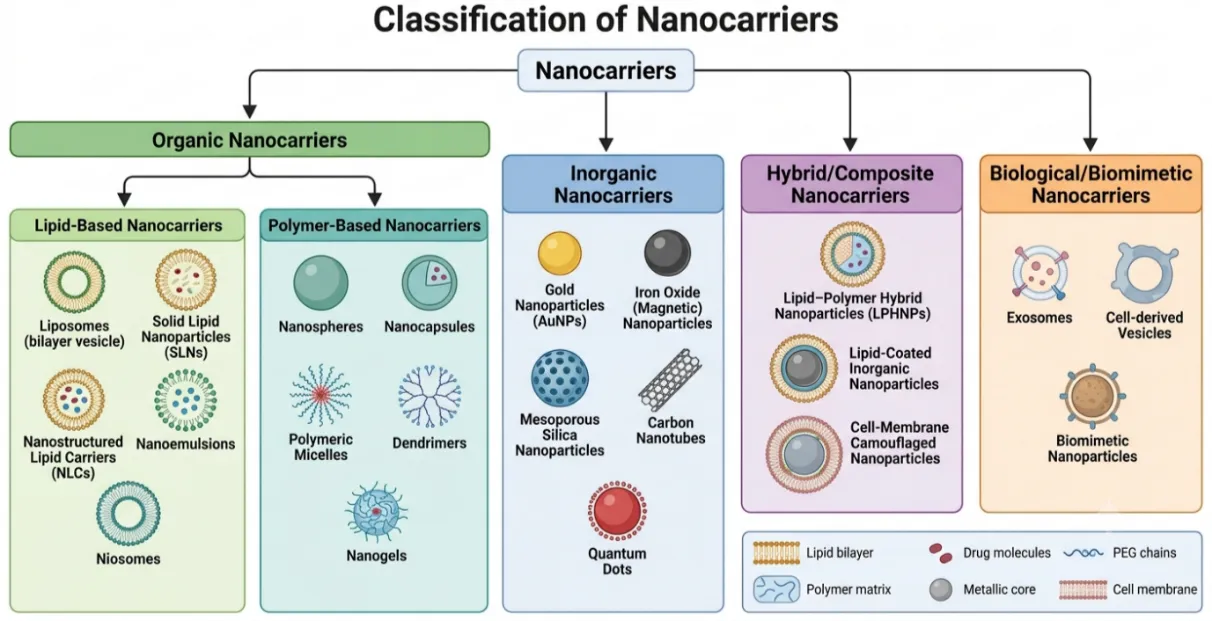
Figure 1: Classification of nanocarriers based on the materials used in their construction. Organic nanocarriers include lipid-based (liposomes, SLNs, NLCs, nanoemulsions, and niosomes) and polymer-based systems (nanospheres, nanocapsules, micelles, dendrimers, and nanogels). Inorganic nanocarriers comprise gold nanoparticles, iron oxide nanoparticles, mesoporous silica nanoparticles, carbon nanotubes, and quantum dots. Hybrid/composite nanocarriers integrate multiple material classes and include lipid–polymer hybrid nanoparticles (LPHNPs), lipid-coated inorganic nanoparticles, and cell-membrane camouflaged systems for enhanced drug delivery performance1,5,18,23,30,31. Created using generative AI.
3. Lipid-Based Nanocarriers
Nanocarriers made of lipids have become a pillar of nanomedicine because of their versatility, safety and their ability to deliver a wide range of therapeutic agent. These systems make use of physiological lipids in order to entrap or conjugate drugs, and then deliver the drugs specifically with minimum side effects. The main types of lipid-based carriers are the liposomes, solid lipid nanoparticles (SLN) and the nanostructured lipid carriers (NLC)5,26,27.
3.1 Liposomes
Liposomes are colloidal vesicles that are self-assembled spherical in structure; the structures are made of a water core and composed of one or more amphiphilic phospholipid bilayers. They are the most widely studied lipid-based system discovered in the 1960s, and have had great clinical success with notable example products such as Doxil (liposomal doxorubicin)27,32,33.
• Structure and Capability: They possess a unique amphiphilic character enabling them to entrap hydrophilic and hydrophobic drugs to the aqueous center and the lipid bilayer respectively. These can be sized and lamellar to form small unilamellar vesicles (SUV), large unilamellar vesicles (LUV) and multilamellar vesicles (MLV) 27,30,33.
• Performance: Liposomes are very biocompatible because they mimic cell membranes that are part of nature and this makes them become easily absorbed by cells. The traditional liposomes, however, have had their faults of lack of physical stability, leakage of drugs and clearance by reticuloendothelial system (RES)5,34.
3.2 Solid Lipid Nanoparticles (SLN)
In the 1990s, SLNs were created as the first generation to replace liposomes and polymeric nanoparticles and merge their benefits. They are made of solid lipid core, which is made of high-melting-point lipids such as triglycerides, fatty acids, or waxes, and is stabilized by surfactants in an aqueous dispersion35.
Structural Models: SLNs are usually characterized by three different structures: the homogeneous matrix model (Type I), drug-enriched shell model (Type II) and drug-enriched core model (Type III)36.
Performance: SLNs Compared to liposomes, the solid matrix enables the encapsulated drugs to be better-protected against chemical degradation and the release of the drugs can be more precisely-controlled and sustained. They are also less hazardous than polymeric particles since they do not require the application of organic solvents in the manufacturing process. Their principal deficiency is a comparatively low drug loading ability and the possibility of loss of drugs expelled by being squandered throughout storage as a result of lipid polymorphic transitions30,36.
3.3 Nanostructured Lipid Carriers (NLC).
NLCs are a type of second-generation lipid nanoparticles, which are intentionally developed to address the drawbacks of SLNs. They consist of a mixture of solid lipids and liquid lipids (oils), usually in the proportion of between 70:30 and 99:1 18,37.
• Structural Innovation: Liquid lipid forms an imperfect and less ordered crystalline structure with increased voids and spaces. NLCs are of three different types: imperfect crystal (Type I), amorphous (Type II), and multiple type (Type III)27,36.
• Performance: This disorderly structure substantially raises the amount of drug loading and the elimination of active substances throughout shelf life38. NLCs are more stable in storage and can even better control drug release profiles than SLNs27.
3.4 The main benefits of fatty Nanocarriers.
Biocompatibility: These carriers consist mainly of physiological, bio-degradable lipids which are considered as Generally Recognized as Safe (GRAS)27. Their biomimetic characteristics guarantee the low systemic toxicity and low immunogenicity, which means that the human body tolerates them well23,39.
Controlled Release: SLNs and NLCs consist of a solid matrix, which effectively fixes the drug molecules and therefore drastically decreases the mobility of such molecules relative to that of liquid emulsions. This allows a slow delivery of the payload during prolonged intervals, which enhances the therapeutic index and lowers the dosing frequency13,38.
Lipid nanocarriers are remarkably efficient at solubilizing drugs that are poorly water-soluble (hydrophobic) in solution. They improve the apparent aqueous solubility and oral bioavailability of potent lipophilic chemotherapeutic agents by encasing them in their lipid cores or bilayers5,6.
Table 1: Comparison of Lipid-Based Nanocarriers
|
Nanocarrier |
Structure |
Advantages |
Limitations |
|
Liposomes |
Spherical vesicles with an aqueous core enclosed by one or more phospholipid bilayers. |
High biocompatibility; mimics cell membranes; can carry both hydrophilic and hydrophobic drugs. |
Low physical stability; potential for drug leakage; high production costs; rapid RES clearance.5,26,27 |
|
Solid Lipid Nanoparticles (SLN) |
Solid colloidal particles with a solid lipid core at room and body temperature. |
Improved drug stability; controlled/prolonged release; absence of organic solvents; easier to scale up. |
Limited drug loading capacity; risk of drug expulsion during storage (polymorphic transition).27,30 |
|
Nanostructured Lipid Carriers (NLC) |
Second-generation lipid particles with an imperfect matrix of solid and liquid lipids. |
Enhanced drug loading capacity; minimized drug expulsion; superior physical stability during storage. |
Potential for particle aggregation; technical complexity in achieving consistent symmetry.23,27 |
4. Polymeric Nanoparticles
Polymeric nanoparticles (NPs) represent very versatile drug delivery systems (DDS) with dimensions that are usually between one and 1000 nm. These systems are made of natural polymers such as chitosan, albumin and hyaluronic acid or synthetic polymers which include poly (lactic-co-glycolic acid) (PLGA), poly (lactic acid) (PLA) and poly (epsilon-caprolactone) (PCL)5,40. They are morphologically classified as nanospheres (a form of matrix systems in which the drug is evenly dispersed) and Nano capsules (reservoir systems in which a polymeric shell is capable of covering a drug-filled core). Their versatility enables them to package a large diversity of therapeutic agents, such as small-molecule drugs, proteins, and nucleic acids21,28.
4.1 The most important Polymeric Nanocarrier Types.
PLGA Nanoparticles: PLGA is considered among the most common polymers used in nanomedicine because of its high biocompatibility and biodegradability. It is an FDA approved copolymer that can degrade through hydrolysis to the constituent monomers lactic and glycolic acid which are readily degraded by the metabolic systems of the body. PLGA NPs are especially useful in maintaining a sustained release of drugs; an example of this is the PLGA (50:50) which has the advantage of having a balance degradation rate that will be compatible with delivery of chemotherapeutics to the tumor cells that are actively growing5,41.
Polymeric Micelles: Micelles are self-assembled colloidal nanostructures (usually 10-100 nm) of amphiphilic block copolymers. When the concentration of polymer is higher than the critical micelle concentration (CMC), they spontaneously assemble to form a hydrophobic core to which poorly water-soluble drugs can be solubilized and a hydrophilic shell (usually PEG), which offers stability in aqueous conditions42. It is an efficient architecture in enhancing the pharmacokinetics of drug lipophilic chemotherapeutics such as paclitaxel5.
Dendrimers: These are mono-disperse, highly branched, 3D synthetic macromolecules. Dendrimers grow out radially around a central core with several layers (generations) that have high densities of surface functional groups. The ligand conjugation and active targeting can be done with these groups43. Dendrimers Polyamidoamine (PAMAM) dendrimers are also of interest due to their potential to condense genetic material and be able to bypass the endosome via the so-called proton-sponge effect4.
4.2 Benefits: Stability and Controlled Release of drugs.
Mechanical and Storage stability: Polymeric nanoparticle uptakes tend to be more physically and mechanically stable than lipid-based systems. They have solid matrix or strong core-shell structure, which offers high structural integrity, which ensures the safe encapsulated payloads against the premature degradation and leakage in storage and systemic circulation22,26,44.
Controlled and Sustained release: It is one of the main advantages of polymeric NPs that it is possible to tunable drug release kinetics. A rate of release is usually controlled by a product of drug diffusion and such rate of degradation/erosion of polymer matrix. As an example, the ratio of lactic to glycolic in PLGA can be modified to produce release profiles of days to weeks to provide steady therapeutic concentration at the site of interest. Moreover, "smart" polymers can be designed as being stimuli-responsive, responsive to ambient conditions, such as acidic tumor pH, or increased enzyme concentration5,45.
4.3 Limitation: Toxicity and RES Clearance.
Cytotoxicity Concerns: Surface charge and composition of polymeric nanocarrier have a significant effect on their safety. Cationic polymers (e.g., polyethylenimine or amine-terminated dendrimers) have been reported to have high toxicity due to their high density of positive charges that is capable of disruption of cell membranes and resultant cell death46,47. Moreover, a potential amount of non-biodegradable fragments of polymer or residual solvents that have toxic consequences in the manufacture can be dangerous in the long term23.
RES and MPS Clearance: When polymeric NPs are injected into the body, the immune system seems to detect them as foreign bodies in the body. Particles larger than about 200 nm and of hydrophobic surface are prone to opsonization, whereby they become covered by plasma proteins and then removed via the mononuclear phagocytic system (MPS), which is mainly located in the liver and spleen48,49. PEGylation (PEG shelling) is employed to form so-called stealth particles, which avoid the MPS but it may cause the so-called PEG dilemma, where the hydrophilic shell prevents interaction of the nanoparticle to target cancerous cells. Moreover, the accumulation of the PEGylated systems can cause accelerating blood clearance (ABC) by the development of anti-PEG antibodies23,26.
5. Lipid–Polymer Hybrid Nanoparticles
Lipid-polymer hybrid nanoparticles (LPHNPs) are an important advancement of nanomedicine, developed with a synergy between the mechanical stability of polymeric nanocarriers and the biomimetic benefits of lipid-based systems 5,50. Allowing these materials to interact, LPHNPs would overcome the shortcomings of their predecessors: the low physical stability and leakage of drugs found in liposomes, and the immunogenicity or low cellular interaction of purely polymeric particles 4,26,44. These novel core-shell structures have high drug-loading efficacy, controlled release kinetics, and a strong platform to deliver therapeutic agents and theranostic utilization18,28,44.
5.1 Structure of LPHNPs
A typical designation of the standard architecture of an LPHNP is that of a three-layer concentric structure with each component playing a specific functional role33,41,51.
Polymer Core: The core is made up of the innermost area that is a hydrophobic polymeric framework, which is typically an FDA-approved, biodegradable polymer which could be either PLGA, PLA, or polycaprolactone (PCL)2,3. This core gives the particle its structural pillar, allows the loading of large amounts of hydrophobic therapeutic drug, and regulates a burst of the cargo whether over time or triggered by stimuli 33,52.
Lipid Layer: The lipid monolayer or bilayer, which coats around the polymeric core, is usually made of phospholipids such as lecithin or phosphatidylcholine52. This layer provides a molecular "resistance to diffusion" that retards the diffusion of water into the core and slows the hydrolysis of polymer and greatly diminishes the burst release effect53. Moreover, such lipid mantle also offers the biomimetic interface to achieve better cellular interaction and biocompatibility54.
PEG Outer Coating: The outermost layer is made up of a hydrophilic layer of polymer, usually PEGylated lipids (e.g., DSPE-PEG)21,23. This type of coating can be considered as a stealth coating which offers steric stabilization of the nanoparticles and prevents aggregation of particles and opsonization by plasma proteins. This layer assures systemic circulation time by preventing the reticuloendothelial system (RES) to recognize it as a foreign body 55,56.
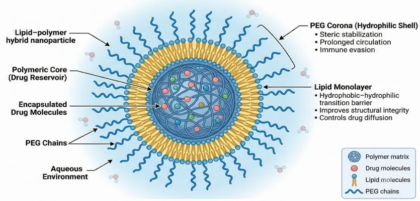
Figure 2. Structure of a lipid–polymer hybrid nanoparticle (LPHNP). The nanoparticle consists of a drug-loaded polymeric core surrounded by a stabilizing lipid monolayer and an outer PEG corona that enhances colloidal stability, prolongs systemic circulation, and reduces immune recognition13,21,57. Created using generative AI.
5.2 Types of LPHNPs
To meet the therapeutic requirements and delivery pathways of particular groups of patients, LPHNPs may be designed to take different forms58.
Polymer Core-Lipid Shell Hybrid Nano-systems: The most frequent and the most studied structure. Under this model, the therapeutic agent is wrapped in the solid polymeric core and it is surrounded by a lipid layer. The structure is perfect in providing drug delivery of hydrophobic drugs, with the polymer maintaining the lipid layer and the lipid giving biocompatible surface reducing the possibility of the polymer to interact with non-specific molecules59.
Monolithic Hybrid Nanoparticles: Unlike the separate core-shell models, monolithic hybrids (or mixed lipid-polymer NPs) have a structure in which lipids are evenly distributed in the polymer core. This architecture generates a rare example of a colloidal vehicle that works exceptionally well to entrap highly lipophilic drug molecules that would be difficult to entrap in a typical polymer matrix. Researchers can stabilize the particles and systemic toxicity of the particle by adjusting the mixing ratios of lipids and polymers 60.
Hollow Core (Lipid-Polymer-Lipid) Structures: This is an A-B-A form of complex with an inner hollow core (filled with water or buffer) encased in an inner lipid layer, a middle hydrophobic polymer shell and an outer PEGylated lipid layer. The lipid layer is often cationically charged, and these particles are ideal in the encapsulation of anionic drugs or genetic materials (siRNA, mRNA). It is possible to use this design to deliver two or more agents at the same time, e.g., a hydrophilic genetic cargo in the core and a hydrophobic small molecule in the polymeric shell61.
Cell Membrane Camouflaged (Biomimetic) Nanoparticles: This is an advanced system that entails the clock wrapping of a polymeric core in a natural cell membrane which is based on red blood cells (RBCs), platelets, or leucocytes. Such biomimetic surfaces confer the synthetic nanoparticles with the innate biological attributes of the source cells including excellent immune evasion and natural targeting abilities. Although RBC-camouflaged systems are highly effective in providing an effective barrier to low drug release and prolonged circulation, leucocyte-camouflaged systems have the capability of actively transporting the nanoparticles to inflammatory or tumor sites57.
The different types of LPHNPs provide the opportunity to make multifunctional platforms, which can be functionalized with targeting ligands (such as antibodies or peptides) and stimuli-responsive components to achieve unmatched precision in cancer therapy.
6. Preparation Methods of Hybrid Nanoparticles
Lipid-polymer hybrid nanoparticles (LPHNPs) synthesis is developed taking into consideration the stable core-shell configuration, which combines the proven benefits of both types of materials. These approaches can be broadly divided into one-step and two-step approaches, with the components being assembled at the same time or in a sequence. Method selection has strong effects on critical quality attributes which include particle size, polydispersity and encapsulation efficiency of drugs 62.
6.1 One-Step Methods
One-step processes are gaining considerable popularity due to their efficiency, time-saving ability, and enhanced prospects of scale-up to industrial dimensions. Such procedures are based on the co-assembly of the lipid shell and the polymeric core.
Nanoprecipitation: This is the most popular one-step method that is commonly known as solvent diffusion. Here, a water-miscible organic solvent (e.g. acetonitrile, ethanol or acetone) is used to dissolve the polymer and hydrophobic drug, and the lipids and PEGylated lipids are dispersed in an aqueous phase. On dropwise addition of the organic phase to the aqueous phase under constant stirring, the solvent quickly diffuses and leads to the precipitation of the polymer as nanospheres.
Self-Assembly: The lipids self-assemble in activity with the precipitation of the polymer. Hydrophobic interactions are responsible for this self-assembly with the non-polar lipid tails sticking to the hydrophobic polymer surface and the hydrophilic heads exposed to the external aqueous environment stabilizing. The process usually gives a uniform and uniformly sized LPHNPs that are less than 100 nm, and is very successful in encapsulating hydrophobic therapeutic agents63,64.
6.2 Two-Step Methods
Two-step strategies involve the synthesis of the polymeric core and lipid shell parts, and then the incorporation of the two to form one hybrid structure. The approaches also enable the control of the properties of each particular layer to a high degree of precision, but such methods are more time-consuming.
Emulsification-Solvent Evaporation: This is a flexible method that can also be carried out as a single (oil-in-water) or a double (water-in-oil-in-water) emulsion. In the case of hybrid synthesis, the polymeric core can easily be pre-formed through emulsification and subsequently combined with already formed lipid vesicles or a thin lipid film. The absorption of the lipid shell onto the polymer surface is aided by the input of external energy, e.g. ultrasonication or vortexing, usually through electrostatic attraction. This approach is especially effective in entrapment of hydrophilic drugs or nucleic acids in the water droplets of the hybrid structure65,66.
High-Pressure Homogenization (HPH): HPH is an efficient and robust technique that is mostly intended to pre-shape the polymeric nanoparticles or to homogenate the final hybrid blend to the final homogenised size distribution. It entails the extrusion of the pre-emulsion using a small orifice under extremely high pressures at which mechanical shear and cavitation forces destruct the microparticles into the nanoscale. The method is highly desirable because of its reproducibility and its usefulness in large-scale manufacturing, but it might not be suitable with highly heat-sensitive payloads because of the heating energy produced during processing27,67.
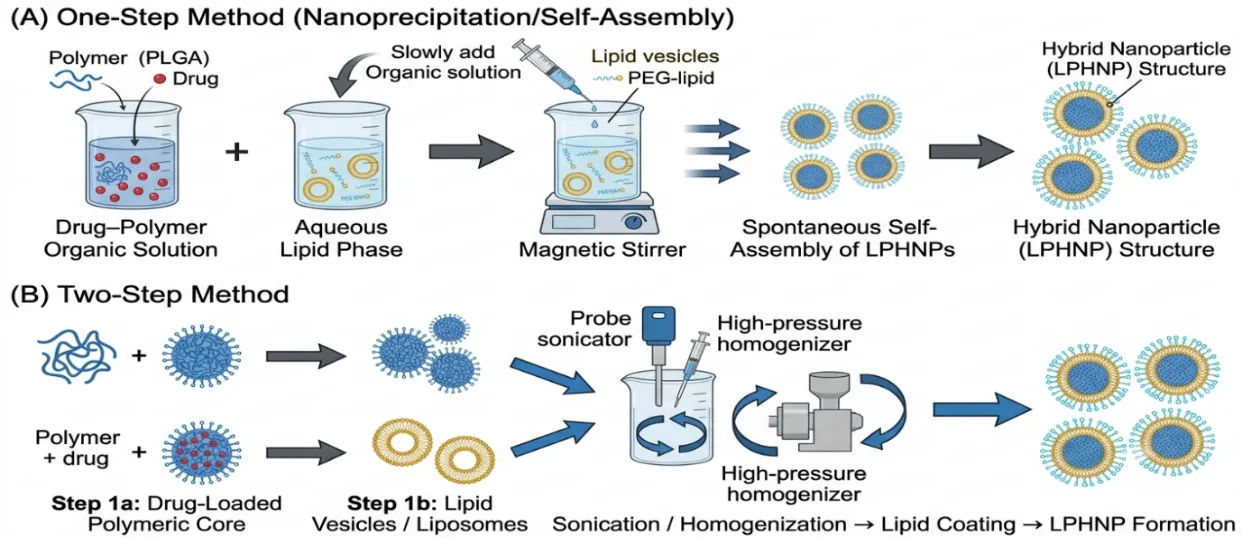
Figure 3. Schematic illustration of lipid–polymer hybrid nanoparticle (LPHNP) synthesis methods. (A) One-step method, where a drug–polymer organic solution is added to an aqueous lipid phase, resulting in spontaneous self-assembly of core–shell hybrid nanoparticles. (B) Two-step method, in which drug-loaded polymeric nanoparticles and lipid vesicles are prepared independently and subsequently combined by sonication or homogenization to produce lipid–polymer hybrid nanoparticles13,18,21. Created using generative AI.
Table 2: Summary of Fabrication Parameters
|
Method |
Phase Composition |
Driving Force |
Key Advantage |
|
Nanoprecipitation |
Water-miscible organic solvent + Aqueous lipid phase. |
Solvent diffusion and hydrophobic interaction. |
Simple, produces small (<100 nm) uniform particles. |
|
Self-Assembly |
Integrated during nanoprecipitation or microfluidics. |
Hydrophobic and electrostatic interactions. |
Rapid formation of stable core-shell units. |
|
Emulsification |
Oil-in-water (o/w) or water-in-oil-in-water (w/o/w). |
Mechanical shear and solvent evaporation. |
High encapsulation for both hydrophobic and hydrophilic drugs. |
|
High-Pressure Homogenization |
Melted lipid/polymer dispersion or pre-emulsion. |
Mechanical shear and cavitation. |
Excellent reproducibility and industrial scalability13,18,23. |
7. Characterisation Techniques
Detailed characterisation of lipid-polymer hybrid nanoparticles (LPHNPs) is needed to confirm their integrity, foresee their behaviour in vivo and to guarantee batch to batch reproducibility. Since they have a complex core-shell structure, they need a multifaceted analysis to investigate their physicochemical properties, drug loading capacity, and reaction to environmental factors65,68.
7.1 Particle Size and Polydispersivity (DLS):
Photon correlation spectroscopy, or Dynamic Light Scattering (DLS) is the commonly used method of measuring hydrodynamic diameter and size distribution of LPHNPs in suspension. The non-destructive technique follows the Brownian movement of the particles in order to measure their size, usually to a range of 50-200 nm in order to take advantage of the Enhanced Permeability and Retention (EPR) effect in tumours. Polydispersity Index (PDI) is also measured and a PDI of less than 0.3 represents a monodisperse and stable system, higher PDI values indicate aggregation or heterogeneity. Stimulus responsive behaviour, i.e. increase in size or disassembly of particles in the presence of a reducing agent or acidic pH, is also monitored using DLS14,50,69.
7.2 Zeta Potential:
Zeta potential is the measure of the electrokinetic potential at the plane slipping of the particle, which gives a quantitative value of the surface charge. This parameter is a key measure of colloidal stability; the absolute values (either positive or negative) are high, which facilitates repulsion on the basis of electrostatic interactions between nanoparticles, avoiding their aggregation. In the case of LPHNPs, the zeta potential usually depends on the polymer core (e.g., deprotonated carboxyl groups of PLGA) or functional lipids employed in the shell. Surface alterations, e.g. PEGylation, generally lower the absolute zeta potential by shielding the surface charge that aids the particles to avoid immune detection44,46.
7.3 Morphological Imaging (TEM / SEM):
Transmission Electron Microscopy (TEM): It is the instrument that can be used to verify the core-shell architecture. As a result of density dissimilarity amid the polymeric core and the lipid shell, TEM has the capacity to frequently differentiate the thin lipid ring (normally less than 5 nm) around the solid core. Cryo-TEM is getting more and more popular because it maintains the nanoparticles in their native, hydrated condition, eliminating the artifacts due to drying or staining33,70.
Scanning Electron Microscopy (SEM): SEM yields images that resemble a topography map of the surface in three dimensions and shape, typically demonstrating that LPHNPs are homogenous, smooth, and round51.
7.4 Encapsulation Efficiency (EE) and Drug loading (DL).
The EE and DL are calculated to identify the ability of the nanocarrier to carry its therapeutic cargo.
Encapsulation Efficiency: This is the ratio of the original drug that gets entrapped in the nanoparticles.
Drug Loading: Refers to the ratio of the mass of the drug to the overall mass of the lipid-polymer carrier. The values of drug loading are usually measured by ultrafiltration-centrifugation to isolate free, unencapsulated drug and the nanoparticles. Quantification of the concentration of the drug in the filtrate or supernatant is then done by using either HPLC, UV-Vis, or fluorescence spectroscopy14,21.
7.5 Drug Release Studies
The dialysis bag is the most common mode of measuring the release kinetics of LPHNPs. In this case, the drug-loaded nanoparticles are introduced into a semi-permeable membrane and put in a release medium (generally, PBS at 37deg C). To compare stimuli-responsiveness, release is sometimes compared at physiological pH (7.4) and at pH 5.5 of tumour/lysosome. The obtained data are modeled to mathematical equations, e.g., the Higuchi (diffusion-controlled) or Korsmeyer-Peppas equations, to explain the drug transport mechanism14,50,71.
7.6 Stability Analysis
Physical stability studies on several months determine the long-term viability of LPHNPs. The size of particles, PDI, and zeta potential are monitored in different storage conditions e.g. 4degC (refrigerated) and 25degC (room temperature). The majority of LPHNP formulations have a higher stability at refrigeration temperature, whereas higher temperatures or humidity can provoke a lipid phase transition, structure, or drug leakage44,72.
Table 3: Characterisation Techniques for LPHNPs
|
Technique |
Purpose |
|
Dynamic Light Scattering (DLS) |
Measures hydrodynamic size, PDI, and monitors stimuli-induced size changes14,33. |
|
Zeta Potential |
Determines surface charge to predict colloidal stability and confirm surface modification21. |
|
TEM / Cryo-TEM |
Visualises internal core-shell morphology, size, and structural integrity. |
|
SEM |
Examines surface topography, 3Dshape and distribution51. |
|
Centrifugation / HPLC |
Quantifies encapsulation efficiency and drug loading capacity50. |
|
Dialysis / Franz Cell |
Evaluates drug release kinetics and stimuli-responsive release profiles31. |
|
DSC / XRD |
Assesses drug crystallinity, polymer interactions, and thermal phase transitions44. |
|
FTIR |
Identifies functional groups and confirms successful chemical conjugation of ligands. |
8. Stimuli-Responsive Nanocarriers
Traditional drug delivery methods are usually characterised by uncontrolled drug release resulting in inefficient therapeutic levels at the tumour with systemic toxicity . In order to defeat these drawbacks, stimuli-responsive nanocarriers (or so-called smart) delivery systems have been invented. These are intelligent platforms designed to be stable and pharmacologically neutral in systemic circulation but to undergo rapid physicochemical changes, e.g., disassembly, swelling, bond cleavage, etc. in response to some triggers. These stimuli can be broadly divided into internal (endogenous) and external (exogenous) stimuli where internal stimuli take advantage of the special pathophysiological characteristics of the tumour microenvironment (TME) and external stimuli are applied extracorporeally to attain the desired spatiotemporal control of drug release31.
8.1 Internal Stimuli
Internal stimuli-responsive systems operate in an autonomous mode and are activated by the presence of hallmark biochemical conditions of the tumour or its intracellular compartments.
pH-Responsive: The most studied endogenous trigger is the pH-responsive as it relies on reliable pH gradients present in tumor tissues. The Warburg effect causes tumour cells to have high glycolytic activity and have low vascular perfusion, resulting in excessive production of lactic acid and subsequent extracellular TME acidification (pH = 6.5-6.9) relative to the normal tissues (pH = 7.4)6. Besides, after being taken over through the process of endocytosis, nanocarriers enter even more acidic conditions in endosomes and lysosomes (pH 4.5-5.5). PH-triggered release mechanisms encompass either the utilization of acid-sensitive chemical bonds (e.g., hydrazone, imine or acetal) to break, or the addition of protonatable functional groups (e.g., tertiary amines or imidazole) to induce a hydrophobic-hydrophilic switch, leading to the swelling/dispersal of nanocarriers8.
Redox-Responsive: Redox imbalance is unique to tumours with high concentration of intracellular reducing agents, which are mainly glutathione (GSH). The tumour cytosol GSH levels are frequently 100-1000 times greater (2-10 mM) than those in the extracellular environment or blood (2-20.5 mM). The most common linkers or cross linkers in redox-sensitive nanocarriers are disulfide (-S-S-) or diselenide (-Se-Se-) bonds1. These bonds are soluble in the blood but cleave rapidly in high-GSH conditions allowing release of site-specific payloads directly into the nucleus or cytosol1,73.
Enzyme-Responsive: Tumour progression is linked to the impaired expression and over-expression of certain proteolytic enzymes. The enzymes that are frequently exploited are matrix metalloproteinases (MMPs), cathepsins and hyaluronidases. Functional groups may be enzyme-sensitive peptide sequences (e.g., GFLG to Cathepsin B or GPLGIAGQ to MMP-2) incorporated into nanocarriers, or these natural polymers can be functionalised (e.g. MMP-2 by incorporating GPLGIAGQ), or degraded (e.g. gelatin by Cathepsin B by inclusion of GFLG) when exposed to an overexpression level of these pathological enzymes, and this leads to localised activation of drugs in the tumour cell1,8,73
8.2 External Stimuli
Exogenous stimuli provide clinicians with the opportunity to directly control the time, place, and amount of drug release and minimize the effects of biological heterogeneity among patients5.
Temperature-Responsive: The temperature of tumour tissues is mildly higher than that of normal tissues based on inflammation and high metabolism. The thermosensitive nanocarriers are designed to change state (e.g. gel-to-sol or gel-to-liquid crystalline) at a defined trigger temperature, usually somewhere between 40 -45degC (mild hyperthermia). Common examples of such materials are polymers such as PNIPAAm, which fold at temperatures above its lower critical solution temperature (LCST), and thermosensitive lipids such as DPPC, which unfold with warming at5,31.
Light-Responsive: Light has been found to be the optimal trigger since it is highly spatial and non-invasive. Although U.V. and visible light have limited penetration, near-infrared (NIR) light (600-1000 nm) is desirable in penetrating deep tissues. Photosensitizer-based photodynamic therapy (PDT), plasmonic-based photothermal therapy (PTT), converting light into heat to warm the carrier and liberate the drug are frequently used as light-activated nanocarriers1,33,74.
Ultrasound-Responsive: Ultrasound has deep tissue penetration and is already a common practice in clinical diagnostics. It causes the release of drugs by thermal mechanisms (localized heating) and non-thermal mechanism, especially acoustic cavitation. The microbubbles formed by cavitation can cause extension and temporary permeabilization of cell membranes (sonoporation) through physical force and cause deep tumour penetration and high levels of cellular uptake by the nanocarrier2,33.
Magnetic Field-Responsive: Magnetic fields show little biological interaction, and no biological constraint on penetration depth of tissue. Such systems usually use superparamagnetic iron oxide nanoparticles (SPIONs) that are encased in the nanocarrier core. These particles produce heat under an alternating magnetic field (AMF) due to the magnetic relaxation process which causes structural disruption of the carrier and release of drugs. Also, magnetic targeting can be performed with the use of magnetic fields, in which case the nanocarriers will be guided to desired locations in the anatomy with the help of external magnets2,9.
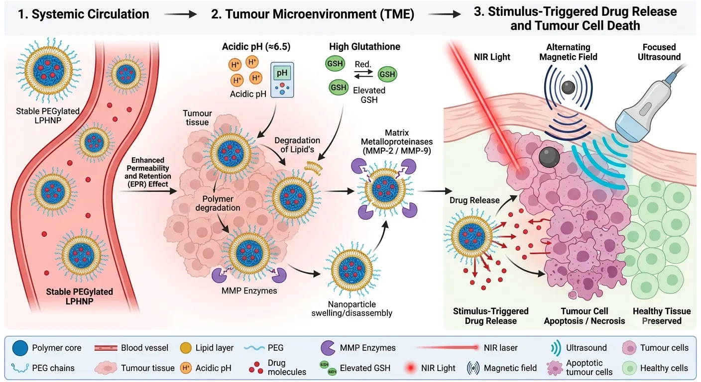
Figure 4. Tumour microenvironment-responsive lipid–polymer hybrid nanoparticles (LPHNPs). Following passive accumulation within the tumour via the enhanced permeability and retention (EPR) effect, endogenous tumour-specific stimuli—including acidic pH, elevated glutathione (GSH), and matrix metalloproteinases (MMPs)—induce nanoparticle disassembly, polymer degradation, or surface charge reversal, resulting in site-specific drug release. External stimuli such as near-infrared (NIR) light, alternating magnetic fields, and focused ultrasound further provide spatiotemporal control over drug release, leading to localized tumour cell apoptosis and necrosis while minimizing damage to surrounding healthy tissues3,8,75. Created using generative AI.
Table 4: Comparison of Stimuli-Responsive Triggers
|
Stimulus |
Type |
Mechanism of Action |
Key Advantage |
|
pH |
Internal |
Protonation of amines; acid-labile bond cleavage. |
Exploits universal tumour acidosis8. |
|
Redox |
Internal |
GSH-mediated disulfide/diselenide bond cleavage. |
High intracellular vs. extracellular gradient1. |
|
Enzyme |
Internal |
Peptide cleavage by MMPs or Cathepsins. |
High biological specificity to disease stage2. |
|
Light |
External |
Photothermal heat; PDT-mediated ROS; photocleavage. |
Exceptional spatial and temporal precision6. |
|
Ultrasound |
External |
Thermal hyperthermia; mechanical cavitation. |
Deep tissue penetration; non-invasive33. |
|
Magnetic |
External |
Magnetothermal energy conversion; mechanical stress. |
No tissue penetration limits; enables MRI9. |
9. Cancer Therapy Applications.
Oncology LPHNPs are a versatile and robust platform that can address the fundamental failures of traditional therapies using a multimodal platform. These nanocarriers combine the structural integrity of the polymers and the biomimetic surfaces of lipids to uniquely target the challenges of complex biological barriers of a human body. Their use in significant types of cancer has shown significant promise in altering the norms of drug delivery in both site-specific activation therapy and synergistic conjunction therapies21,22.
9.1 Breast Cancer
Breast cancer is a leading cause of morbidity and mortality among women worldwide, and usually complicated by tumour aggressiveness, including Triple-Negative Breast Cancer (TNBC) and tumours with HER2-positive.
Targeted Drug Delivery: LPHNPs are often functionalised to include ligands such as trastuzumab or EGFR antibodies to enhance the specificity of cancer cells but not healthy tissue. Indicatively, EGFR-based nanoparticles that were loaded with siXBP1 yielded a 75 percent decrease in gene expression, which in this case was under hypoxia conditions common in TNBC.
Improved efficacy: Hybrid systems that deliver chemotherapeutic agents and RNA therapeutics (siRNA/miRNA) have proved effective in overcoming multidrug resistance (MDR). Telomeric DNA damage induced with miR-182-3p was effective in hybrid systems, which decreased tumour volume by half in TNBC models.
Combination Strategies: It has been shown that LPHNPs can be used in chemo-radiotherapy sensitisation; silencing of the RAD50 protein with siRNA-loaded hybrid particles enhanced considerably the effectiveness of ionising radiation on resistant breast cancer cells10.
9.2 Pancreatic Cancer
The high interstitial fluid pressure and dense stroma (desmoplasia) of pancreatic ductal adenocarcinoma (PDAC) can be regarded as potent physical obstacles to drug penetration.
TME Modulation: Smart hybrid systems are aimed at remodelling tumour microenvironment. Deep tumour penetration of encapsulated payload can be achieved by LPHNPs functionalised with hyaluronidase to degrade the extracellular matrix.
Enhanced Pharmacokinetics: Standard schedules such as FOLFIRINOX have a high systemic toxicity. Drugs such as irinotecan have been shown to have their half-life extended using bilayer-coated mesoporous silica or bilayer-cores made of PLGA that reduce off-target adverse side effects and cause a significant increase in tumoral drug concentration.
Theranostic Potential: Therapies SPIONs can be engineered into therapeutic hybrids to enable real-time monitoring of therapeutic efficacy through the use of MRI contrast enhancement. Cancer of the liver (hepatocellular carcinoma) is a type of malignancy that impacts the liver and may cause liver failure in severe cases. The stage-of-diagnosis and non-response to general multikinase inhibitors frequently restrict the treatment of liver cancer.
Active Targeting: Hybrid Targets Hybrid systems that make use of the asialoglycoprotein receptor by using GalNAc ligands have shown an extreme specificity to HCC cells. Likewise, it has been demonstrated that iRGD-functionalized hybrid particles can also be targeted to HCC cells and tumour vasculature simultaneously.
Biomimetic Approaches: New approaches include the coating of the PLGA cores with homotypic membranes of HepG2 cells, giving the particles natural immune avoidance and homing properties. Such biomimetic systems have so far achieved a tumour volume reduction of up to 90 per cent in preclinical models.
Overcoming Resistance: It has been demonstrated that the co-delivery of sorafenib and selumetinib using targeted hybrid nanosystems can enhance the programmed cell death in malignant hepatocytes without causing harm to non-tumour cells2.
9.3 Lung Cancer
Lung cancer is the most prevalent cancer-related cancer in the world, and some new delivery channels are needed to enhance the quality of life of the patients.
Pulmonary Delivery: LPHNPs are especially applicable to inhalable preparations, which provide direct delivery to the lungs, low system exposure, and high compliance with patients.
Redox-Responsive Release: Lung tumours have elevated fractions of glutathione (GSH) in the cell. Hybrid particles containing disulfide bonds are stable when circulating and dissociate quickly when they enter the reductive microenvironment of lung cancer cells, guaranteeing the accurate delivery of cargo.
Synergistic Outcomes: Paclitaxel and triptolide loaded into LPHNPs resulted in complete tumour remission in resistant NSCLC models at combinations (77.4 per cent) that is synergistic, indicating that the platform can overcome conventional chemoresistance8,22.
9.4 Mechanistic Benefits of LPHNPs.
Targeted Drug Delivery: LPHNPs take advantage of active targeting by conjugation of ligands (folate, aptamers, peptides) to specific tumour cell receptors excessively created on the tumour cell surface, including FRa, EGFR, and CD44. This is complemented by passive targeting through the EPR effect, which is characterized by the small size (usually 100-200 nm) of the particles, which leads to accumulation in the leaky tumour vasculature6,18.
Enhanced Pharmacokinetics: PEGylated lipid shell forms a so-called stealth layer that reduces opsonization and avoids the mononuclear phagocytosis system (MPS), which significantly prolongs systemic circulation time. Pharmacokinetic investigations have revealed that hybrid formulations are able to enhance the half-life of a drug three to eightfold of free drug solutions26.
Improved Therapeutic Potency: LPHNPs can be used to achieve pharmacological synergy by favoring the simultaneous co-delivery of particularly hydrophobic drugs (core) and hydrophilic RNA (shell). Moreover, stimuli-responsive procedures (pH, redox, enzymes) make sure that the highest dose of the therapeutic is not released until the tumour site is reached, radically enhancing the therapeutic index and decreasing the side effects on the systemic level Advances in Lipid-Polymer Hybrid Nanoparticles Design4,23.
Table 5. Summary of LPHNP Applications in Targeted Cancer Therapy
|
Cancer Type |
Therapeutic Cargo |
Targeting Strategy / Stimulus |
Key Therapeutic Outcome |
|
Breast |
Paclitaxel + ABCB1-siRNA |
Trastuzumab (HER2) |
Reversed MDR; synergistic inhibition of tumour growth10. |
|
Breast |
Docetaxel |
pH-sensitive (Acidic TME) |
Improved cell uptake via charge reversal; 5–6x longer half-life22. |
|
Pancreatic |
Gemcitabine + pGem |
MMP-2 / Hypoxia-responsive |
Deep penetration into dense stroma; softened tumour ECM. |
|
Pancreatic |
Irinotecan |
Lipid-coated Silica NP |
Reduced systemic toxicity; enhanced tumoral drug concentration2. |
|
Liver |
Sorafenib + Selumetinib |
GalNAc Ligand (ASGPR) |
Precise targeting to HCC; increased programmed cell death. |
|
Liver |
Adriamycin (DOX) |
Homotypic HepG2 Membrane |
90% tumour volume reduction; superior immune evasion18. |
|
Lung |
Erlotinib + Bevacizumab |
Hyaluronic Acid (CD44) |
Synergistic anti-angiogenic effect; improved survival rate22. |
|
Lung |
Paclitaxel (Dimeric) |
GSH-responsive / Inhalable |
44.9% drug loading; superior safety vs. free Taxol8. |
10. Challenges and Limitations
Even though lipid-polymer hybrid nanoparticles (LPHNPs) have the potential to transform the current landscape in oncology, there are a number of scientific, technical, and regulatory bottlenecks that are still impeding their transition of laboratory research to large-scale clinical use. These multi-layered systems are associated with an inherent complexity, which introduces a special set of challenges that should be overcome to make them safe and reproducible2,8.
10.1 The issue of large-scale production:
LPHNPs fabrication is a complex process and it is notoriously hard to be able to attain batch-to-batch reproducibility and uniform quality characteristics at an industrial scale. Although nanoprecipitation and microfluidics, which are used within the laboratory, offer very good control over size and poly-dispersivity of the particles, these systems can often become challenging when scaled to Good Manufacturing Practice (GMP)-compliant scale. The efficiency of drug loading and uniformity in lipid coating a high quantity of batch drug is a key consideration since even small differences in the rate of mixing or solvent evaporation can result in structural flaws of the particle or other undesired aggregation. Moreover, these hybrid architectures can be difficult to prepare, typically they demand a variety of ligands and stimuli-responsive components which also significantly adds to the cost of production and the overall success of making high-quality formulations3,31.
10.2 Nanoparticle Toxicity and Immunogenicity:
Although LPHNPs are meant to be biocompatible, some synthetic ingredients especially cationic lipids or polymers such as polyethylenimine (PEI) may initiate acute cytotoxicity and immune response. Major positive surface charges have been demonstrated to interfere with the membranes of the cells, thereby causing oxidative stress, mitochondrial dysfunction, and even the damage of DNA. Also, the chronic bioaccumulation of inorganic cores or metallic materials in the body that cannot be biodegraded, in key organs of the body as the liver and spleen will lead to serious chronic safety issues. The primary immunological challenge is the PEG dilemma in which recurring use of PEGylated nanoparticle can generate anti-PEG antibodies and leads to faster blood clearance (ABC) and potentially deadly hypersensitivity responses23,46.
10.3 Stability and Storing Issues:
A major prerequisite of clinical application is to assure long-term physicochemical stability of hybrid nanostructures. LPHNPs are prone to physical instability, i.e., aggregation, fusion, or drug leeks, especially in liquid dispersions throughout transportation and storage. The structural integrity of the carrier can be destabilized by chemical degradation such as oxidation of phospholipids or hydrolysis of the polymeric core to modify the kinetics of drug release. In addition, although there are formulae that are stable at refrigerated temperatures (4degC), a large number of these hybrids utilise either costly cold-chain logistics or complex lyophilisation procedures to preserve their activity in the long term5,14,26.
10.4 Regulatory and Translational Issue
Regulatively, the multi-component nature of LPHNPs is quite ambiguous since they occupy a gray area. The fact that no single regulatory framework is present, specifically designed to address hybrid systems, makes it difficult to develop the standard characterization processes and safety levels. The nanomedicines are frequently regarded as drug-device combination products, which must be approved by pharmaceutical and medical device authorities, which greatly increases the process of approval. In addition, the majority of the present preclinical validations are based on animal models that do not recapitulate the heterogeneous human tumour microenvironment and therefore, it is extremely difficult to predict the in vivo efficacy and responses of human patients7,23,31.
FUTURE PERSPECTIVES
The future of lipid-polymer hybrid nanoparticles (LPHNPs) is taking a new turn of paradigm shift in which intelligent design, patient biology, and simplified production come together to rebrand oncology. Although the generation of such nanocarriers has reached several important milestones at the preclinical stage, the new generation of these nanocarriers will be characterized by the possibility to go beyond the classical one-size-fits-all paradigm using computational optimization and biomimetic engineering2,8.
Nanoparticles Design with AI.
Combining machine learning (ML) and Artificial Intelligence (AI) is transforming the LPHNP design by dissolving the constraints of the traditional, experience-oriented design. Nanocarrier-complex biological systems interactions can be predicted through AI algorithms that analyze large data sets to accurately forecast such parameters as encapsulation efficiency, release kinetics, and cellular uptake. Convolutional Neural Networks (CNNs) are advanced deep learning systems that are utilized to process imaging data to predict the response of tumors to certain nanoformulations. Moreover, AI makes it easy to optimize the synthesis parameters like flow rates in microfluidic systems to allow the structural stability and reproducibility demanded by industrial-scale production2,5.
Personalized Nanomedicine
In order to respond to the severely heterogeneous nature of patients, the future of nanomedicine is in the form of personalized, precision-engineered therapeutics. New approaches include the application of AI-based multi-omics data integration (genomics, proteomics, and transcriptomics) to determine patient-specific biomarker patterns, which has offered a molecular basis of individualized LPHNP targeting. The next-generation tumor-organoid models can be used to assess the safety and efficacy of nanotherapy on patient-derived tissues that closely mimic the human tumor microenvironment. Also, real-time condition of such parameters as pH and enzyme activity, with the addition of nanosensors and pattern recognition based on neural networks, allows "closed-loop" control of drug release, depending on the dynamic situation of a tumor in an individual2,28.
Clinical Translation
The increased mRNA is necessary to reduce the complexity of the architectures and assure the scalability of the LPHNPs to clinical reality that complies with the GMP standards. Although lipid-based systems such as liposomes have been successful in vaccines, canonical LPHNPs (e.g., PLGA-core lipid-shell) have yet to reach widespread clinical trials, because they are complicated to manufacture and have different batches1,2,23. To bridge this gap between translation and the translatologic methodology, one needs:
Interdisciplinary Collaboration: Developing combined pipelines between materials scientists, clinicians and regulatory experts to match design with clinical trial models8.
Regulatory Harmonization: Creating more sensible classification systems of hybrid nanostructures to homogenize the safety thresholds and quality control.
Safety Refinement: To address the PEG dilemma of accelerated clearance, focus on the development of biodegradable, non-immunogenic, and other forms of the so-called stealth coating with alternative biodegradable coats23.
Finally, with more routine manufacturing methods, such as microfluidics, being established and regulatory mechanisms becoming increasingly established, LPHNPs will become the foundation of precision oncology, which will greatly improve the life expectancy and life quality of cancer patients5,18.
CONCLUSION
Importance of Hybrid Nanoparticles Lipid-polymer hybrid nanoparticles (LPHNPs) are a revolutionary improvement in nanomedicine that provide a significant bridge between polymeric and lipid nanoparticles. The core-shell nanostructures combine in a synergistic way the mechanical strength and sustained-release potential of polymer with its high biocompatibility and drug loading. The hybrid design uniquely avoids some of the major drawbacks of its separate components, including structural instability and drug leakage from liposomes, as well as potential immunogenicity or fast clearance from the circulation of purely polymeric particles. Thus, LPHNPs provide strong structural integrity and high serum stability as well as a well-established platform for providing various therapeutic payloads18,23,57.
Role in Targeted Cancer Therapy
LPHNPs have become a leading way to target cancer therapy in a multimodal fashion. They take advantage of the Enhanced Permeability and Retention (EPR) effect for passive accumulation within tumor tissue and they use active targeting by functionalization on the surface of the nanocarriers with specific ligands such as antibodies, aptamers, peptides (e.g., RGD), and folate that bind to the tumor tissue through a high affinity binding with the receptor that is overexpressed in the tumor. Their high level of sophistication enables them to be activated at the site, by recognising the specific characteristics of the tumor microenvironment, such as the pH, GSH levels or the presence of enzymes in excess, or by external stimuli such as near-infrared light and magnetic fields. Moreover, synchronous co-delivery of multiple agents, e.g., hydrophobic chemotherapeutics and hydrophilic genetic material (siRNA/mRNA), can be achieved by LPHNPs to enable synergistic effects that can overcome multidrug resistance and thereby improve overall therapeutic efficacy with reduced systemic toxicity5,21.
Future Clinical Potential:
The potential of the LPHNPs is being realized as they evolve from experimental proof of concept experiments into personalized medicine. New technologies growing in prominence, such as AI-driven design and computational modelling, will be added into the mix, promising to speed up the optimization of these carriers for each individual tumour profile. But there are significant challenges to overcome before these can be successfully clinically translated, such as the need for large-scale production of these products to GMP standards, reproducibility of these products in the long term, and rigorous regulatory guidance for these multi-component products. With the advent of new manufacturing technologies such as microfluidics and regulatory pathways harmonization, LPHNPs have the potential to change the standard of care and greatly improve both longevity and quality of life for patients suffering with cancer worldwide5,13,22
REFERENCES




Jayaprakash K, Kamaleshwari B, Sankar C, Cathrien J, Lipid–Polymer Hybrid Nanoparticles for Targeted Cancer Therapy: Recent Advances, Stimuli-Responsive Strategies, and Clinical Translation, Int. J. of Pharm. Sci., 2026, Vol 4, Issue 7, 3353-3378, https://doi.org/10.5281/zenodo.21405127
 10.5281/zenodo.21405127