
View Article

Abstract

: Drug delivery through the skin provides various benefits which includes, avoiding the first-pass metabolism in the liver, which maintain the steady plasma level concentration, ensuring safety, and improve the patient compliances as compared to other dosage form like oral and parentals. A major difficulty in the drug delivery through the skin is that only limited number of drugs with suitable physical and chemical properties which can passively pass through the skin's layers to reach the required therapeutic levels. Various approaches have been developed to enhanced the drug penetration through the skin. Among which microneedles are an approach which increase the rate of drug delivery through the skin, which are tiny structures which help to increase the range of drugs that can be delivered through or into the skin. These microneedles are typically between 0.025 to 2 millimetre in length. This review covers the different materials used to make microneedles, which includes silicon, stainless steel, and various polymers, as well as the ways they are made and their applications for skin drug delivery. The review also discusses the role of these microneedles in drug delivery, vaccine administration, cancer therapy and cosmetic applications. But there are still challenges like maintaining steady drug release, ensuring effectiveness, making the process cost-efficient, and producing microneedles on a large scale. It also explores the potential impact of microneedles on the future of drug delivery, vaccines, and cosmetics.

Keywords

Microneedle technology, Transdermal Drug Delivery, Skin Permeation Enhancers, Drug Release, Vaccine Delivery, Polymer-based microneedles, Therapeutic Applications

Introduction

× Popup Image

Microneedle is a technology which has been developed to facilitates drug delivery through the skin without causing pain or significant tissue damage. Microneedles are tiny projections usually between 25 μm to 2000 μm in length, which are designed to overcome the skin barrier and allow drug delivery directly into the epidermal or dermal layer of the skin. Microneedle provide the accuracy of the injections with easy use of transdermal patches, which offers a safer and more acceptable option for patients. Various microneedle designs such as solid, coated, dissolvable, hollow, and hydrogel-forming enable the administration of different drug classes, including small molecules, biologics, and vaccines. The Improvement in microneedle fabrication technologies and biocompatible materials have increased the application of microneedles in clinical applications, making them more useful in vaccine delivery, chronic disease treatment, and cosmetic therapy.[1]

Transdermal Drug Delivery (TDD)

Transdermal drug delivery involves applying medication directly to the skin to facilitate drug absorption. The drug must cross the outermost skin barrier, known as the stratum corneum, and pass through the epidermis to reach the dermis layer [2]. Once the drug reaches the dermal layer, it can be absorbed and enter into the bloodstream [3]. This method controls the movement of drug molecules across the skin, helping deliver the medication into the systemic circulation [4].
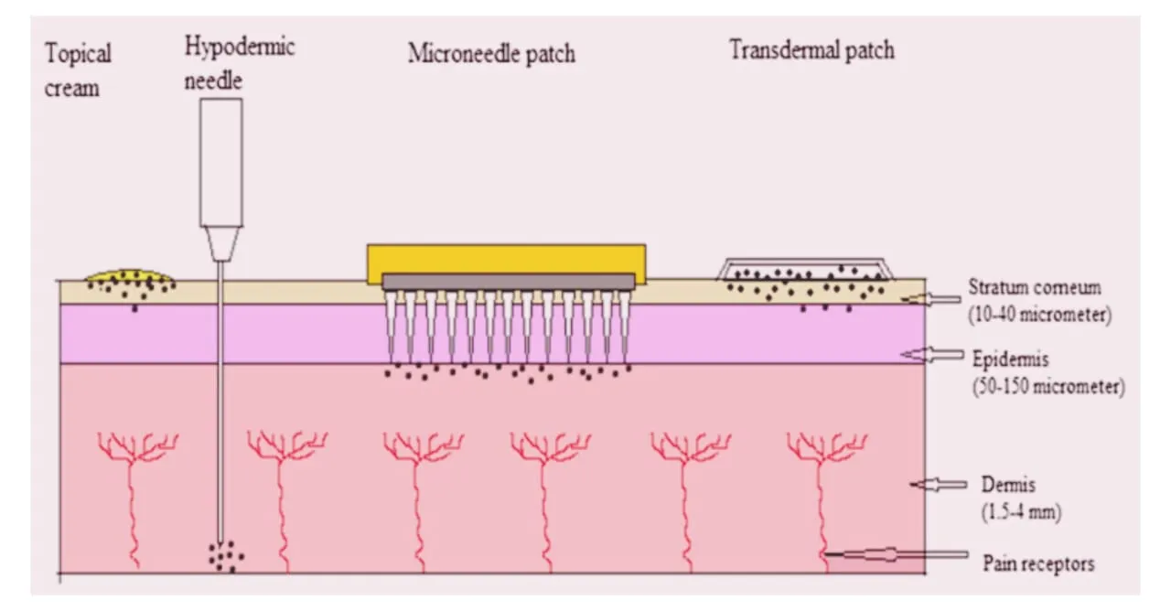
figure

Figure 1: Comparison of Topical Cream, Hypodermic Needle, Microneedle Patch and Transdermal Patch.[5].

Comparing the transdermal drug delivery (TDD) with alternative drug delivery systems gives a number of benefits. TDD has the capacity to deliver the medication to the blood in a regulated and sustained way at the appropriate dosage [6]. Reducing drug adverse effects by keeping medications from getting to vital organs like the liver and kidneys is another benefit of the transdermal method [7]. Furthermore, the transdermal drug delivery system can be used to address the limited bioavailability of many oral medications, particularly for the administration of proteins, peptides, and macromolecules that are generally poorly bioavailable when taken orally [8],[9].

Microneedle (MN) for Transdermal Drug Delivery

MN technology is used to delivered the drug directly through the skin, which replace the regular needle shots. The MN array delivered the drug in the layer of the skin with a small amount of pain [9]. These arrays of microneedles have the height from 25 to 2000 μm [10]. MNs have been used for jobs like giving medicine and vaccines for beauty and for checking for diseases. MNs have shapes, forms, materials and ways to make them, which are shown in this paper. Donnelly et al. Said that 30% of the recent scientific papers in "transdermal delivery technology" were about MN studies [11].

Microneedle History

The first-time microneedles were mentioned in a paper was in 1921 by Chambers when he put a needle into the nucleus of an egg [12]. In the 1960s the drug delivery through the skin started to get attention [13]. The concept of microneedles was introduced in the 1970s [14]. In 1979 the first transdermal system was approved to use for treating motion sickness by putting a patch on the skin for three days [15]. In 1994 a surgery called subcision which was done by Orentreich where he used a needle to cut under the skin to fix marks and wrinkles [16]. This surgery was for the skin issues under the surface which caused scars and lines. The first microneedle for sending drugs through the skin was made in 1998. Was made from silicon wafers using a process called ion etching and photolithography [17]. The study talked about using needles to help drugs get through the skin. Different materials are used in the manufacturing of microneedles like glass, ceramic, metal, polymer and plastics. In 2004 a group of microneedles was used to make holes in the skin for drug delivery which led to ways to make the microneedles. There are five types of microneedles which are Solid, coated, hollow, dissolving and hydrogel-forming microneedles [18]. Also, the manufacturing methods which was used are - laser cutting, lithography and micro-molding. These discoveries led to the reports of a dissolving microneedle being used for drug delivery in 2005 [19]. According to clinicalTrials.gov 43 studies have been done using microneedles with the first study done in 2007. Recently special ways to make molds for microneedles were developed to give a low-cost way to make molds [20,21].

Mechanism of Drug Delivery

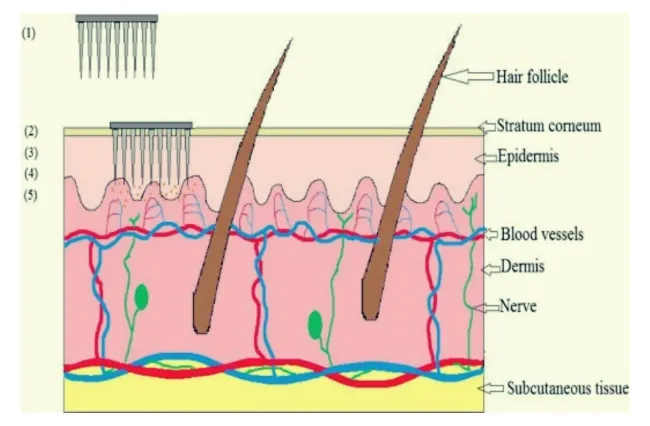
The medication is delivered topically using the diffusion process. The skin is momentarily disturbed by the microneedle drug delivery method. In order to administer enough medication to provide the necessary therapeutic response, a microneedle device is created by arranging hundreds of microneedles in arrays on a tiny patch, similar to a typical transdermal patch on the market. By penetrating the stratum corneum, it avoids the barrier layer. When the medication reaches the site of action, it exhibits a therapeutic reaction after being injected straight into the epidermis or higher dermis layer and entering the systemic circulation [22,23]. Figure 2 illustrates the medication delivery mechanism using microneedles.[5]
figure

Figure 2: Mechanism of Drug Delivery by Microneedle Device

(1) Microneedle device with drug solution; (2) Device inserted into the skin; (3) Temporary mechanical disruption of the skin; (4) Releasing the drug in the epidermis; (5) Transport of drug to the site of action.

Classification of Microneedles

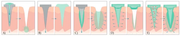
Types of microneedles made and looked at for using in sending medicine are solid, coated, dissolving, hollow and hydrogel microneedles. Each type of microneedle has its way to send the drug into the skin. Some are just to make holes in the outer layer some have the drug on the outside some dissolve and some have the drug inside [24] [25].
figure

Figure 3: A Schematic Representation of Drug Release Methods with Five Different Types of Microneedles [26]

A) Hollow MNs, B) Solid MNs, C) Coated MNs, D) Dissolving MNs, E) Hydrogel MNs

Hollow MNs

Hollow microneedles have a space inside which is filled with the drug dispersion or solution. They have holes at the tips. On inserting into the skin, the drug is directly deposited into the epidermis or the upper dermis layer. Mostly it is used for molecular weight compounds such as proteins, vaccines and oligonucleotides [27]. The drug flow rate and release pressure can be adjusted if the drug is to be given by a bolus injection. These microneedles are capable of administering a dose of the drug as more amount of drug can be accommodated into the empty space inside the needle. Maintaining a flow rate is essential here [28]. Increase in the microneedle bore can increase flow rate. Lead to reduced strength and sharpness. Sometimes a metal coat is applied on the microneedle to increase the strength of the microneedle. This can make the needles sharp [27].

Solid microneedles

Solid microneedles are mostly used for pre-treating the skin by forming pores. Pointed tips of the needles penetrate into the skin; create channels of micron size through which the drug directly enters the skin layers on the application of a drug patch thus increasing the permeation. The drug is taken up by the capillaries to show an effect. [29]. Solid microneedles deliver the drug with diffusion to skin layers [30]. Narayanan et al fabricated silicon long and tapered microneedles using tetramethylammonium hydroxide etching process. Microneedles with a height of 158 μm and base width of 110.5 μm were successfully fabricated [31]. Stainless steel microneedles are also studied by researchers. Enhanced delivery of captopril and metoprolol tartrate was studied after application of stainless-steel MN arrays [32].

Coated microneedles

The microneedles are surrounded with the drug solution or drug dispersion layer [27]. Subsequent dissolution of drug from the layer takes place. The drug is delivered quickly. The amount of drug that can be loaded depends on the thickness of the coating layer and the size of the needle which's usually very less [30]. Baek et al loaded lidocaine on l-lactide (PLLA) microneedle arrays. The loaded lidocaine released rapidly in phosphate buffer saline. Was found to be stable for 3 weeks [33]. Coated microneedle also explored for delivery of agents through same formulation. Li et al coated each microneedle with formulations and drugs thus allowing co-delivery of multiple agents with different properties. These delivered water insoluble dyes simultaneously [34].

Dissolving microneedles

Dissolving microneedles are fabricated with polymers by encapsulating the drug into the polymer. After inserting microneedle in the skin dissolution takes place which releases the drug. The application involves a single step as the microneedle is not to be removed out after insertion as in other cases. The polymer gets degraded inside the skin. Controls the drug release. The bio-acceptability and dissolution of the polymer inside the skin make it one of the choices for long-term therapy with improved patient compliance [27]. Effective needle drug distribution is a factor which faces problems while developing dissolving microneedles. Hence polymer-drug mixing is a step in such fabrication [30].

Hydrogel-forming microneedles

This type of microneedle is recently developed. Super-swelling polymers are used to make microneedles. The polymers constitute the structure which makes it capable of taking up a large amount of water into their three-dimensional polymeric network. These polymers swell when inserted into the skin due to the presence of the fluid. This leads to the formation of channels between the circulation and the drug patch. Before needling these microneedles are just used to disrupt the skin barrier. On swelling they behave as a rate controlling membrane. They have flexibility in size and shape. Easy sterilization and intact removal from the skin are the properties of such microneedles [35].

Material used in Fabrication of Microneedles

Table 1: Material for the Fabrication of Microneedles

S.no Fabrication Material Properties References
1. Silicon Anisotropic in nature, crystalline and possesses crystal alignment lattice, Fabrication process is time-consuming so limited use in the formation of microneedles. Due to brittle nature, it has biocompatibility issues. 36
2. Metal Alloys for example palladium-nickel and palladium-cobalt are the majorly used metal used for fabrication. It has good mechanical properties and robust to withstand breaking. 37
3. Ceramic Calcium sulfate dihydrate commonly known as Gypsum, calcium phosphate dihydrate and Al2O3 are major example of ceramics which are used in fabrication of microneedles. Organic modified ceramics are gaining interest in recent time due to its good biocompatibility. 38
4. Silica Glass Borosilicate glass composed of silica and boron trioxide used for experimental purpose in the laboratory due to its elastic nature. Silica glass is inert but it exhibits brittle properties. Therefore, it is not suitable for use as it causes the breaking of the skin. 39
5. Carbohydrates Mannitol Maltose, sucrose, xylitol are the certain examples of carbohydrates which are used in fabrication of microneedles. Silicone or metal templates are used to shape the carbohydrates slurries. Carbs are inexpensive, non-toxic but it gets degrade at the higher temperature which results in difficulty in fabrication. 40
6. Polymers Various polymers such as polyglycolic acid, poly lactic acid, polyvinylpyrrolidone, polyurethanes, poly methyl methacrylate are used in fabrication of microneedles as they are biodegradable in nature. 41

MN manufacturing method

There are several ways to create MN arrays. The most common methods include laser ablation, micromolding, additive manufacturing, injection molding, isotropic chemical etching, surface/volume micromachining, lithography, electroforming, and replication.

Laser ablation
figure

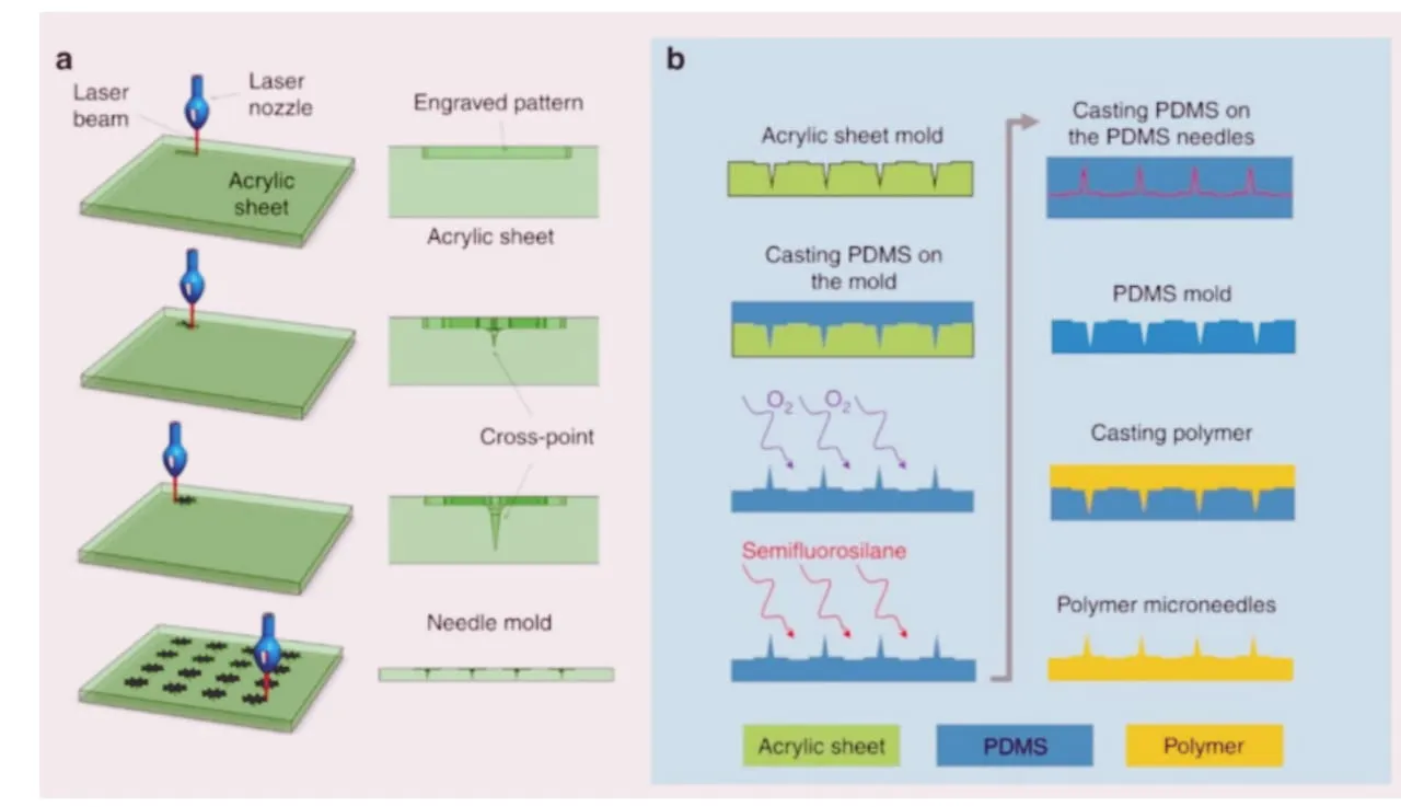
Figure 4: Fabrication of MN Mold [44]

In order to produce MN arrays, laser ablation combines the removal of material from a substrate using a focused optical light beam. For a variety of applications, lasers have been utilized to treat materials at the micro and nanoscale. Several types of lasers have been investigated for the production of MN arrays. They include femtosecond laser machines and CO2 UV excimer. The laser ablation technique is thought to be a quick and efficient way to create MNs. The material sheet's burn point is reached by the laser beam in 10 to 100 nanoseconds [42]. Any metal could be shaped using a laser. This process is linked to heat processes at the cutting surface that change the mechanical properties and structure of MN [43].

(a) CO2 laser cutter was used to fabricate MN acrylic mold using the proposed cross-over lines (COL) technique. (b) The acrylic mold was used to fabricate polydimethylsiloxane (PDMS) MNs mold, which can be used to fabricate a variety of polymer-based MNs.

Lithography
figure

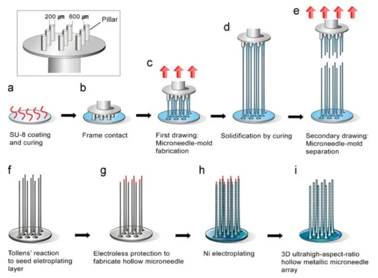
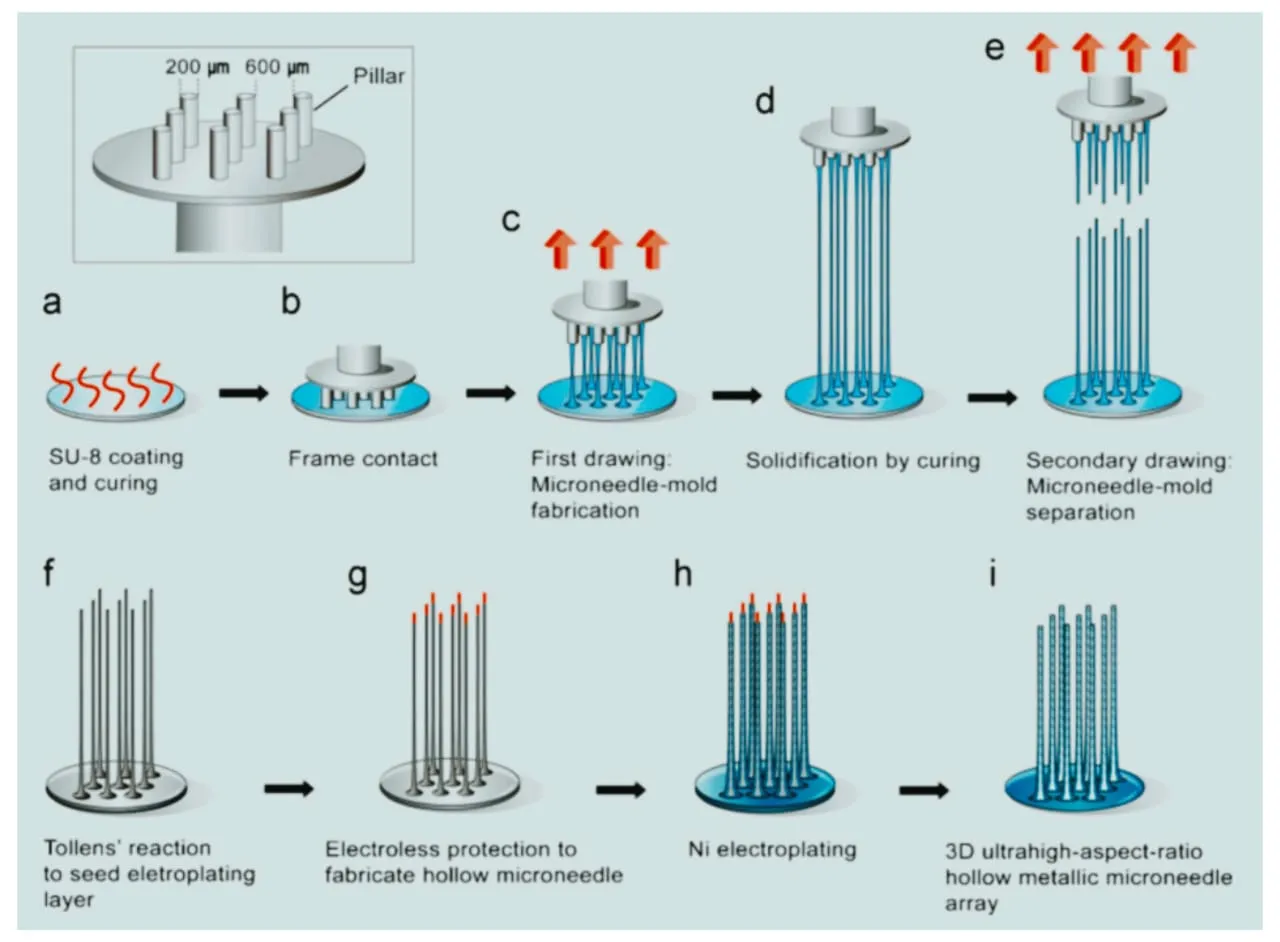
The master design of the geometric shapes is transferred onto a substrate's surface using the lithography technique [45]. Because photolithography has so many applications in the realm of microelectronics, it is mostly employed for pattern transfer [46]. Lithography is the initial stage in the fabrication of an MN in other methods including microelectronic and micromachining. Precise photoresist processing is necessary for lithography [47]. About 30–35% of integrated circuit manufacturing expenses are attributed to this method [48]. Glass, metal, ceramics, and plastics are only a few of the materials from which lithography may produce goods [49].
figure

Figure 5: Drawing Lithography to Produce a 3D UHAR MN. the Inset Shows a Drawing System with Patterned Pillars for Drawing Lithography. Stainless Drills with a Diameter of 200mm and a Length of 3 mm Were Used as Pillars and Fixed in a 3 × 3 Array on a PDMS Frame. [50]

(a) The SU-8 2050 photoresist was spin coated and cooled.

(b) After the photoresist contacted the patterned pillar, drawing lithography was performed.

(c) Drawing caused the appearance of an extended conical-shaped bridge between the substrate and pillar.

(d) The desired UHAR micro-needle mold was cured to generate a rigid structure.

(e) The separation of the 3D microstructure bridge produced a solid MN mold.

(f) Chemical deposition on the solid MN molds.

(g) The upper portion of the MN mold was coated with electroless material using a drawing system.

(h) Nickel electroplating on conducted solid MN molds.

(i) The hollow metallic MN array was created upon elimination of the electroless protection and the photoresist.

Micro -molding

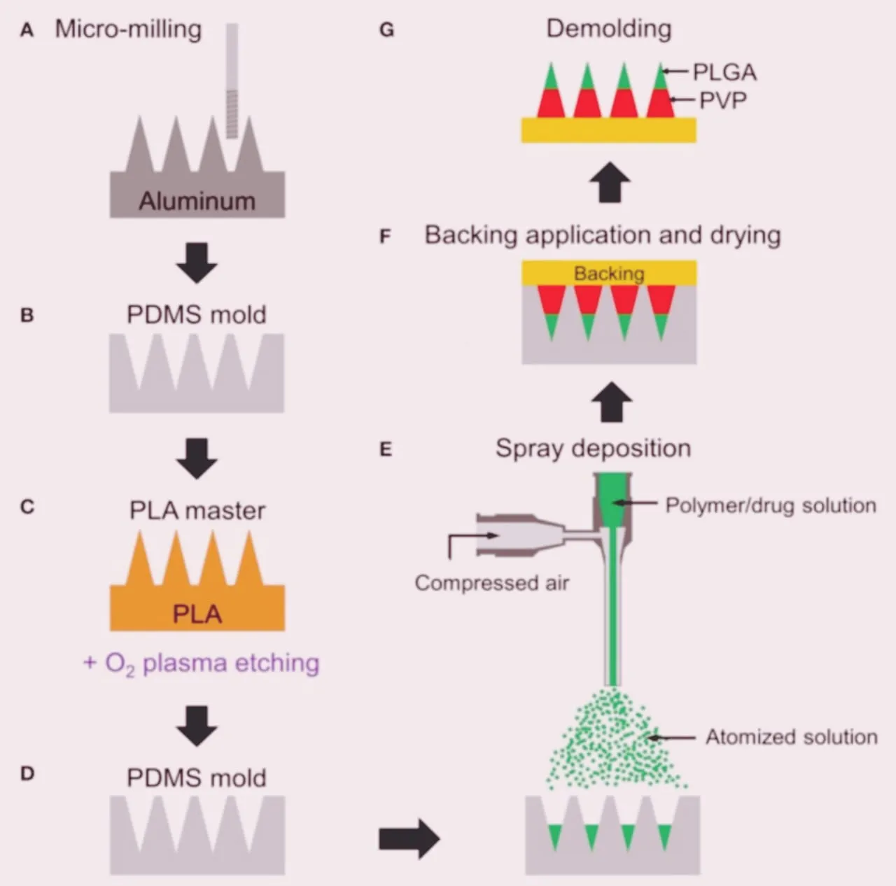
The technique of micro-molding involves creating copies of the master mold. A solution containing a polymer and active medicinal ingredients is poured into the mold. Micro-molding is utilized for mass production and is regarded as an economical technology. For MN manufacture, polymer material is frequently employed with micro-molding [51, 52]. Low cost, convenience of usage, low surface energy, and thermal stability are some of the benefits of PDMS in micro-molding processes [53]. Controlling the drug load capacity, mechanical behavior of the polymer, and depth of penetration are the technique's drawbacks [54]
figure

Figure 6: Schematic Fabrication Process of Multilayer MNs [52]

(A) Aluminium master fabrication using micro-milling.

(B) Replication of PDMS mold from the master.

(C) Fabrication of PLA master by micromolding and tip-sharpening using oxygen plasma.

(D) Replication of PDMS mold from the PLA master.

(E) Spray deposition of drug-containing polymer solution to fill the mold cavity. Multilayer MN is formed by sequential deposition of polymer solutions.

(F) Application of backing material (yellow) on the mold and drying at room temperature for polymer solidification.

(G) Demolding solidified multilayer MN array from the mold. Green and red represent PLGA and PVP layers, respectively.

Injection Molding

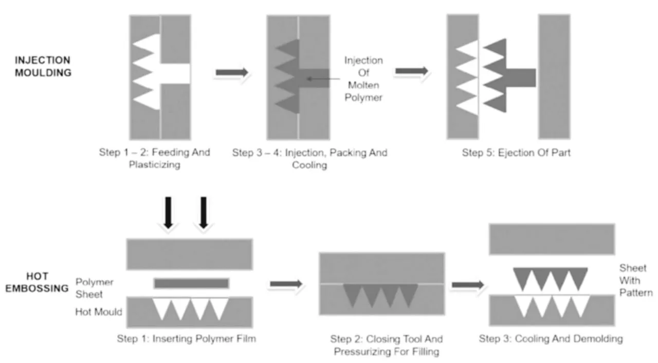
Another MN fabrication technique is injection molding. Lhernould et al. employed poly carbonate (PC) material to create a 4 × 4 hollow polymer MN array using injection molding and the hot embossing technique [55]. The MNs were utilized for several insertions without blunting the needle and were demonstrated to be able to sustain high force. Another study created a solid MN using a micro-injection molding technique [56]. High molecular weight, hydrophilic compounds could be delivered using these needles. Sammoura et al. molded plastic to create a polymeric MN [57]. The suggested technique makes it possible to produce MNs in large quantities at a minimal cost. Micro-injection molding provides excellent repeatability, precise dosing, and elevated injection flow rates by decoupling the plasticization process from the polymer melt injection [58].
figure

Figure 7: Process Steps of Standard Injection Molding and Step-And-Repeat Hot Embossing [59].

Additive Manufacturing

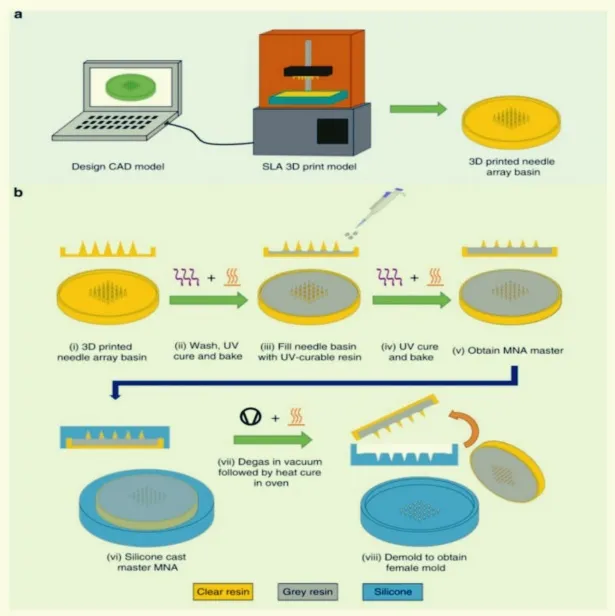
In recent years, additive manufacturing (3D printing) has rapidly gained attention as a means of manufacturing MN arrays. 3D printers build objects by depositing the desired material layer by layer. In recent years, the biomedical device industry has seen rapid growth in 3D printing technologies for tissue implants [60,61]. In 2019, Johnson et al. fabricated the first MN master using a commercial 3D printer [62]. Krieger et al. introduced a two-step called “print and fill” method to fabricate a MN mold [63]. Another study used a stereolithography technique to fabricate MN patches [64]. The advantages of using a 3D printer to fabricate MN arrays are flexibility in design parameters and reduced processing time [62].
figure

Figure 8: Overview of “Print and Fill” Fabrication Method [65]

(a) Needle array basin design followed by 3D printing of the design using a Form 2 SLA printer.

(b) MNA master mold fabrication method (i) take 3D printed needle array basin; (ii) washing followed by UV curing and baking of printed needle array basin; (iii) filing of needle array basin with UV-curable resin; (iv) second UV curing and baking; (v) obtain MNA master; (vi) silicone casting of MNA master; (vii) silicone mold is degassed followed by heat cure in oven; (viii) demoulding to obtain usable MN mold.

Applications

Drug Delivery

A dissolvable MN patch was used to deliver growth hormone for transdermal delivery to hairless rat skin [66]. A dissolvable caffeine loaded MN patch was able to control the weight of mice and work as an anti-obesity treatment plan [67]. A coated MN patch was used to deliver salmon calcitonin [68]. A solid microneedle was used to deliver a protein antigen (ovalbumin) into guinea pig skin [69]. Furthermore, MNs have been used for permeation for several drugs such as ibuprofen, ketoprofen and paracetamol [70]. Other drugs administered via microneedles include L-Ascorbic acid, riboflavin, aspirin, docetaxel, pilocarpine, lidocaine, hydrochloride, ketoprofen and glycerol [71]. Despite the fact that most studies used microneedle array for drug delivery into mice, pig, human skin there were studies which successfully demonstrated microneedle injection into chicken thigh [72].

Vaccine therapy

DNA vaccine was delivered using microneedle. Immune responses seen were much better than that of the injections [73]. An attempt to develop a microneedle patch which can be used for administering vaccine was also made [74]. A less dose is required when the drug is administered using microneedles as compared to intramuscular injection. Ogai and colleagues fabricated microneedles from poly-glycolic acid to enhance the vaccination efficiency by the intradermal route. The precise delivery of the drug in the dermis provides enhanced immunity. After the vaccination the antibody titers were significantly higher with vaccination with microneedles as compared to subcutaneous injection on 15th day [75]. Dissolving microneedles were also investigated for vaccination [76].

Cancer therapy

Cancer affects people every year in the world and cancer treatment faces lots of challenges. Microneedles have been investigated for anticancer drugs delivery. Self-degradable microneedles were investigated for melanoma treatment by delivering -PD-1 (aPD1) in a sustained manner. Anti-PD-1 and glucose oxidase loaded pH-sensitive dextran nanoparticles were delivered through microneedle [77]. A topical cream containing 5-fluorouracil is used to treat cell carcinoma. The permeability of 5-fluorouracil was enhanced up to 4.5 times when the cream was applied on the skin treated with microneedles. Significant inhibition of tumour growth further confirmed efficacy using microneedles [78]. Bhatnagar et al investigated the delivery of agents- tamoxifen and gemcitabine through microneedles for the treatment of breast cancer. Localized delivery of these drugs would help to reduce the side effects [79]. Polymeric microneedles were also investigated for skin cancer and localized delivery of anticancer drugs [80].

Cosmetic Application

MNs have widely been used in applications such as skin treatment and hair growth. Kim et al. Developed an acid-based dissolvable MN patch for the intradermal delivery of ascorbic acid and retinyl retinoate [81]. Kumar et al. Showed an enhancement of delivery of eflornithine (used to reduce facial hirsutism) in vitro and in vivo using a solid MN [82]. Further MN technology was able to treat two patients suffering from alopecia disease [83]. These patients experienced hair growth after treatment. Effective clinical trials have been conducted in facial scarring atrophic acne scars and hypertrophic burn scars using a MN. [84,85] Microneedles are considered as a treatment for cosmetic applications related to aging, skin lesions, vulgaris and wrinkles [86]. With an increasing demand of products, microneedles (patches and rollers) have a high potential in the future [87].

Advantages Disadvantages

Table 1: Approved Microneedle Products [92,93].

Product name Company name Description of the product Use
Dermaroller® Dermaroller® Germany, White Lotus A cylindrical roller with solid or metal microneedles, 0.2–2.5 mm in length. Improve skin texture, treat scars and hyperpigmentation.
C-8 (Cosmetic type) The Dermaroller Series by Anastassakis K. A needle length of only 0.13 mm (130 μm) Used to enhance penetration of topical agents.
CIT-8 (Collagen Induction Therapy The Dermaroller Series by Anastassakis K. A needle length of 0.5 mm (500 μm) Used in collagen induction and skin remodeling.
MF-8 type The Dermaroller Series by Anastassakis K. A needle length of 1.5 mm (1500 μm) Treat scars.
MS-4 The Dermaroller Series by Anastassakis K. A Small cylinder, 1 cm length, 2 cm diameter, and 4 circular arrays of needles which are 1.5 mm in length Used on facial acne scars
MicroHyala® CosMed transdermal drug delivery Dissolving microneedle patch with hyaluronic acid Wrinkle treatment
LiteClear® Nanomed skincare Solid silicon microneedles are used as pre-treatment and then drug applied topically. Treats acne and skin blemishes
Soluvia® Sanofi Pasteur Europe Hollow microneedle attached to a syringe Influenza vaccination
h-patch Valeritas Small adhesive machine-like patch is used To deliver drugs in subcutaneous tissue(insulin)
Microstructured transdermal system 3M Hollow microneedle To deliver biologics and other small molecules

CONCLUSION

Microneedle technology can be used to enhance the delivery of drugs through the transdermal and intradermal layers of the skin. The studies mentioned in this article indicate that microneedles are able to easily pass through stratum corneum which is main barrier for drug delivery in the skin and make it possible to deliver therapeutic agents with little or no discomfort and with minimal damage to the skin. The microneedles are classified into five types, which are solid, coated, hollow, dissolving, and hydrogel-forming, which works on the different mechanisms and can be appropriate for a variety of medical treatments and to meet the different patient needs. The review also points out the potential of microneedles in a number of areas, including drug delivery, vaccination, cancer treatment and cosmetic uses. Advances in materials and manufacturing techniques have increased their potential for effective and user-friendly drug delivery. Still, a number of challenges exist, such as limitations in drug-loading capacity, mechanical durability, variations in skin characteristics, safety concerns, manufacturing difficulties and difficulties in adapting these systems for clinical use. In conclusion, microneedles have great potential as an alternative to conventional methods of transdermal and injectable drug delivery. Future research and development should seek to improve drug loading, control drug release, enhance the mechanical properties and penetration depth, ensure safety and compatibility with the body, and establish large-scale manufacturing processes. If these challenges can be overcome, the large use of microneedle-based delivery systems in clinical settings could be promoted.

REFERENCES

  1. Parhi R. Recent advances in microneedle designs and their applications in drug and cosmeceutical delivery. J Drug Deliv Sci Technol. 2022; 75:103639. doi: 10.1016/j.jddst.2022.103639.
  2. Alkilani AZ, McCrudden MTC, Donnelly RF. Transdermal drug delivery: innovative pharmaceutical developments based on disruption of the barrier properties of the stratum corneum. Pharmaceutics. 2015;7 (4):438-70.
  3. Donnelly RF, Singh TRR, Morrow DIJ. Microneedle-mediated intradermal drug delivery. Hoboken (NJ): John Wiley & Sons; 2012.
  4. Han T, Das DB. Potential of ultrasound and microneedles for enhanced transdermal drug permeation: a review. Eur J Pharm Biopharm. 2015; 89:312-28.
  5. Waghule T, Singhvi G, Dubey SK, Pandey MM, Gupta G, Singh M, et al. Microneedles: a smart approach and increasing potential for transdermal drug delivery system. Biomed Pharmacother. 2019; 109:1249-58.
  6. Rastogi V, Yadav P. Transdermal drug delivery system: an overview. Asian J Pharm. 2012;6 (3):161-70.
  7. Verma NK, Panda P, Mishra JN, Vishwakarma DK, Singh AP, Alam G. Advances and development in drug delivery system: a review. Int J Adv Pharm. 2017; 6:49-62.
  8. Prausnitz MR, Langer R. Transdermal drug delivery. Nat Biotechnol. 2008;26 (11):1261-8. doi:10.1038/nbt.1504.
  9. Goodman MP. Are all estrogens created equal? A review of oral vs. transdermal therapy. J Womens Health. 2012;21 (2):161-9.
  10. Singh T, McMillan H, Mooney K, Alkilani A, Donnelly R. Microneedles for drug delivery and monitoring. In: Microfluidic devices for biomedical applications. 2013. p.185-230.
  11. Donnelly RF, Singh TRR, Larrañeta E, McCrudden MTC. Microneedles for drug and vaccine delivery and patient monitoring. Hoboken (NJ): John Wiley & Sons; 2018.
  12. Larrañeta E, Lutton REM, Woolfson AD, Donnelly RF. Microneedle arrays as intradermal drug delivery systems: materials science, manufacture and commercial development. In: Advances in biomembranes and lipid self-assembly. Vol. 24. Amsterdam: Elsevier; 2016. p.1-32.
  13. Gerstel MS, Place VA. Drug delivery device. US patent US3964482A. 1976 Jun 22.
  14. Reed M, Lye WK. Microsystems for drug and gene delivery. Proc IEEE. 2004; 92:56-75. doi:10.1109/JPROC.2003.820542.
  15. Pastore MN, Kalia YN, Horstmann M, Roberts MS. Transdermal patches: history, development and pharmacology. Br J Pharmacol. 2015;172 (9):2179-209. doi:10.1111/bph.13059.
  16. Orentreich DS, Orentreich N. Subcutaneous incisionless (subcision) surgery for the correction of depressed scars and wrinkles. Dermatol Surg. 1995; 21:543-9. doi:10.1111/j.1524-4725. 1995.tb00259. x.
  17. Henry S, McAllister DV, Allen MG, Prausnitz MR. Microfabricated microneedles: an approach to transdermal drug delivery. J Pharm Sci. 1998; 87:922-5.
  18. Prausnitz MR. Microneedles for transdermal drug delivery. Adv Drug Deliv Rev. 2004; 56:581-7. doi: 10.1016/j.addr.2003.10.023.
  19. Dang N, Liu TY, Prow TW. Microtechnology in skin delivery of vaccines. In: Micro and nanotechnology in vaccine development. Norwich (NY): William Andrew Publishing; 2017.
  20. Wang QL, Zhu DD, Chen Y, Guo XD. A fabrication method of microneedle molds with controlled microstructures. Mater Sci Eng C Mater Biol Appl. 2016; 65:135-42. doi: 10.1016/j.msec.2016.03.097.
  21. Chen Z, Lin Y, Lee W, Ren L, Liu B, Liang L, et al. Additive manufacturing of honeybee-inspired microneedle for skin insertion and difficult removal. ACS Appl Mater Interfaces. 2018; 10:29338-46. doi:10.1021/acsami.8b09563.
  22. Sharma D. Microneedles: an approach in transdermal drug delivery: a review. Pharma Tutor. 2018;6 (1):7-15. doi:10.29161/PT. v6.i1.2018.7.
  23. Akhtar N. Microneedles: an innovative approach to transdermal delivery—a review. Int J Pharm Sci. 2014;6 (4):18-25.
  24. Prausnitz MR. Engineering microneedle patches for vaccination and drug delivery to skin. Annu Rev Chem Biomol Eng. 2017; 8:177-200.
  25. Suzuki M, Takahashi T, Aoyagi S. 3D laser fabrication of hollow microneedle mimicking mosquitos and its characterisation. Int J Nanotechnol. 2018;15 (1-3):157-73.
  26. Tucak A, Sirbubalo M, Hindija L, Rahić O, Hadžiabdić J, Muhamedagić K, et al. Microneedles: characteristics, materials, production methods and commercial development. Micromachines (Basel). 2020;11 (11):961. doi:10.3390/mi11110961.
  27. Ita K. Transdermal delivery of drugs with microneedles—challenges. Pharmaceutics. 2015;7 (3):90-105. doi:10.3390/pharmaceutics7030090.
  28. Narayanan SP, Raghavan S. Solid silicon microneedles for drug delivery applications. Int J Adv Manuf Technol. 2017;93 (1-4):407-22.
  29. Prausnitz MR. Engineering microneedle patches for vaccination and drug delivery to skin. Annu Rev Chem Biomol Eng. 2017; 8:177-200.
  30. Suzuki M, Takahashi T, Aoyagi S. 3D laser fabrication of hollow microneedle mimicking mosquitos and its characterisation. Int J Nanotechnol. 2018;15 (1-3):157-73.
  31. Li QY, Zhang JN, Chen BZ, Wang QL, Guo XD. A solid polymer microneedle patch pretreatment enhances the permeation of drug molecules into the skin. RSC Adv. 2017;7 (25):15408-15.
  32. Baek SH, Shin JH, Kim YC. Drug-coated microneedles for painless local anesthesia. Biomed Microdevices. 2017;19 (1):2.
  33. Li S, Li W, Prausnitz M. Individually coated microneedles for co-delivery of compounds with different properties. Drug Deliv Transl Res. 2018;8 (5):1043-52.
  34. Donnelly RF, Singh TRR, Alkilani AZ, McCrudden MTC, O'Neill S, O'Mahony C, et al. Hydrogel-forming microneedle arrays exhibit antimicrobial properties: potential for enhanced patient safety. Int J Pharm. 2013;451 (1-2):76-91. doi: 10.1016/j.ijpharm.2013.04.045
  35. Hong X, Wei L, Wu F, Wu Z, Chen L, Liu Z, et al. Dissolving and biodegradable microneedle technologies for transdermal sustained delivery of drug and vaccine. Drug Des Devel Ther. 2013; 7:945-952. doi:10.2147/DDDT.S44401.
  36. Williams AC, Barry BW. Penetration enhancers. Adv Drug Deliv Rev. 2004;56 (5):603-618. doi: 10.1016/j.addr.2003.10.025.
  37. Gupta J, Felner EI, Prausnitz MR. Minimally invasive insulin delivery in subjects with type 1 diabetes using hollow microneedles. Diabetes Technol Ther. 2009;11 (6):329-337. doi:10.1089/dia.2008.0103.
  38. Miyano T, Tobinaga Y, Kanno T, Matsuzaki Y, Takeda H, Wakui M, et al. Sugar micro needles as transdermic drug delivery system. Biomed Microdevices. 2005;7 (3):185-188. doi:10.1007/s10544-005-3024-7.
  39. Gill HS, Prausnitz MR. Coated microneedles for transdermal delivery. J Control Release. 2007;117 (2):227-237. doi: 10.1016/j.jconrel.2006.10.017.
  40. Chu LY, Choi SO, Prausnitz MR. Fabrication of dissolving polymer microneedles for controlled drug encapsulation and delivery: bubble and pedestal microneedle designs. J Pharm Sci. 2010;99 (10):4228-4238. doi:10.1002/jps.22140.
  41. Menazea A, El-Newehy MH, Thamer BM, El-Naggar ME. Preparation of antibacterial film-based biopolymer embedded with vanadium oxide nanoparticles using one-pot laser ablation. J Mol Struct. 2021; 1225:129163. doi: 10.1016/j.molstruc.2020.129163.
  42. Tu KZ, Chung CK. Fabrication of biodegradable polymer microneedle array via CO2 laser ablation. In: 2015 IEEE 10th International Conference on Nano/Micro Engineered and Molecular Systems (NEMS); 2015. p.494-7. doi:10.1109/NEMS.2015.7147476.
  43. Chen YT, Ma KJ, Tseng AA, Chen PH. Projection ablation of glass-based single and arrayed microstructures using excimer laser. Opt Laser Technol. 2005; 37:271-80. doi: 10.1016/j.optlastec.2004.04.007.
  44. Jeggy C. Micro-injection moulding: from process to modelling. Louvain-la-Neuve (Belgium): Presses Univ de Louvain; 2004.
  45. Lutton R, Larrañeta E, Kearney MC, Boyd P, Woolfson A, Donnelly RF. A novel scalable manufacturing process for the production of hydrogel-forming microneedle arrays. Int J Pharm. 2015; 494:417-29. doi: 10.1016/j.ijpharm.2015.08.049.
  46. Nejad HR, Sadeqi A, Kiaee G, Sonkusale S. Low-cost and cleanroom-free fabrication of microneedles. Microsyst Nanoeng. 2018; 4:17073. doi:10.1038/micronano.2017.73.
  47. Gaikwad A, Desai S. Molecular dynamics investigation of the deformation mechanism of gold with variations in mold profiles during nanoimprinting. Materials. 2021; 14:2548. doi:10.3390/ma14102548.
  48. Madou MJ. Fundamentals of microfabrication: the science of miniaturization. Boca Raton (FL): CRC Press; 2002.
  49. Khuen HW, Lay LL, Schaper C. On control of resist film uniformity in the microlithography process. IFAC Proc Vol. 2002; 35:19-24. doi:10.3182/20020721-6-ES-1901.01154.
  50. Plummer JD, Deal MD, Griffin PB. Silicon VLSI technology: fundamentals, practice and modelling. India: Pearson Education; 2009.
  51. Tran KT, Nguyen TD. Lithography-based methods to manufacture biomaterials at small scales. J Sci Adv Mater Devices. 2017; 2:1-14. doi: 10.1016/j.jsamd.2016.12.001.
  52. Lee K, Lee HC, Lee DS, Jung H. Drawing lithography: three-dimensional fabrication of an ultrahigh-aspect-ratio microneedle. Adv Mater. 2010; 22:483-6. doi:10.1002/adma.200902418.
  53. Griffiths CA. Micro injection moulding: tooling and process factors. Cardiff: Cardiff University; 2008.
  54. Kim MJ, Park SC, Rizal B, Guanes G, Baek SK, Park JH, Betz AR, Choi SO. Fabrication of circular obelisk-type multilayer microneedles using micro-milling and spray deposition. Front Bioeng Biotechnol. 2018; 6:54. doi:10.3389/fbioe.2018.00054.
  55. Armani D, Liu C, Almu N. Re-configurable fluid circuits by PDMS elastomer micromachining. In: Proceedings of the IEEE International Conference on Micro Electro Mechanical Systems; 1999 Jan 21; Orlando, FL, USA. p.222-7.
  56. Lhernould MS, Deleers M, Delchambre A. Hollow polymer microneedles array resistance and insertion tests. Int J Pharm. 2015; 480:152-7. doi: 10.1016/j.ijpharm.2015.01.019.
  57. Sammoura F, Kang J, Heo YM, Jung T, Lin L. Polymeric microneedle fabrication using a microinjection molding technique. Microsyst Technol. 2006; 13:517-22. doi:10.1007/s00542-006-0204-1.
  58. Juster H, van der Aar B, de Brouwer H. A review on microfabrication of thermoplastic polymer-based microneedle arrays. Polym Eng Sci. 2019; 59:877-90. doi:10.1002/pen.25078.
  59. Nair KJ. Micro-injection moulded microneedles for drug delivery. Bradford (UK): University of Bradford; 2014.
  60. Parupelli SK, Desai S. Hybrid additive manufacturing (3D printing) and characterization of functionally gradient materials via in situ laser curing. Int J Adv Manuf Technol. 2020; 110:543-56. doi:10.1007/s00170-020-05884-9.
  61. Bidanda B, Desai S. Strategic planning models for prototyping and product development centres. Int J Prod Dev. 2004; 1:133. doi:10.1504/IJPD.2004.005701.
  62. Pere CPP, Economidou SN, Lall G, Ziraud C, Boateng JS, Alexander BD, et al. 3D printed microneedles for insulin skin delivery. Int J Pharm. 2018; 544:425-32. doi: 10.1016/j.ijpharm.2018.03.031.
  63. Johnson AR, Procopio AT. Low-cost additive manufacturing of microneedle masters. 3D Print Med. 2019; 5:2. doi:10.1186/s41205-019-0039-x.
  64. Krieger KJ, Bertollo N, Dangol M, Sheridan JT, Lowery MM, O’Cearbhaill ED. Simple and customizable method for fabrication of high-aspect ratio microneedle molds using low-cost 3D printing. Microsyst Nanoeng. 2019; 5:42. doi:10.1038/s41378-019-0088-8.
  65. Lee JW, Choi SO, Felner EI, Prausnitz MR. Dissolving microneedle patch for delivery of human growth hormone. Small. 2011; 7:531-9. doi:10.1002/smll.201001091.
  66. Dangol M, Kim S, Li CG, Lahiji SF, Jang M, Ma Y, et al. Anti-obesity effect of a caffeine-loaded dissolving microneedle patch in high-fat diet-induced obese C57BL/6J mice. J Control Release. 2017; 265:41-7. doi: 10.1016/j.jconrel.2017.03.400.
  67. Tas C, Mansoor S, Kalluri H, Zarnitsyn VG, Choi SO, Banga AK, et al. Delivery of salmon calcitonin using a microneedle patch. Int J Pharm. 2012; 423:257-63. doi: 10.1016/j.ijpharm.2011.11.046.
  68. Matriano JA, Cormier M, Johnson J, Young WA, Buttery M, Nyam K, et al. Macroflux® microprojection array patch technology: an efficient approach for intracutaneous immunization. Pharm Res. 2002; 19:63-70. doi:10.1023/A:1013607400040.
  69. Donnelly RF, Morrow DIJ, McCarron P, Woolfson AD, Morrissey A, Juzenas P, et al. Microneedle arrays permit enhanced delivery of a preformed photosensitizer. Photochem Photobiol. 2009; 85:195-204. doi:10.1111/j.1751-1097.2008.00417. x.
  70. Stahl J, Wohlert M, Kietzmann M. Microneedle pretreatment enhances the permeation of hydrophilic compounds with high melting points. BMC Pharmacol Toxicol. 2012; 13:5. doi:10.1186/2050-6511-13-5.
  71. Nayak S, Suryawanshi S, Bhaskar V. Microneedle technology for drug delivery: applications and combination with other enhancing techniques. J Drug Deliv Ther. 2016;6 (5):65-83. doi:10.22270/jddt. v6i5.1285.
  72. Stoeber B, Liepmann D. Fluid injection through out-of-plane microneedles. In: Proceedings of the Annual International IEEE-EMBS Special Topic Conference on Microtechnologies in Medicine and Biology; 2000 Oct 12-14; Lyon, France. 2000. p.224-8.
  73. Mikszta JA, Alarcon JB, Brittingham JM, Sutter DE, Pettis RJ, Harvey NG. Improved genetic immunization via disruption of skin-barrier function and targeted epidermal delivery. Nat Med. 2002;8 (4):415-9.
  74. Mistilis MJ, Bommarius AS, Prausnitz MR. Development of a thermostable microneedle patch for influenza vaccination. J Pharm Sci. 2015;104 (2):740-9. doi:10.1002/jps.24283.
  75. Ogai N, Nonaka I, Toda Y, Ono T, Minegishi S, Inou A, et al. Enhanced immunity in vaccination by novel hollow microneedles. Skin Res Technol. 2018;24 (4):630-5. doi:10.1111/srt.12576.
  76. Rodgers AM, Courtenay AJ, Donnelly RF. Dissolving microneedles for intradermal vaccination: manufacture, formulation and stakeholder considerations. Expert Opin Drug Deliv. 2018;15 (11):1039-43. doi:10.1080/17425247.2018.1522301.
  77. Wang C, Ye Y, Hochu GM, Sadeghifar H, Gu Z. Enhanced cancer immunotherapy by microneedle patch-assisted delivery of anti-PD1 antibody. Nano Lett. 2016;16 (4):2334-40. doi: 10.1021/acs.nanolett.5b05030.
  78. Naguib YW, Kumar A, Cui Z. The effect of microneedles on the skin permeability and antitumor activity of topical 5-fluorouracil. Acta Pharm Sin B. 2014;4 (1):94-9.
  79. Bhatnagar S, Kumari P, Pattarabhiran SP, Venuganti VVK. Zein microneedles for localized delivery of chemotherapeutic agents to treat breast cancer: drug loading, release behavior, and skin permeation studies. AAPS PharmSciTech. 2018;19 (4):1818-26. doi:10.1208/s12249-018-1004-5.
  80. Ye Y, Wang C, Zhang X, Hu Q, Zhang Y, Liu Q, et al. A melanin-mediated cancer immunotherapy patch. Sci Immunol. 2017;2 (17): eaan5692. doi:10.1126/sciimmunol. aan5692.
  81. Park YH, Ha SK, Choi I, Kim KS, Park J, Choi N, et al. Fabrication of carboxymethyl cellulose (CMC) microneedle with laser writing and replica molding process for enhancement of transdermal drug delivery. Biotechnol Bioprocess Eng. 2016; 21:110-8. doi:10.1007/s12257-015-0634-7.
  82. Kumar A, Naguib Y, Shi YC, Cui Z. A method to improve the efficacy of eflornithine hydrochloride cream. Drug Deliv. 2016; 23:1495-501. doi:10.3109/10717544.2014.951746.
  83. Mysore V, Chandrashekar B, Yepuri V. Alopecia successful outcome with microneedling and triamcinolone acetonide. J Cutan Aesthet Surg. 2014; 7:63-4. doi:10.4103/0974-2077.129989.
  84. Majid I. Microneedling therapy in facial scars: an objective assessment. J Cutan Aesthet Surg. 2009; 2:26-30. doi:10.4103/0974-2077.53096.
  85. El-Domyati M, Barakat M, Awad S, Medhat W, El-Fakahany H, Farag H. Microneedling therapy for acne scars: an objective evaluation. J Clin Aesthet Dermatol. 2015; 8:36-42.
  86. Sharma S, Hatware K, Bhadane P, Sindhikar S, Mishra DK. Recent advances in composites for biomedical applications: advanced drug delivery technologies. Mater Sci Eng C Mater Biol Appl. 2019; 103:109717. doi: 10.1016/j.msec.2019.05.002.
  87. Pattani A, McKay PF, Garland MJ, Curran RM, Migalska K, Cassidy CM, et al. Microneedle mediated delivery of adjuvanted recombinant HIV-1 CN54gp140 effectively primes mucosal boost inoculations. J Control Release. 2012; 162:529-37. doi: 10.1016/j.jconrel.2012.07.039.
  88. Bora P, Kumar L, Bansal A. Microneedle technology for advanced drug delivery: evolving vistas, 2008
  89. Li J, Zeng M, Shan H, Tong C. Microneedle patches as drug and delivery platform. Curr Med Chem. 2017;24 (22):2413-2422.
  90. Kim YC, Park JH, Prausnitz MR. Microneedles for drug and vaccine delivery. Drug Deliv Transl Res. 2015;5 (4):311-2.
  91. Amsden BG, Goosen MFA. Transdermal delivery of peptide and protein drugs: an overview. AIChE J. 1995;41 (8):1972-97.
  92. Giudice EL, Campbell JD. Needle-free vaccine delivery. Adv Drug Deliv Rev. 2006;58 (1):68-89.
  93. Jeong HR, Lee HS, Choi IJ, Park JH. Considerations in the use of microneedles: pain, convenience, anxiety and safety. J Drug Target. 2017;25 (1):29-40.

Reference

  1. Parhi R. Recent advances in microneedle designs and their applications in drug and cosmeceutical delivery. J Drug Deliv Sci Technol. 2022; 75:103639. doi: 10.1016/j.jddst.2022.103639.
  2. Alkilani AZ, McCrudden MTC, Donnelly RF. Transdermal drug delivery: innovative pharmaceutical developments based on disruption of the barrier properties of the stratum corneum. Pharmaceutics. 2015;7 (4):438-70.
  3. Donnelly RF, Singh TRR, Morrow DIJ. Microneedle-mediated intradermal drug delivery. Hoboken (NJ): John Wiley & Sons; 2012.
  4. Han T, Das DB. Potential of ultrasound and microneedles for enhanced transdermal drug permeation: a review. Eur J Pharm Biopharm. 2015; 89:312-28.
  5. Waghule T, Singhvi G, Dubey SK, Pandey MM, Gupta G, Singh M, et al. Microneedles: a smart approach and increasing potential for transdermal drug delivery system. Biomed Pharmacother. 2019; 109:1249-58.
  6. Rastogi V, Yadav P. Transdermal drug delivery system: an overview. Asian J Pharm. 2012;6 (3):161-70.
  7. Verma NK, Panda P, Mishra JN, Vishwakarma DK, Singh AP, Alam G. Advances and development in drug delivery system: a review. Int J Adv Pharm. 2017; 6:49-62.
  8. Prausnitz MR, Langer R. Transdermal drug delivery. Nat Biotechnol. 2008;26 (11):1261-8. doi:10.1038/nbt.1504.
  9. Goodman MP. Are all estrogens created equal? A review of oral vs. transdermal therapy. J Womens Health. 2012;21 (2):161-9.
  10. Singh T, McMillan H, Mooney K, Alkilani A, Donnelly R. Microneedles for drug delivery and monitoring. In: Microfluidic devices for biomedical applications. 2013. p.185-230.
  11. Donnelly RF, Singh TRR, Larrañeta E, McCrudden MTC. Microneedles for drug and vaccine delivery and patient monitoring. Hoboken (NJ): John Wiley & Sons; 2018.
  12. Larrañeta E, Lutton REM, Woolfson AD, Donnelly RF. Microneedle arrays as intradermal drug delivery systems: materials science, manufacture and commercial development. In: Advances in biomembranes and lipid self-assembly. Vol. 24. Amsterdam: Elsevier; 2016. p.1-32.
  13. Gerstel MS, Place VA. Drug delivery device. US patent US3964482A. 1976 Jun 22.
  14. Reed M, Lye WK. Microsystems for drug and gene delivery. Proc IEEE. 2004; 92:56-75. doi:10.1109/JPROC.2003.820542.
  15. Pastore MN, Kalia YN, Horstmann M, Roberts MS. Transdermal patches: history, development and pharmacology. Br J Pharmacol. 2015;172 (9):2179-209. doi:10.1111/bph.13059.
  16. Orentreich DS, Orentreich N. Subcutaneous incisionless (subcision) surgery for the correction of depressed scars and wrinkles. Dermatol Surg. 1995; 21:543-9. doi:10.1111/j.1524-4725. 1995.tb00259. x.
  17. Henry S, McAllister DV, Allen MG, Prausnitz MR. Microfabricated microneedles: an approach to transdermal drug delivery. J Pharm Sci. 1998; 87:922-5.
  18. Prausnitz MR. Microneedles for transdermal drug delivery. Adv Drug Deliv Rev. 2004; 56:581-7. doi: 10.1016/j.addr.2003.10.023.
  19. Dang N, Liu TY, Prow TW. Microtechnology in skin delivery of vaccines. In: Micro and nanotechnology in vaccine development. Norwich (NY): William Andrew Publishing; 2017.
  20. Wang QL, Zhu DD, Chen Y, Guo XD. A fabrication method of microneedle molds with controlled microstructures. Mater Sci Eng C Mater Biol Appl. 2016; 65:135-42. doi: 10.1016/j.msec.2016.03.097.
  21. Chen Z, Lin Y, Lee W, Ren L, Liu B, Liang L, et al. Additive manufacturing of honeybee-inspired microneedle for skin insertion and difficult removal. ACS Appl Mater Interfaces. 2018; 10:29338-46. doi:10.1021/acsami.8b09563.
  22. Sharma D. Microneedles: an approach in transdermal drug delivery: a review. Pharma Tutor. 2018;6 (1):7-15. doi:10.29161/PT. v6.i1.2018.7.
  23. Akhtar N. Microneedles: an innovative approach to transdermal delivery—a review. Int J Pharm Sci. 2014;6 (4):18-25.
  24. Prausnitz MR. Engineering microneedle patches for vaccination and drug delivery to skin. Annu Rev Chem Biomol Eng. 2017; 8:177-200.
  25. Suzuki M, Takahashi T, Aoyagi S. 3D laser fabrication of hollow microneedle mimicking mosquitos and its characterisation. Int J Nanotechnol. 2018;15 (1-3):157-73.
  26. Tucak A, Sirbubalo M, Hindija L, Rahi? O, Hadžiabdi? J, Muhamedagi? K, et al. Microneedles: characteristics, materials, production methods and commercial development. Micromachines (Basel). 2020;11 (11):961. doi:10.3390/mi11110961.
  27. Ita K. Transdermal delivery of drugs with microneedles—challenges. Pharmaceutics. 2015;7 (3):90-105. doi:10.3390/pharmaceutics7030090.
  28. Narayanan SP, Raghavan S. Solid silicon microneedles for drug delivery applications. Int J Adv Manuf Technol. 2017;93 (1-4):407-22.
  29. Prausnitz MR. Engineering microneedle patches for vaccination and drug delivery to skin. Annu Rev Chem Biomol Eng. 2017; 8:177-200.
  30. Suzuki M, Takahashi T, Aoyagi S. 3D laser fabrication of hollow microneedle mimicking mosquitos and its characterisation. Int J Nanotechnol. 2018;15 (1-3):157-73.
  31. Li QY, Zhang JN, Chen BZ, Wang QL, Guo XD. A solid polymer microneedle patch pretreatment enhances the permeation of drug molecules into the skin. RSC Adv. 2017;7 (25):15408-15.
  32. Baek SH, Shin JH, Kim YC. Drug-coated microneedles for painless local anesthesia. Biomed Microdevices. 2017;19 (1):2.
  33. Li S, Li W, Prausnitz M. Individually coated microneedles for co-delivery of compounds with different properties. Drug Deliv Transl Res. 2018;8 (5):1043-52.
  34. Donnelly RF, Singh TRR, Alkilani AZ, McCrudden MTC, O'Neill S, O'Mahony C, et al. Hydrogel-forming microneedle arrays exhibit antimicrobial properties: potential for enhanced patient safety. Int J Pharm. 2013;451 (1-2):76-91. doi: 10.1016/j.ijpharm.2013.04.045
  35. Hong X, Wei L, Wu F, Wu Z, Chen L, Liu Z, et al. Dissolving and biodegradable microneedle technologies for transdermal sustained delivery of drug and vaccine. Drug Des Devel Ther. 2013; 7:945-952. doi:10.2147/DDDT.S44401.
  36. Williams AC, Barry BW. Penetration enhancers. Adv Drug Deliv Rev. 2004;56 (5):603-618. doi: 10.1016/j.addr.2003.10.025.
  37. Gupta J, Felner EI, Prausnitz MR. Minimally invasive insulin delivery in subjects with type 1 diabetes using hollow microneedles. Diabetes Technol Ther. 2009;11 (6):329-337. doi:10.1089/dia.2008.0103.
  38. Miyano T, Tobinaga Y, Kanno T, Matsuzaki Y, Takeda H, Wakui M, et al. Sugar micro needles as transdermic drug delivery system. Biomed Microdevices. 2005;7 (3):185-188. doi:10.1007/s10544-005-3024-7.
  39. Gill HS, Prausnitz MR. Coated microneedles for transdermal delivery. J Control Release. 2007;117 (2):227-237. doi: 10.1016/j.jconrel.2006.10.017.
  40. Chu LY, Choi SO, Prausnitz MR. Fabrication of dissolving polymer microneedles for controlled drug encapsulation and delivery: bubble and pedestal microneedle designs. J Pharm Sci. 2010;99 (10):4228-4238. doi:10.1002/jps.22140.
  41. Menazea A, El-Newehy MH, Thamer BM, El-Naggar ME. Preparation of antibacterial film-based biopolymer embedded with vanadium oxide nanoparticles using one-pot laser ablation. J Mol Struct. 2021; 1225:129163. doi: 10.1016/j.molstruc.2020.129163.
  42. Tu KZ, Chung CK. Fabrication of biodegradable polymer microneedle array via CO2 laser ablation. In: 2015 IEEE 10th International Conference on Nano/Micro Engineered and Molecular Systems (NEMS); 2015. p.494-7. doi:10.1109/NEMS.2015.7147476.
  43. Chen YT, Ma KJ, Tseng AA, Chen PH. Projection ablation of glass-based single and arrayed microstructures using excimer laser. Opt Laser Technol. 2005; 37:271-80. doi: 10.1016/j.optlastec.2004.04.007.
  44. Jeggy C. Micro-injection moulding: from process to modelling. Louvain-la-Neuve (Belgium): Presses Univ de Louvain; 2004.
  45. Lutton R, Larrañeta E, Kearney MC, Boyd P, Woolfson A, Donnelly RF. A novel scalable manufacturing process for the production of hydrogel-forming microneedle arrays. Int J Pharm. 2015; 494:417-29. doi: 10.1016/j.ijpharm.2015.08.049.
  46. Nejad HR, Sadeqi A, Kiaee G, Sonkusale S. Low-cost and cleanroom-free fabrication of microneedles. Microsyst Nanoeng. 2018; 4:17073. doi:10.1038/micronano.2017.73.
  47. Gaikwad A, Desai S. Molecular dynamics investigation of the deformation mechanism of gold with variations in mold profiles during nanoimprinting. Materials. 2021; 14:2548. doi:10.3390/ma14102548.
  48. Madou MJ. Fundamentals of microfabrication: the science of miniaturization. Boca Raton (FL): CRC Press; 2002.
  49. Khuen HW, Lay LL, Schaper C. On control of resist film uniformity in the microlithography process. IFAC Proc Vol. 2002; 35:19-24. doi:10.3182/20020721-6-ES-1901.01154.
  50. Plummer JD, Deal MD, Griffin PB. Silicon VLSI technology: fundamentals, practice and modelling. India: Pearson Education; 2009.
  51. Tran KT, Nguyen TD. Lithography-based methods to manufacture biomaterials at small scales. J Sci Adv Mater Devices. 2017; 2:1-14. doi: 10.1016/j.jsamd.2016.12.001.
  52. Lee K, Lee HC, Lee DS, Jung H. Drawing lithography: three-dimensional fabrication of an ultrahigh-aspect-ratio microneedle. Adv Mater. 2010; 22:483-6. doi:10.1002/adma.200902418.
  53. Griffiths CA. Micro injection moulding: tooling and process factors. Cardiff: Cardiff University; 2008.
  54. Kim MJ, Park SC, Rizal B, Guanes G, Baek SK, Park JH, Betz AR, Choi SO. Fabrication of circular obelisk-type multilayer microneedles using micro-milling and spray deposition. Front Bioeng Biotechnol. 2018; 6:54. doi:10.3389/fbioe.2018.00054.
  55. Armani D, Liu C, Almu N. Re-configurable fluid circuits by PDMS elastomer micromachining. In: Proceedings of the IEEE International Conference on Micro Electro Mechanical Systems; 1999 Jan 21; Orlando, FL, USA. p.222-7.
  56. Lhernould MS, Deleers M, Delchambre A. Hollow polymer microneedles array resistance and insertion tests. Int J Pharm. 2015; 480:152-7. doi: 10.1016/j.ijpharm.2015.01.019.
  57. Sammoura F, Kang J, Heo YM, Jung T, Lin L. Polymeric microneedle fabrication using a microinjection molding technique. Microsyst Technol. 2006; 13:517-22. doi:10.1007/s00542-006-0204-1.
  58. Juster H, van der Aar B, de Brouwer H. A review on microfabrication of thermoplastic polymer-based microneedle arrays. Polym Eng Sci. 2019; 59:877-90. doi:10.1002/pen.25078.
  59. Nair KJ. Micro-injection moulded microneedles for drug delivery. Bradford (UK): University of Bradford; 2014.
  60. Parupelli SK, Desai S. Hybrid additive manufacturing (3D printing) and characterization of functionally gradient materials via in situ laser curing. Int J Adv Manuf Technol. 2020; 110:543-56. doi:10.1007/s00170-020-05884-9.
  61. Bidanda B, Desai S. Strategic planning models for prototyping and product development centres. Int J Prod Dev. 2004; 1:133. doi:10.1504/IJPD.2004.005701.
  62. Pere CPP, Economidou SN, Lall G, Ziraud C, Boateng JS, Alexander BD, et al. 3D printed microneedles for insulin skin delivery. Int J Pharm. 2018; 544:425-32. doi: 10.1016/j.ijpharm.2018.03.031.
  63. Johnson AR, Procopio AT. Low-cost additive manufacturing of microneedle masters. 3D Print Med. 2019; 5:2. doi:10.1186/s41205-019-0039-x.
  64. Krieger KJ, Bertollo N, Dangol M, Sheridan JT, Lowery MM, O’Cearbhaill ED. Simple and customizable method for fabrication of high-aspect ratio microneedle molds using low-cost 3D printing. Microsyst Nanoeng. 2019; 5:42. doi:10.1038/s41378-019-0088-8.
  65. Lee JW, Choi SO, Felner EI, Prausnitz MR. Dissolving microneedle patch for delivery of human growth hormone. Small. 2011; 7:531-9. doi:10.1002/smll.201001091.
  66. Dangol M, Kim S, Li CG, Lahiji SF, Jang M, Ma Y, et al. Anti-obesity effect of a caffeine-loaded dissolving microneedle patch in high-fat diet-induced obese C57BL/6J mice. J Control Release. 2017; 265:41-7. doi: 10.1016/j.jconrel.2017.03.400.
  67. Tas C, Mansoor S, Kalluri H, Zarnitsyn VG, Choi SO, Banga AK, et al. Delivery of salmon calcitonin using a microneedle patch. Int J Pharm. 2012; 423:257-63. doi: 10.1016/j.ijpharm.2011.11.046.
  68. Matriano JA, Cormier M, Johnson J, Young WA, Buttery M, Nyam K, et al. Macroflux® microprojection array patch technology: an efficient approach for intracutaneous immunization. Pharm Res. 2002; 19:63-70. doi:10.1023/A:1013607400040.
  69. Donnelly RF, Morrow DIJ, McCarron P, Woolfson AD, Morrissey A, Juzenas P, et al. Microneedle arrays permit enhanced delivery of a preformed photosensitizer. Photochem Photobiol. 2009; 85:195-204. doi:10.1111/j.1751-1097.2008.00417. x.
  70. Stahl J, Wohlert M, Kietzmann M. Microneedle pretreatment enhances the permeation of hydrophilic compounds with high melting points. BMC Pharmacol Toxicol. 2012; 13:5. doi:10.1186/2050-6511-13-5.
  71. Nayak S, Suryawanshi S, Bhaskar V. Microneedle technology for drug delivery: applications and combination with other enhancing techniques. J Drug Deliv Ther. 2016;6 (5):65-83. doi:10.22270/jddt. v6i5.1285.
  72. Stoeber B, Liepmann D. Fluid injection through out-of-plane microneedles. In: Proceedings of the Annual International IEEE-EMBS Special Topic Conference on Microtechnologies in Medicine and Biology; 2000 Oct 12-14; Lyon, France. 2000. p.224-8.
  73. Mikszta JA, Alarcon JB, Brittingham JM, Sutter DE, Pettis RJ, Harvey NG. Improved genetic immunization via disruption of skin-barrier function and targeted epidermal delivery. Nat Med. 2002;8 (4):415-9.
  74. Mistilis MJ, Bommarius AS, Prausnitz MR. Development of a thermostable microneedle patch for influenza vaccination. J Pharm Sci. 2015;104 (2):740-9. doi:10.1002/jps.24283.
  75. Ogai N, Nonaka I, Toda Y, Ono T, Minegishi S, Inou A, et al. Enhanced immunity in vaccination by novel hollow microneedles. Skin Res Technol. 2018;24 (4):630-5. doi:10.1111/srt.12576.
  76. Rodgers AM, Courtenay AJ, Donnelly RF. Dissolving microneedles for intradermal vaccination: manufacture, formulation and stakeholder considerations. Expert Opin Drug Deliv. 2018;15 (11):1039-43. doi:10.1080/17425247.2018.1522301.
  77. Wang C, Ye Y, Hochu GM, Sadeghifar H, Gu Z. Enhanced cancer immunotherapy by microneedle patch-assisted delivery of anti-PD1 antibody. Nano Lett. 2016;16 (4):2334-40. doi: 10.1021/acs.nanolett.5b05030.
  78. Naguib YW, Kumar A, Cui Z. The effect of microneedles on the skin permeability and antitumor activity of topical 5-fluorouracil. Acta Pharm Sin B. 2014;4 (1):94-9.
  79. Bhatnagar S, Kumari P, Pattarabhiran SP, Venuganti VVK. Zein microneedles for localized delivery of chemotherapeutic agents to treat breast cancer: drug loading, release behavior, and skin permeation studies. AAPS PharmSciTech. 2018;19 (4):1818-26. doi:10.1208/s12249-018-1004-5.
  80. Ye Y, Wang C, Zhang X, Hu Q, Zhang Y, Liu Q, et al. A melanin-mediated cancer immunotherapy patch. Sci Immunol. 2017;2 (17): eaan5692. doi:10.1126/sciimmunol. aan5692.
  81. Park YH, Ha SK, Choi I, Kim KS, Park J, Choi N, et al. Fabrication of carboxymethyl cellulose (CMC) microneedle with laser writing and replica molding process for enhancement of transdermal drug delivery. Biotechnol Bioprocess Eng. 2016; 21:110-8. doi:10.1007/s12257-015-0634-7.
  82. Kumar A, Naguib Y, Shi YC, Cui Z. A method to improve the efficacy of eflornithine hydrochloride cream. Drug Deliv. 2016; 23:1495-501. doi:10.3109/10717544.2014.951746.
  83. Mysore V, Chandrashekar B, Yepuri V. Alopecia successful outcome with microneedling and triamcinolone acetonide. J Cutan Aesthet Surg. 2014; 7:63-4. doi:10.4103/0974-2077.129989.
  84. Majid I. Microneedling therapy in facial scars: an objective assessment. J Cutan Aesthet Surg. 2009; 2:26-30. doi:10.4103/0974-2077.53096.
  85. El-Domyati M, Barakat M, Awad S, Medhat W, El-Fakahany H, Farag H. Microneedling therapy for acne scars: an objective evaluation. J Clin Aesthet Dermatol. 2015; 8:36-42.
  86. Sharma S, Hatware K, Bhadane P, Sindhikar S, Mishra DK. Recent advances in composites for biomedical applications: advanced drug delivery technologies. Mater Sci Eng C Mater Biol Appl. 2019; 103:109717. doi: 10.1016/j.msec.2019.05.002.
  87. Pattani A, McKay PF, Garland MJ, Curran RM, Migalska K, Cassidy CM, et al. Microneedle mediated delivery of adjuvanted recombinant HIV-1 CN54gp140 effectively primes mucosal boost inoculations. J Control Release. 2012; 162:529-37. doi: 10.1016/j.jconrel.2012.07.039.
  88. Bora P, Kumar L, Bansal A. Microneedle technology for advanced drug delivery: evolving vistas, 2008
  89. Li J, Zeng M, Shan H, Tong C. Microneedle patches as drug and delivery platform. Curr Med Chem. 2017;24 (22):2413-2422.
  90. Kim YC, Park JH, Prausnitz MR. Microneedles for drug and vaccine delivery. Drug Deliv Transl Res. 2015;5 (4):311-2.
  91. Amsden BG, Goosen MFA. Transdermal delivery of peptide and protein drugs: an overview. AIChE J. 1995;41 (8):1972-97.
  92. Giudice EL, Campbell JD. Needle-free vaccine delivery. Adv Drug Deliv Rev. 2006;58 (1):68-89.
  93. Jeong HR, Lee HS, Choi IJ, Park JH. Considerations in the use of microneedles: pain, convenience, anxiety and safety. J Drug Target. 2017;25 (1):29-40.

Photo
Rutika Gandhe
Corresponding author

Research Scholar, P.R. Pote Patil College of Pharmacy, Kathora Road, Amravati - 444604, Maharashtra, India.

Photo
Amit Jaiswal
Co-author

Research Scholar, P.R. Pote Patil College of Pharmacy, Kathora Road, Amravati - 444604, Maharashtra, India.

Photo
Arpit Hadole
Co-author

Research Scholar, P.R. Pote Patil College of Pharmacy, Kathora Road, Amravati - 444604, Maharashtra, India.

Photo
Sanjivani Khawashi
Co-author

Research Scholar, P.R. Pote Patil College of Pharmacy, Kathora Road, Amravati - 444604, Maharashtra, India.

Rutika Gandhe, Amit Jaiswal, Sanjivani Khawashi, Arpit Hadole, Microneedles – A Novel Approach to Drug Delivery: A Review, Int. J. of Pharm. Sci., 2026, Vol 4, Issue 11, 19-34. https://doi.org/10.5281/zenodo.23876290

More related articles
Synthesis, Characterization, DFT, in Silico ADME, ...
Smriti Raizada, Seema Nagar, Neha Tripathee...
Ocular in Situ Gel-Based Drug Delivery System: Rec...
Pragya Rani, Rozila Rai, Priya Pandey, Pempo Lhamu Bhutia, Debdat...
Probiotics: Therapeutic Applications and Drug Delivery Approaches...
Vedika Pawar, Sanskruti Pawar, Bhagyashree Potdar, Shruti Patil, Trunali Ghude...
Acmella Oleracea in Herbal Cosmeceuticals: A Comprehensive Review...
Aakanksha Bhawal, Shrutika Patil, Sahil Mishra, Prince Yadav, Janhavi Patil, Roshani Shah...
More related articles
Ocular in Situ Gel-Based Drug Delivery System: Recent Development...
Pragya Rani, Rozila Rai, Priya Pandey, Pempo Lhamu Bhutia, Debdatta Sarkar, Pragya Baghel...
Ocular in Situ Gel-Based Drug Delivery System: Recent Development...
Pragya Rani, Rozila Rai, Priya Pandey, Pempo Lhamu Bhutia, Debdatta Sarkar, Pragya Baghel...
