View Article

  • Microsponge Drug Delivery Systems: A Comprehensive Review of Formulation, Characterization and Therapeutic Applications.

  • Pioneer Pharmacy College, Vadodara, 390019, Gujarat, India..

Abstract

Microsponge Drug Delivery Systems (MDDS) represent an innovative class of polymeric drug carriers composed of highly cross-linked, porous microspheres that range from 5 to 300 ?m in diameter. These systems function as versatile reservoirs capable of encapsulating a broad spectrum of active pharmaceutical ingredients (APIs), encompassing both hydrophilic and lipophilic compounds. The primary objectives of MDDS include achievement of controlled and sustained drug release, minimization of adverse effects, enhancement of physicochemical stability and improvement of patient compliance. This review provides a comprehensive account of the fundamental aspects of MDDS, encompassing structural characteristics, physicochemical properties and eligibility criteria for drug entrapment. Preparation methodologies including quasi-emulsion solvent diffusion, liquid-suspension polymerization, water-in-oil-in-water emulsion, oil-in-oil emulsion and emerging electrohydrodynamic atomization are critically evaluated. Stimulus-responsive drug release mechanisms triggered by pH, temperature, pressure and solubility are discussed in detail. Key characterization parameters such as particle size, entrapment efficiency, porosity, zeta potential and in vitro drug release kinetics are examined. Critical formulation variables, including polymer type and concentration, drug polymer ratio, solvent selection and stirring speed, are analysed for their influence on system performance. Therapeutic applications spanning topical dermatology, oral drug delivery and cosmeceuticals are highlighted along with comparative evaluation against microspheres and microbeads

Keywords

Microsponge; Controlled Drug Delivery; Novel Drug Delivery System; Porous Microspheres; Polymer Matrix; Sustained Release; Encapsulation

Introduction

× Popup Image

Growing interest in the development of novel drug delivery platforms has driven substantial research efforts toward systems capable of modulating and sustaining drug release behaviour. The incorporation of therapeutic agents into carrier systems enables modification of their pharmacokinetic profiles thereby improving their therapeutic index and prolonging the duration of pharmacological activity. Concurrently, the rising consumer demand for advanced dermatological and cosmetic products has accelerated interest in skin-compatible ingredients such as α-hydroxy acids and vitamins, which have demonstrated clinically appreciable benefits particularly in aged or photodamaged skin [1].

Microsponge systems are patented polymeric drug carriers composed of porous microspheres capable of entrapping diverse active substances including emollients, fragrances, essential oils, sunscreens, antimicrobial, antifungal, and anti-inflammatory agents [2]. Resembling the architecture of natural sponges, each microsphere within these systems contains a vast network of interconnected internal voids enclosed within a non-collapsible polymeric framework, resulting in an exceptionally large porous surface area. The Microsponge Drug Delivery System (MDDS) is characterized by this highly porous microsphere architecture supported by interconnected void channels. First conceptualized and developed by Won in 1987, with the original intellectual property assigned to Advanced Polymer Systems Inc. [3] the technology was initially introduced for topical acne management to alleviate the cutaneous irritation associated with benzoyl peroxide [10]. Microsponge particles are inert spherical entities that govern drug release exclusively at the skin surface without breaching the dermal barrier. MDDS achieves reduction in systemic adverse effects, enhancement of API stability, and modulation of release kinetics by encapsulating water-soluble drugs, thereby broadening its scope across therapeutic applications [4].

As an advanced drug delivery platform, MDDS exhibits a characteristic porous, sponge-like architecture that supports controlled and sustained drug release profiles. The system functions by immobilizing APIs within a polymeric matrix, enabling enhanced physicochemical stability, improved bioavailability, and superior patient compliance through mitigation of undesirable side effects and promotion of optimal therapeutic outcomes.[5] Formulated within the microscopic size range, MDDS-encapsulated APIs can be incorporated into gels, creams, liquids, or powders intended for topical administration. [6,7] The system can also facilitate delivery of hydrophilic therapeutic agents, contributing to the advancement of individualized pharmacotherapy through safer and more effective treatment strategies [8].

Microsponge particles are porous polymeric microspheres with diameters spanning 5 to 300 μm constructed from a highly cross-linked polymer network forming an interconnected system of pores that serve as drug reservoirs [2]. Since their inception, microsponge technology has progressively expanded into oral, transdermal, and cosmetic dosage forms. These systems facilitate site-specific and sustained drug release while simultaneously preserving drug integrity and reducing the likelihood of adverse reactions [9]. The growing interest in microsponge platforms is attributable to their ability to accommodate both hydrophilic and lipophilic drugs, compatibility with a wide variety of dosage form types, and a markedly superior safety profile when compared to conventional delivery systems [5]. Researchers have further extended applications of collagen-based microsponge scaffolds to tissue regeneration in bone repair and cardiovascular applications [10].

Recent pharmaceutical research has focused on integrating plant-derived excipients such as herbal mucilage and gums into controlled-release microsponge formulations. These biopolymers offer advantages including enhanced formulation stability, cost-effectiveness, and reduced toxicity, though challenges such as microbial contamination and inter-batch variability continue to warrant systematic attention [11].

The dimensions of microsponge particles may be varied between 5 and 300 μm depending upon the desired surface texture and sensory characteristics of the final formulation. A representative microsponge particle measuring 25 μm in diameter may contain up to 250,000 individual pores with an internal pore network equivalent to approximately 10 feet in total length, yielding a total pore volume of approximately 1 mL/g. This configuration provides each microsponge with an internal reservoir capacity sufficient to load up to its own weight in active substance. Since microsponge particles are too large to permeate through the skin, they inherently provide an additional margin of safety. Furthermore, the pore diameter of these particles is smaller than the size range of bacteria (0.007 to 0.2 μm) which prevents microbial penetration into the internal tunnel architecture of the microsponge matrix [2].
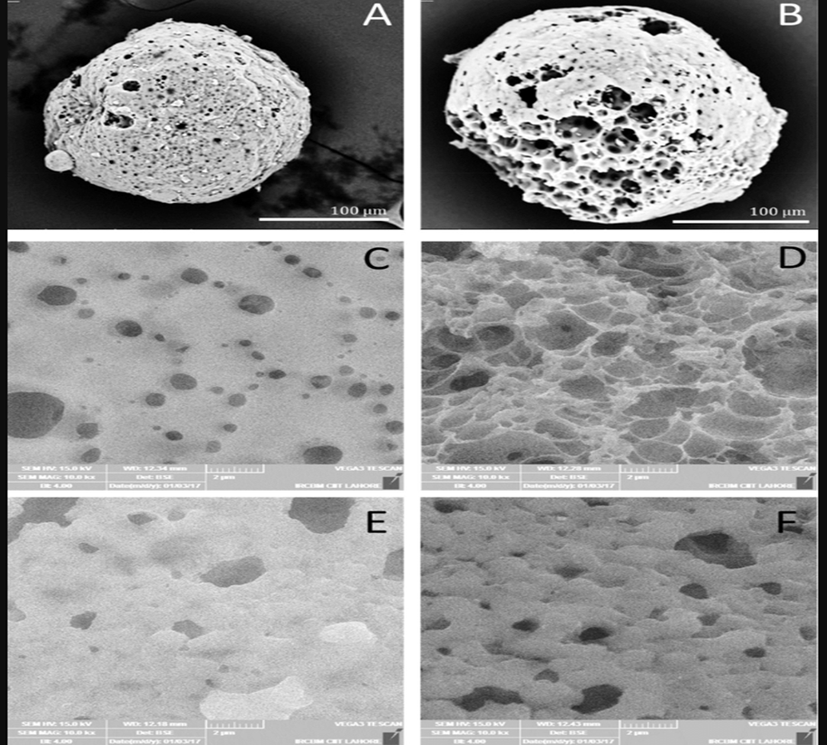
 

Figure 1 SEM images of microsponge A) 600-X magnification of blank PVA microsponge, B) 600-X magnification of blank Tween 80 microsponge, C) 10k-X magnification of PVA based microsponge, D) 10K-X magnification of Tween80 based microsponge, E) 10k-X magnification [12]


Advantages of the Microsponge Drug Delivery System

Microsponge systems offer several clinically and formulation-relevant advantages over conventional drug carriers:

  • Unlike microcapsules, which release their entire API payload upon wall rupture without any further modulation, MDDS provides programmable and sustained drug release kinetics, offering superior therapeutic control.
  • In contrast to liposomes, which are hindered by low drug payload capacity, complex manufacturing processes, limited chemical stability, and susceptibility to microbial degradation, microsponge systems exhibit broad chemical stability across a wide pH range (1–11) and high resistance to physical and biological degradation.
  • MDDS demonstrates thermal stability at temperatures up to 130°C.
  • The system supports a drug payload of up to 50–60% by weight.
  • Microsponge formulations are free-flowing in nature and economically viable in terms of production costs.
  • The porous microsphere architecture enables effective absorption of skin secretions, resulting in reduction of cutaneous oiliness and surface shine [2].

Properties of the Actives for the Entrapment into Microsponge

Active pharmaceutical ingredients or excipients intended for entrapment within microsponge systems must satisfy the following physicochemical criteria:

  • The active substance must be either fully miscible with the monomer or capable of being rendered miscible through the addition of a small quantity of a water-immiscible organic solvent.
  • The substance must be water-immiscible or exhibit only negligible aqueous solubility.
  • It must be chemically inert with respect to the monomers and must not cause a significant increase in the viscosity of the reaction mixture during the formulation process.
  • It must remain stable in the presence of the polymerization catalyst and under the conditions employed during the polymerization reaction.
  • The structural integrity of the spherical microsponge architecture must not be compromised [2].
  • The active ingredient must demonstrate adequate compatibility with the polymer matrix to facilitate efficient drug loading and controlled release.
  • A relatively low molecular weight is preferred to promote diffusion through the porous channels of the microsponge network.
  • The substance must not induce premature rupture or mechanical deformation of the microsponge particles.
  • The API must retain its pharmacological activity following entrapment and throughout the storage period.
  • An appropriate partition coefficient is necessary to ensure retention within the porous structure and facilitate gradual, controlled release.
  • The active agent must not adversely alter the porosity or pore size distribution of the loaded microsponge.
  • The substance must be amenable to release in a controlled and reproducible manner from the microsponge delivery system.
  • The ingredient must exhibit chemical and thermal stability during both the manufacturing process and storage conditions.

Benefits of microsponge tech Microsponge offers following

Microsponge technology confers a spectrum of pharmaceutical and formulation benefits:

  • Enhanced therapeutic performance of the active ingredient
  • Extended and controlled drug release profiles
  • Reduced local irritation and improved patient compliance
  • Superior product elegance and cosmetic acceptability
  • Improved oil-control, with capacity to absorb oily secretions up to six times the weight of the microsponge without generating a dry or occlusive effect
  • Greater formulation flexibility for development of novel dosage forms
  • Improved thermal, physical, and chemical stability of the encapsulated active
  • Microsponge systems are non-irritating, non-mutagenic, non-allergenic and non-toxic in nature [12].

Characteristics of the material entrapped in microsponge

Most liquid or soluble ingredients are amenable to entrapment within microsponge particles; however, such materials must satisfy the following essential requirements:
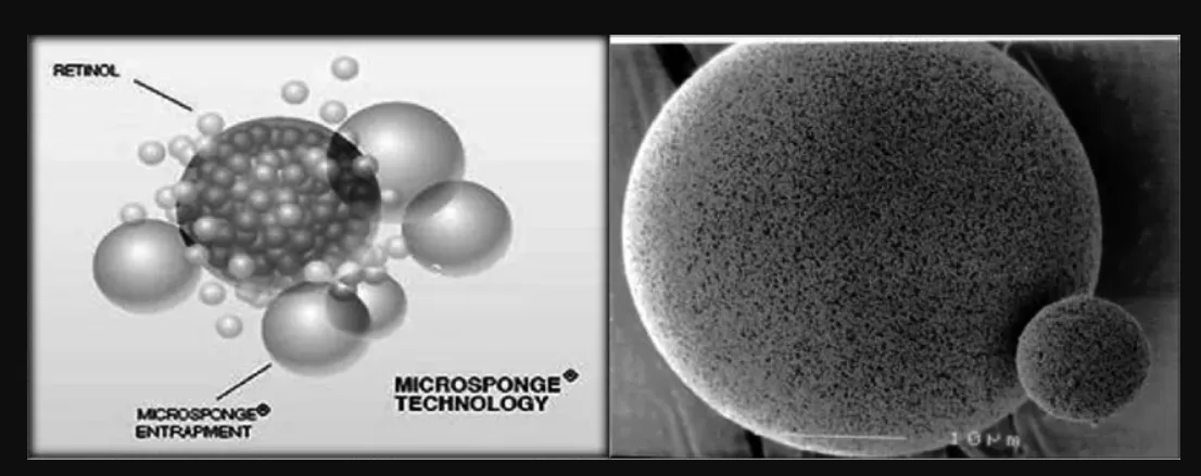
  • They should be either fully miscible with the monomer or capable of being rendered miscible through addition of a small quantity of a water-immiscible solvent.
  • They must be chemically inert with respect to the monomers used in fabrication.
  • They must be stable under the conditions of polymerization, including exposure to the initiator or catalyst [12,13].

Figure 1 CHARACTERISTICS OF MICROSPONGE in microscopic view [14]

Methodology for preparation of the Microsponge

Drug loading into microsponge can be accomplished through either a one-step or a two-step process, depending on the physicochemical properties of the drug substance. When the drug is an inert, non-polar compound, it serves as a porogen, generating the porous internal structure during polymerization. Such drugs, which do not interfere with or become activated by the polymerization reaction and remain stable to free radical species, are typically incorporated using a one-step process [12].

The principal methods employed for microsponge preparation are as follows:
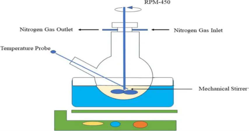
  1. Liquid- suspension polymerization: This technique is based on a free radical suspension polymerization approach. As described by Grochowicz et al., the reaction is conducted in a round-bottomed, three-necked flask equipped with a mechanical stirrer a water-cooled condenser and a thermometer. A homogeneous solution of the non-polar drug and the selected monomer(s) is prepared and subsequently combined with an aqueous phase containing a surfactant and a dispersant. Polymerization is initiated through catalysis, elevation of temperature, or addition of a chemical initiator. Water-insoluble pore-forming diluents (porogen) may also be incorporated into the reaction mixture to enhance internal porosity. When the drug is sensitive to polymerization conditions, a two-step procedure is employed. Polymerization results in the formation of cross-linked ladder-like polymer chains; folding of these chains generates spherical particles, and their subsequent agglomeration produces clusters of microspheres which ultimately bind to form the microsponge architecture. Upon completion of polymerization, the liquid porogen diffuses out, leaving behind the porous microsponge matrix. Although operationally convenient, the principal limitation of this method is the potential entrapment of unreacted monomeric residues within the microsponge structure [15,16,17].

Figure 2 Liquid-- suspension polymerization [15]

Quasi-Emulsion Solvent Diffusion: This method involves the formation of a quasi-emulsion comprising two immiscible phases. The internal phase, consisting of a drug-polymer solution prepared in a volatile solvent such as ethanol or acetone, is introduced into an external phase of aqueous polyvinyl alcohol (PVA) solution under vigorous mechanical stirring. The resulting emulsion globules, termed quasi-emulsion droplets, undergo phase separation as the organic solvent diffuses into the surrounding aqueous medium, yielding insoluble microparticulate structures. Following adequate stirring the dispersion is filtered to isolate the microsponge particles which are subsequently dried in a thermostatically controlled oven. Mechanistically finely dispersed droplets of the drug-polymer solution solidify within the aqueous phase through counter-diffusion of the organic solvent and water resulting in co-precipitation of both drug and polymer progressive solidification to form matrix-type porous microspheres. This method offers several advantages over liquid-suspension polymerization particularly reduced drug exposure to ambient conditions and minimal residual solvent levels in the final product[15,16,17].
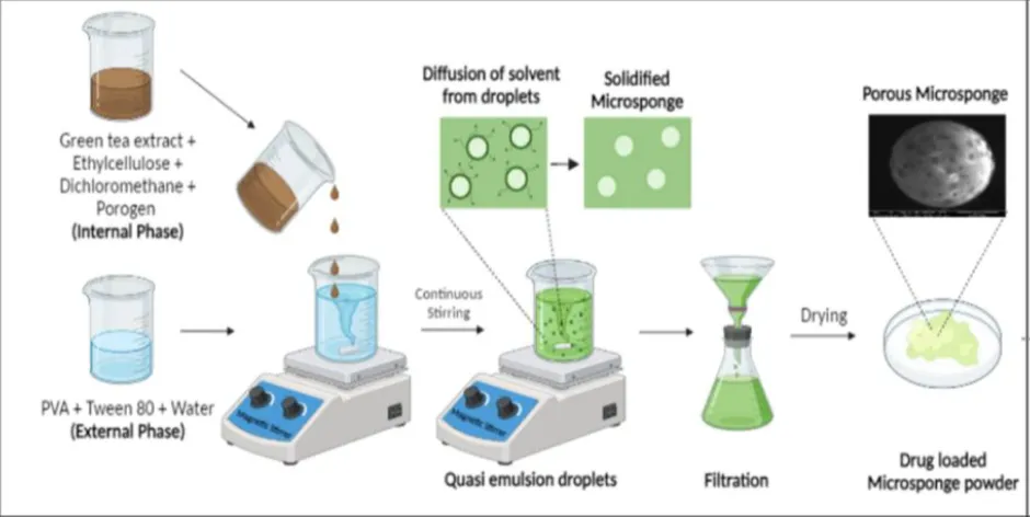
Figure 3 Quasi-emulsion solvent diffusion [15]

Water in oil in water (w/o/w) emulsion: This novel approach was developed to fabricate biodegradable porous microspheres from a double emulsion system. In this method, an internal aqueous phase containing an emulsifying agent such as Span, polyethyleneimine, or stearyl amine is dispersed within an organic polymeric solution to form a primary water-in-oil (w/o) emulsion. This primary emulsion is then re-dispersed into an external aqueous phase containing PVA, yielding a double (w/o/w) emulsion. A key advantage of this technique is its ability to entrap both water-soluble and water-insoluble drugs, as well as thermolabile materials such as proteins. Several investigators have also reported the use of xanthan gum as an emulsifier to stabilize the internal w/o emulsion in this process. [15,16,17]

  1. Addition of Porogen: In this approach, the internal aqueous phase of the conventional w/o/w double emulsion is replaced by a chemical porogen such as hydrogen peroxide or sodium bicarbonate. The porogen is uniformly dispersed within the polymeric solution to prepare a homogeneous dispersion, which is then re-dispersed in an external aqueous PVA phase. An initiator is subsequently added to the w/o/w emulsion, and the organic solvent is permitted to evaporate, leaving behind the solid microparticles. The use of hydrogen peroxide as the porogen yields evenly distributed and well-interconnected pores with diameters ranging from 5 to 20 μm [15,16,19].


Figure 4 Addition of Porogen [21]


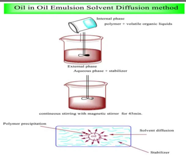
  1. Oil in oil emulsion solvent diffusion: Unlike the w/o/w method, this technique employs an oil-in-oil emulsion in which a volatile organic liquid constitutes the internal phase and is permitted to evaporate gradually under controlled conditions with continuous stirring. This method has been reported using dichloromethane as the internal phase solvent, polylactic-co-glycolic acid (PLGA) as the encapsulating polymer, and a mixture of fixed oil (corn or mineral oil) with dichloromethane containing Span 85 as the external phase. The internal phase is added dropwise to the dispersion medium under constant agitation to form the microsponge particles. This technique has also been applied to the development of hydroxyzine HCl-loaded Eudragit RS-100 microsponge using acetone as the dispersing solvent and liquid paraffin as the continuous medium. The selection of the organic solvent and external phase is dictated by the physicochemical properties of the drug and the polymer chosen for fabrication [15,19].


Figure 5 Oil in oil emulsion solvent diffusion [15]

Vibrating orifice aerosol generator method: The VOAG method was originally reported for the synthesis of lipid bilayer-coated mesoporous silica particles. The technique is based on evaporation-driven surfactant templating within aerosol microdroplets. Core particles are prepared by refluxing tetraethyl orthosilicate, ethanol, water, and dilute hydrochloric acid to yield a stock solution, which is subsequently diluted with a surfactant-containing solvent and subjected to VOAG to generate monodisperse droplets. The resulting microspheres are encapsulated within liposomes for targeted drug delivery applications [19,20].

  1. Lyophilization: Lyophilization has been employed as a secondary step to convert gelation-prepared microspheres into porous particles. In this approach, pre-formed microspheres are incubated in a chitosan hydrochloride solution and subsequently freeze-dried. Rapid solvent removal during the lyophilization cycle promotes pore formation within the microsphere matrix. Although this approach is rapid, it carries the disadvantage of producing structurally compromised particles exhibiting cracking or shrinkage as a result of abrupt solvent elimination [15,20].
  2. Ultrasound-assisted production: This method was developed through modification of the liquid-liquid suspension polymerization technique, employing β-cyclodextrin (β-CD) as the monomer and diphenyl carbonate as the cross-linking agent for nano sponge synthesis. Particle size control is achieved through a combination of thermal treatment and sonication of the reaction mixture. Following cooling, the resulting product is milled to generate irregularly shaped particles, which are then successively washed with distilled water and ethanol. The cross-linked β-CD porous microparticles serve as effective drug carriers; however, this method presents the disadvantage of potential entrapment of toxic cross-linking agent residues within the matrix [15,20,21].
  3. Electro-hydrodynamic Atomization Method: Electrohydrodynamic Atomization (EHDA) utilizes a high-voltage electric field to disperse a polymer solution into fine charged droplets, enabling the formation of uniform micro- or nanoparticles upon solvent evaporation. The apparatus consists of a syringe pump, a metallic nozzle a high-voltage power supply and an earthed collector, enclosed within an airflow-regulated chamber to control solvent evaporation and ensure particle uniformity. Various spray regimes—including dripping, cone-jet and multi-jet modes—are employed for droplet generation, with the choice governed by the interplay among electric stress, surface tension and liquid flow rate. The cone-jet mode is particularly suited for producing uniform particles of less than 10 μm diameter. High-viscosity biopolymers such as chitosan present processing challenges due to their tendency to form larger, polydisperse particles; however, this limitation can be addressed by reducing surface tension through appropriate formulation modifications. By systematically optimizing viscosity and surface tension, monodisperse spherical particles suitable for controlled drug release can be reliably produced [15,22].

Mechanism of Drug Release

Microsponge systems can be engineered to release predetermined quantities of active substances over a defined period in response to various external stimuli. The principal drug release mechanisms are as follows:

1. pH Triggered release: The surface coating of microsponge particles can be modified to initiate drug release in response to changes in environmental pH. This mechanism has considerable utility in pH-sensitive drug delivery applications across multiple anatomical sites[23].

2. Temperature Triggered Release: Elevated temperature represents another stimulus capable of activating drug release from the microsponge matrix. At ambient temperatures, certain substances entrapped within the microsponge—such as emollients and sunscreens—may exhibit high viscosity that impedes natural diffusion onto the skin surface. Upon exposure to body heat, solar radiation, or an external heat source, viscosity decreases, thereby enhancing the flow rate of the active substance and facilitating its release [23,24].

3. Pressure Triggered Release: Physical compression or mechanical pressure applied to the microsponge matrix results in the release of liquid or active constituents, replenishing the cutaneous supply of the entrapped material. The extent of pressure-induced release is also governed by the resilience and water-retention capacity of the microsponge [8].

4. Solubility Triggered Release: In the presence of aqueous media, microsponge systems loaded with water-soluble active substances—such as antimicrobials or deodorants—release their contents through diffusion-mediated processes. The partition coefficient of the active ingredient between the microsponge matrix and the surrounding medium further governs the rate and extent of release [8,24–27]..
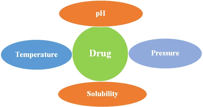
Figure 6Mechanism of drug release [28]

CHARACTERIZATION AND EVALUATION PARAMETERS

Comprehensive characterization of microsponge systems is indispensable to ensure quality, reproducibility, drug loading capacity, and controlled release behaviour. A range of physicochemical and performance-based evaluation parameters are employed to determine the formulation suitability for pharmaceutical applications [1,2].

Particle Size Analysis: Particle size exerts a significant influence on drug release rate, physical stability and dermal penetration characteristics in topical formulations. Microsponge particle size typically falls within the range of 5–300 μm; smaller particles facilitate faster release due to the greater surface area available for diffusion, whereas larger particles provide more prolonged release profiles. Particle size can be determined using optical microscopy, laser diffraction, or dynamic light scattering (DLS). A narrow particle size distribution indicates uniform emulsification conditions during preparation [32].

Surface Morphology: The surface morphology of microsponge particles is examined by Scanning Electron Microscopy (SEM), which typically reveals spherical particles with characteristic porous, sponge-like surfaces. The presence of well-defined interconnected pores confirms successful microsponge formation, while surface smoothness and structural integrity are indicative of formulation stability [11].

Entrapment Efficiency (EE%): Entrapment efficiency quantifies the proportion of drug successfully incorporated into the microsponge matrix and is calculated using the standard formula:

Elevated EE values reflect favourable drug–polymer compatibility and optimized preparation parameters. Key factors influencing EE% include polymer concentration, drug aqueous solubility, and the stirring speed applied during emulsification [1].

Drug Content Analysis: Drug content analysis ensures the homogeneous distribution of API within the microsponge matrix. An accurately weighed sample of microsponge is dissolved in a suitable solvent, filtered, and subjected to spectrophotometric or HPLC analysis. Uniform drug content across production batches is essential for ensuring therapeutic consistency and reproducibility [33].

Porosity Determination: Porosity is a critical parameter governing both drug loading capacity and release kinetics; higher porosity facilitates more rapid diffusion of drug molecules.

Porosity can be measured using:

  • Mercury intrusion porosimetry
  • Liquid displacement method
  • Gas adsorption techniques

Controlled and reproducible porosity is a prerequisite for predictable release kinetics [34].

In Vitro Drug Release Studies:  In vitro drug release studies are conducted using USP dissolution apparatus (Type I or II), selected according to the dosage form. The microsponge formulation is placed in the appropriate dissolution medium, and aliquots are withdrawn at predetermined time intervals for quantification by UV-Visible spectrophotometry or HPLC. Drug release data are fitted to mathematical kinetic models

  • Zero-order kinetics
  • First-order kinetics
  • Higuchi model
  • Korsmeyer–Peppas model

These models help to elucidate the underlying release mechanism (diffusion-controlled, erosion-based, or combined) [35].


Production Yield: Production yield indicates the efficiency of preparation method. It is calculated as: add image in between

High production yield is indicative of minimal material losses during formulation processing.[36]

Compatibility Studies:Drug–polymer compatibility is evaluated using

  • Fourier Transform Infrared Spectroscopy (FTIR)
  • Differential Scanning Calorimetry (DSC)
  • X-ray Diffraction (XRD)

FTIR identifies potential chemical interactions between the API and excipients; DSC detects thermally associated transitions; and XRD characterizes the crystalline or amorphous state of the drug within the microsponge matrix. The absence of significant physicochemical interactions confirms formulation stability [37].

True Density and Bulk Density: Density measurements are employed to assess the flow properties and compressibility of microsponge-containing formulations intended for incorporation into tablet or capsule dosage forms. Bulk density, tapped density, Carr’s Compressibility Index, and Hausner Ratio are determined to evaluate powder flow characteristics [38].

Zeta Potential: Zeta potential provides an indication of the surface charge of microsponge particles in suspension. High absolute zeta potential values are associated with strong electrostatic repulsion between particles, which confers good physical stability and prevents aggregation. [48]

Stability Studies: Stability studies are conducted in accordance with ICH guidelines under accelerated and intermediate storage conditions (e.g., 25°C/60% RH and 40°C/75% RH). Formulations are periodically evaluated for changes in drug content, particle size, morphology, and release behaviour. Stability data are essential for establishing the shelf-life and safety profile of the product [39].

Loading Efficiency

The loading efficiency (%) of the microsponge can be calculated according to the following equation:

Loading efficiency = Actual drug content in microsponge / Theoretical drug content × 100 ......(1)

The production yield is determined by comparing the initial total weight of raw materials used in fabrication against the final recovered weight of microsponge (Kilicarslan and Baykara, 2003).

Characterization of Pore Structure [12]

The presence and characteristics of internal pores are defining features of microsponge systems and therefore require thorough characterization. Pore volume and pore diameter are critical determinants of the intensity and duration of the release of active substances, as well as of the rate of migration of active ingredients from the microsponge into the surrounding vehicle. Mercury intrusion porosimetry is the preferred technique for investigating the relationship between pore dimensions and drug release kinetics.

Pore diameter

It is calculated by using Washburn equation (Washburn, 1921).

D=-4γcosθP
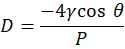
 

Where D is the pore diameter (µm); γ the surface tension of mercury (485 dyne cm⁻¹); θ the contact angle (130°); and P is the pressure (psi).


Total pore area (Atot)

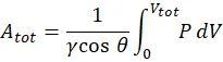
It is calculated by using equation,

Atot=1γcosθ0VtotPdV


Where, P is the pressure (psia); V the intrusion volume (ml g⁻¹); Vtot is the total specific intrusion volume (ml g⁻¹).

Average pore diameter (Dm)

It is calculated by using equation,

Dm=4VtotAtot


Where, Vtot is the total specific intrusion volume (ml g⁻¹); Atot is the total pore area.


Envelope (bulk) density (ρse)

It is calculated by using equation,

ρse=WsVp-VHg
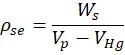
 

Where, Ws is the weight of the microsponge sample (g); Vp the volume of empty penetrometer (ml); VHg is the volume of mercury (ml).


Absolute (skeletal) density (ρsa)

It is calculated by using equation,

ρsa=WsVse-Vtot
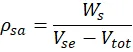
 

Where, Vse is the volume of the penetrometer minus the volume of the mercury (ml).

Percent porosity

It is calculated by equation

Porosity (%)=1ρseρsa×100

 

Where, ρse is the bulk density; ρsa is the absolute density (Orr, 1969).

FACTORS AFFECTING MICROSPONGE FORMULATION

The pharmaceutical performance of a microsponge drug delivery system is governed by a complex interplay of formulation and process variables. Systematic optimization of these parameters is essential to achieve the desired particle size, porosity, entrapment efficiency, and controlled drug release behaviour.

  1. Polymer Type: The selection of polymer is a fundamental determinant of the structural integrity, porosity, and drug release kinetics of the microsponge system. Polymers such as Eudragit RS 100, Eudragit RL 100, ethyl cellulose, and polymethacrylates are widely employed owing to their established biocompatibility and to able permeability. Hydrophobic polymers typically provide sustained drug release, whereas polymers with greater permeability facilitate faster drug diffusion. The molecular weight and intrinsic permeability of the selected polymer exert direct influence on the drug release rate and the mechanical robustness of the microsponge matrix [40].
  2. Polymer Concentration: Polymer concentration profoundly influences particle size, entrapment efficiency, and drug release profile. Increasing polymer concentration elevates the viscosity of the internal phase, producing larger emulsion droplets and consequently larger microsponge particles. Higher polymer content enhances entrapment efficiency due to the formation of a denser polymeric network; however, excessive polymer may excessively retard drug release. Judicious optimization is therefore required to balance sustained release and adequate therapeutic efficacy [41].
  3. Drug–Polymer Ratio: The drug-to-polymer ratio directly governs drug loading capacity and release characteristics. Excessively high drug loading may promote an initial burst release attributable to superficial drug deposition on the particle surface. An optimized drug–polymer ratio ensures uniform drug distribution throughout the porous matrix and facilitates controlled diffusion through the interconnected pore channels [42].
  4. Drug Properties: Physicochemical properties of the drug including aqueous solubility, molecular weight, and chemical stability critically influence microsponge formulation outcomes. Highly water-soluble drugs are prone to significant leaching during preparation, resulting in reduced entrapment efficiency. Lipophilic drugs, by contrast, are generally better suited for microsponge encapsulation due to their stronger affinity for hydrophobic polymer matrices [43]. Drug stability in the selected organic solvent system must also be carefully evaluated to prevent degradation during processing [1].
  5. Type of Solvent: The choice of organic solvent influences droplet formation, polymer precipitation kinetics, and the resulting porosity of the microsponge. Volatile solvents such as dichloromethane and ethanol are commonly employed in quasi-emulsion solvent diffusion methods. The rate of solvent diffusion governs pore formation and internal structural characteristics; rapid solvent removal promotes the formation of highly porous microsponge while slow evaporation tends to yield denser, less porous particles. [1]
  6. Stirring Speed: Stirring speed during the emulsification step controls emulsion droplet size and, consequently, the final particle size of the microsponge. Higher agitation speeds produce finer droplets, yielding smaller particles with greater surface area and faster drug release rates. Lower stirring speeds generate larger particles associated with more prolonged release profiles. Optimization of stirring speed is therefore essential for achieving the target particle size distribution [43].
  7. Surfactant Type and Concentration: Surfactants such as polyvinyl alcohol (PVA) function as emulsion stabilizers in the microsponge preparation process. Insufficient surfactant concentration results in particle aggregation, whereas an excess may adversely affect porosity and drug loading. Appropriate surfactant concentrations ensure the formation of uniform well-dispersed particles with consistent physical stability [44].
  8. Volume of Internal and External Phases: The volumetric ratio of the internal organic phase to the external aqueous phase governs the rate of solvent diffusion and the efficiency of particle formation. A larger external phase volume enhances the solvent diffusion driving force and promotes the formation of highly porous microsponge. An inappropriate phase ratio may result in irregular particle size distribution and reduced production yield. [1].
  9. Temperature: Processing temperature affects the rate of solvent evaporation and polymer solidification. Elevated temperatures accelerate solvent removal, potentially increasing porosity; however, excessive heat may cause degradation of thermolabile APIs. Controlled and reproducible temperature conditions during formulation are therefore imperative [1].
  10. Cross-Linking Density: In polymerization-based microsponge preparation, cross-linking density determines the mechanical strength and porosity of the resulting structure. Higher degrees of cross-linking produce rigid, compact architectures with slower drug release, whereas reduced cross-linking yields more porous but structurally less robust particles [1].
  11. Drying Method: Post-preparation drying conditions—including air drying, vacuum drying, or oven drying—significantly affect the physicochemical properties of the final microsponge product. Rapid or harsh drying may collapse the porous structure, whereas controlled drying conditions preserve the integrity of the pore network. Appropriate drying procedures also prevent particle agglomeration and ensure long-term storage stability [45] .

APPLICATION OF MICROSPOGNE DRUG DELIVERY SYSTEM

  • The versatile nature of microsponge technology has facilitated its integration into a diverse range of dosage forms including creams, gels, lotions, and oral formulations, thereby demonstrating its broad adaptability across drug delivery requirements. MDDS has been extensively utilized in the management of dermatological disorders such as acne, psoriasis, seborrhoeic dermatitis (dandruff), eczema, scleroderma, alopecia and skin cancer. [46]
  • These delivery platform enables effective prolonged retention of therapeutics at the application site, enhancing dermal efficacy while minimizing the risks of over- or under-medication that are frequently associated with conventional topical delivery systems. [47]
  • MDDS also supports the development of gastro retentive systems providing controlled, prolonged drug release with consequent improvements in oral bioavailability and therapeutic outcomes. The technology has been employed for colonic delivery of drugs such as flurbiprofen using pH-responsive coatings, and for enhancing the bioavailability of poorly water-soluble drugs including paracetamol and indomethacin through increased effective surface area. Additionally, MDDS reduces gastrointestinal irritation associated with drugs such as ketoprofen and dicyclomine. Specialized applications include site-specific anticancer drug delivery and regenerative medicine through programmable growth factor release. [48,49]
  • In the cosmeceutical sector, microsponge technology has transformed the development of sunscreen formulations by enabling encapsulation of UV filters, providing extended photoprotection and improved non-greasy aesthetics. Anti-ageing formulations have also benefited from microsponge-mediated sustained release of retinoids and antioxidants. Coloured cosmetics such as lipsticks and foundation products leverage microsponge encapsulation of pigments for prolonged wear and a matte finish. Consumer products such as moisturizers and anti-acne preparations utilize this platform to provide sustained hydration and reduction of skin irritation. Key clinical and formulation advantages include superior physicochemical stability of sensitive active compounds, enhanced patient compliance, and compatibility with diverse carrier systems, collectively establishing MDDS as a transformative technology for both therapeutic and cosmetic drug delivery innovation. [49,50]

Table 1 Comparison table between formulations

Formulation

Microsponge

Microspheres

Microbeads

Morphology

Porous, spongy like microsphere.

Solid, spherical particles.

Small spherical polymeric beads with matrix structure

Size

5-300 μm

1 – 1000 μm

10- 2000μm

Drug loading

Up to 50- 60%

Up to 40- 50 %

Up to 40- 60 %

Stability

Stable at basic medium up to 130º C

Variable stability.

Good physicochemical stability.

Cost

Lower cost compared to other all delivery system.

Moderate cost.

Economic.

Advantages

Controlled drug release.

Sustained released.

Sustained and control release.


Enhanced stability.

Targeted drug delivery.

Improved patient compliance.


Reduces side effect and improve aesthetics.

Protection of drug from degradation.

Easy to manufacture and have better flexibility.

Disadvantages

Organic solvent is used.

Burst drug release.

Possible of dose dumping and polymer related toxicity.


Potential residual monomer toxicity.

Specific speed required for preparation.

Difficult in size uniformity.


Application and Advantages of MDDS


Table 2 Types of formulations with active agent [22,51-58]

Sr No.

Formulation types

Active agent

Advantages

1.

Sunscreens

Aloin, proanthocyanidin, quercetin

Provides long-lasting efficacy with enhanced sunburn protection, even at

higher concentrations, while minimizing irritation and sensitization.

2.

Anti fungal

Terbinafine

Ensures sustained release of active ingredients.

3.

Skin depigmenting

Hydroquinone

Hydroquinone for skin depigmentation provides enhanced stability against

oxidation, improved efficacy, and a more aesthetically appealing

appearance.

4.

Antipruritic

Sertaconazole nitrate

Provides prolonged and enhanced activity.

5.

Anti dandruff shampoos

Zinc pyrithione, selenium sulphide

Reduces unpleasant odour, minimizes irritation and ensures prolonged

safety and efficacy

6.

Rubefacients

-

Ensures prolonged activity with reduced irritancy, greasiness, and odour. [

7.

Anit acne

Benzoyl peroxide

Sustains efficacy with less irritation and sensitization.

8

Anti inflammatory

Hydroquinone 4% and retinol 0.15%

Prolonged activity with reduced allergic response and dermatoses.


Examples of Microsponge Drug Delivery, Their Formulations, and Associated Brands/Trademarks


Table 3 Marketed formulation along with brand and manufacture name. [22,59-74]

Formulation Type

Brand Name

Manufacturer

Active Drug

Application

Gels

Melanin Microsponge

Advanced Polymer Systems Inc., USA

Melanin

Hyperpigmentation disorders

Gels

Clozole Gel 15g

Psyco Remedies

Fluconazole

Antifungal infection treatment

Gels

NA

Terbinafine

Antifungal

Gels

NA

Terbinafine HCl

Antifungal

Gels

NA

Oxiconazole Nitrate

Antifungal

Gels

Ertaczo

Glenmark Pharmaceuticals Ltd.

Sertaconazole nitrate

Antifungal

Gels

Retin-A Micro

Ortho-McNeil Pharmaceutical, Inc., USA

Tretinoin

Acne vulgaris

Gels

NA

Tazarotene

Facial acne vulgaris

Gels

NA

Oxybenzone

Sunscreen agent

Gels

NA

Diclofenac sodium

Inflammation

Gels

NA

Silver sulfadiazine

Reduces irritation with low dermal cytotoxicity

Gels

NA

Hydroxyzine HCl

Urticaria and atopic dermatitis

Gels

NA

Acyclovir

Viral infections

Gels

NA

Nebivolol

Diabetic rash

Gels, Lotions, and Creams

Brevoxyl

Unicure India Pvt. Ltd.

Benzoyl peroxide

Reduces irritation, sensitization, and acne

Gels, Lotions, and Creams

EpiQuin Micro

SkinMedica Inc., USA

Hydroquinone and retinol

Improved oxidation resistance, efficacy, and aesthetics for hyperpigmentation

Lotions

Oil-Free Matte Block SPF 20

Dermalogica, LLC, USA

Zinc gluconate

Sunscreen

Creams

Carbopol Gel

Scott Paper Company

Miconazole

Microsponge gel for diaper dermatitis; treats dermatitis, acne, and topical infections

Creams

Line Eliminator

Avon Products, Inc., UK

Retinol

Anti-wrinkles

Creams

Lactrex 12%

SDR Pharmaceuticals Pvt. Ltd., India

Ammonium lactate

Moisturizer containing lactic acid, ammonium lactate, water, and glycerin

Creams and Lotions

Ultra Guard

Scott Paper Company, USA

Dimethicone

Protective barrier for babies

Gel and Ointments

Salicylic Peel 20 and 30

Biophora Medical Skin Care, Ontario, Canada

Salicylic acid

Effective chemical exfoliant

Creams and Gels

Retinol 15 Night Cream

Biomedical Emporium, South Africa

Retinol

Anti-wrinkle skin supplement

Gels, Creams, and Solid Particles

Carac Cream, 0.5%

Dermik Laboratories, Inc., USA

5-Fluorouracil

Lesion reduction, actinic keratosis, and colon cancer treatment

Tablets

NA

NA

Ketoprofen

Musculoskeletal pain

Tablets

NA

NA

Meloxicam

Arthritis

Tablets

NA

NA

Nicorandil

Cardiovascular application

Tablets

NA

NA

Indomethacin

Inflammation

Tablets

NA

NA

Lafutidine

Anti-ulcer

Tablets

NA

NA

5-Amino salicylic acid

Inflammatory Bowel Disease

Tablets

NA

NA

Telmisartan

Cardiovascular applications

Tablets

NA

NA

Cinnarizine

Treats vertigo, motion sickness, and vomiting

Tablets

NA

NA

Carbamazepine

Sustained drug release

Tablets

NA

NA

Pantoprazole sodium

Management of Gastroesophageal Reflux Disease (GERD)

Implants

NA

NA

Poly(DL-lactic-co-glycolic acid)

Skin tissue engineering

Implants

NA

NA

Ketorolac Tromethamine

Inflammation

Capsules

NA

NA

5-Fluorouracil calcium

Colorectal cancer

Capsules

NA

NA

Ketoconazole

Antifungal

Grafts / Injection

NA

NA

Fibroblast growth factor

Delivery of growth factors for tissue repair

Others

NA

NA

Ibuprofen

Pain and inflammation management using NSAIDs

Others

NA

NA

Erythromycin

Skin infection

Others

NA

NA

Dexamethasone

Ulcerative Colitis

Others

NA

NA

Fluocinolone acetonide

Inflammation


CONCLUSION

The concept of Microsponges Drug Delivery Systems has proven to be an advanced scientific innovation that promises significant commercial benefits within the pharmaceutical field. The highly porous nature of these delivery systems, which consists of a wide variety of channels capable of carrying active pharmaceutical substances, makes them highly superior to other traditional drug delivery systems. Advantages offered by this technology include programmed and stimulus responsive drug release, increased chemical stability, greater patient compliance, and minimized side effects, both locally and systemically. Microsponge Drug Delivery Systems originated in the year 1987, initially designed to address the issue of acne through topical administration. However, over time, their scope has expanded beyond topical application, covering dermatological therapy, oral drug delivery, gastro-intestinal disorders and even regenerative medicine.

Different approaches can be used to prepare microsponge-based drug delivery systems depending on their intended use, including quasi-emulsion solvent diffusion, liquid-suspension polymerization, double emulsions, and electrohydrodynamic atomization. Various formulation variables must be considered during formulation, such as choice of polymer and concentration, drug-polymer ratio, solvent type, stirring speed, and the procedure of drying. The properties of the developed systems can be characterized using various analytical approaches including SEM, FTIR, DSC, mercury intrusion porosimetry and in-vitro dissolution. It ensures the quality, reproducibility, and efficacy of the microsponge systems. The incorporation of natural excipients extracted from plants into microsponge further brings the use of these systems into line with current developments in sustainable and patient centric drug production. With ongoing progress in the field, microsponge technology shows great potential for meeting important challenges related to various types of drug delivery systems.

REFERENCES

  1. Jaiswal N, Shivastava RK. Microsponge drug delivery system: A comprehensive review on formulation, characterization, applications and future perspectives. World J Pharm Res. 2026;15(5):1628-1644. doi:10.5281/zenodo.18884674.
  2. Nacht S, Kantz M. The microsponge: A novel topical programmable delivery system. Top Drug Deliv Syst. 1992;42:299-325.
  3. Won R. Method for delivering an active ingredient by controlled time release utilizing a novel delivery vehicle which can be prepared by a process utilizing the active ingredient as a porogen. US Patent 4690825; 1987.
  4. Nidhi K, Verma S, Kumar S. Microsponge. J Clin Sci Res. 2021;10(2):108-111.
  5. Shaha V, Jain H, Krishna J, Patel P. Microsponge drug delivery: A review. Int J Res Pharm Sci. 2010;1(2):212-218.
  6. Zawar N. Microsponge as a strategy for effective drug delivery system. Al-Mustansiriyah J Pharm Sci. 2023;23(3):322-335.
  7. Anderson DL, Chung-Heng C, Nacht S. Flow characteristics of loosely compacted macroporous microsponge polymeric systems. Powder Technol. 1994;78(1):15-18.
  8. Jadhav N, Patel V, Mungekar S, Bhamare G, Kadams V. Microsponge delivery system: An updated review, current status and future prospects. J Sci Innov Res. 2013;2(6):1097-1110.
  9. Orlu M, Cevher E, Araman A. Design and evaluation of colon-specific drug delivery system. Int J Pharm. 2006;318:103-117.
  10. Kaity S, Maiti S, Ghosh A, Pal D, Ghosh A, Banerjee S. Microsponges: A novel strategy for drug delivery system. J Adv Pharm Technol Res. 2010;1(3):283-290.
  11. Natural polymers in microsponge preparation: An excipients review [Internet]. 2025 [cited 2026 May 20]. Available from: WisdomLib
  12. Shahzad Y, Saeed S, Ghori MU, Mahmood T, Yousaf AM, Jamshaid M, et al. Influence of polymer ratio and surfactants on controlled drug release from cellulosic microsponges. Int J Biol Macromol. 2017;109:963-970. doi:10.1016/j.ijbiomac.2017.11.089.
  13. Aloorkar NH, Kulkarni AS, Ingale DJ, Patil RA. Microsponges as innovative drug delivery systems. Int J Pharm Sci Nanotechnol. 2012;5(1):1597-1606.
  14. Emanuele AD, Dinarvand R. Preparation, characterization and drug release from thermo responsive microspheres. Int J Pharm. 1995;118:237-242.
  15. Ambekar AW. Microsponge: A novel topical drug delivery system. J Drug Deliv Res. 2014;3(1):1-7.
  16. Golechha PA, Atram SC, Wankhade VP, Bobade NN, Pande SD. Role of microsponge drug delivery systems in improving therapeutic outcomes in rheumatoid arthritis. Asian J Pharm Res Dev. 2026;14(1):57-64. doi:10.22270/ajprd.v14i1.1701.
  17. D’Souza JI, Harinath N. Topical anti-inflammatory gels of fluocinolone acetonide entrapped in Eudragit-based microsponge delivery system. Res J Pharm Technol. 2008;1(4):502-506.
  18. Çomoğlu T, Gönül N, Baykara T. Preparation and in vitro evaluation of modified release ketoprofen microsponges. Farmaco. 2003;58(2):101-106.
  19. Patil S, Dandekar V. Microsponge drug delivery system: An overview. Eur J Pharm Med Res. 2016;3(8):212-221.
  20. Dean RC, Silver FH. Weighted collagen microsponges for immobilizing bioactive agents. US Patent US4863856; 1989.
  21. Schaefer H. Cosmetic or pharmaceutical composition containing microspheres of polymers of fatty substances filled with at least one active product. US Patent US5292512; 1989.
  22. Katz M. Porous particles in preparations involving immiscible phases. US Patent US5135740; 1992.
  23. Verma M, Burman A, Majhi S, Chauhan I. Recent advances in microsponge drug delivery system: Patents and future prospects. Curr Indian Sci. 2026;4:e2210299X390762. doi:10.2174/012210299X390762251111050757.
  24. Valluru R, Ravi G, Bose SP, Damineni S. Microsponges-a comprehensive review: Success and challenges. Indo Am J Pharm Res. 2019;9(7):3056-3067.
  25. Chadawar V, Shaji J. Microsponge delivery system. Curr Drug Deliv. 2007;4(2):123-129.
  26. Panwar AS, Yadav CS, Yadav P, Darwhekar GN, Jain DK, Panwar MS, et al. Microsponge: A novel carrier for cosmetics. J Glob Pharma Technol. 2011;3(7):15-24.
  27. Mahant S, Kumar S, Nanda S, Rao R. Microsponges for dermatological applications: Perspectives and challenges. Asian J Pharm Sci. 2020;15(3):273-291.
  28. Mohite PB, Khanage SG, Harishchandre VS, Yogita S. Recent advances in microsponges drug delivery system. J Crit Rev. 2016;3(1):9-16.
  29. Rajeswari S, Swapna V. Microsponges as a neoteric cornucopia for drug delivery systems. Int J Curr Pharm Res. 2019;11(3):4-12.
  30. Jain V, Singh R. Development and characterization of Eudragit RS 100 loaded microsponges and its colonic delivery using natural polysaccharides. Acta Pol Pharm. 2010;67(4):407-415.
  31. Shaha V, Jain H, Krishna A, Patel P. Microsponge drug delivery: A review. Int J Res Pharm Sci. 2010;1(2):212-218.
  32. Patel A, Patel M, Yang X, Mitra AK. Recent advances in protein and peptide drug delivery: A special emphasis on polymeric nanoparticles. Protein Pept Lett. 2014;21(11):1102-1120.
  33. Higuchi T. Mechanism of sustained-action medication: Theoretical analysis of rate of release of solid drugs dispersed in solid matrices. J Pharm Sci. 1963;52(12):1145-1149.
  34. Kawashima Y, Niwa T, Takeuchi H, Hino T, Itoh Y. Control of prolonged drug release and compression properties of ibuprofen microsponges. Chem Pharm Bull (Tokyo). 1992;40(1):196-201.
  35. Silverstein RM, Webster FX. Spectrometric identification of organic compounds. 6th ed. New York: Wiley; 1998.
  36. Carr RL. Evaluating flow properties of solids. Chem Eng. 1965;72:163-168.
  37. Hunter RJ. Zeta potential in colloid science: Principles and applications. London: Academic Press; 1981.
  38. International Council for Harmonisation (ICH). Q1A(R2): Stability testing of new drug substances and products. Geneva: ICH; 2003.
  39. Jain N, Sharma PK, Banik A. Recent advances on microsponge delivery system. Int J Pharm Sci Rev Res. 2011;8(2):13-23.
  40. Vyas SP, Khar RK. Controlled drug delivery: Concepts and advances. 1st ed. New Delhi: Vallabh Prakashan; 2002.
  41. Srivastava R, Pathak K. Microsponges: A futuristic approach for oral drug delivery. Expert Opin Drug Deliv. 2012;9(7):863-878.
  42. Martin A. Physical pharmacy. 4th ed. Philadelphia: Lippincott Williams & Wilkins; 1993.
  43. Lieberman HA, Lachman L, Schwartz JB. Pharmaceutical dosage forms: Tablets. 2nd ed. New York: Marcel Dekker; 1989.
  44. Aulton ME. Pharmaceutics: The design and manufacture of medicines. 4th ed. London: Churchill Livingstone; 2013.
  45. Rahman M, Almalki WH, Panda SK, Das AK, Alghamdi S, Soni K, et al. Therapeutic application of microsponges-based drug delivery systems. Curr Pharm Des. 2022;28(8):595-608.
  46. Mahant S, Kumar S, Nanda S, Rao R. Microsponges for dermatological applications: Perspectives and challenges. Asian J Pharm Sci. 2020;15(3):273-291.
  47. Vidya KP, Gopinath E, Ganesh NS, Jino Nesalin JA, Chandy V. Unveiling the potential of microsponges: Enhancing oral bioavailability. J Biol Pharm Health Sci. 2024;2024(02):405-414.
  48. Saboo S, Bhise YO. Microsponge: An innovative and novel strategy for drug delivery system. Pharma Times. 2024;16(1):211-223.
  49. Muruganantham V, Venkateswarlu BS, Chinraj V. Design and evaluation of isolated herbal extracts of aloin, proanthocyanidin, quercetin loaded microsponge as sunscreen gel. Asian J Biol Life Sci. 2022;11(2):356-366. doi:10.5530/ajbls.2022.11.48.
  50. Dutta D, Goyal N, Sharma DK. Formulation and development of herbal microsponge sunscreen gel. J Cosmet Dermatol. 2022;21(4):1675-1687. doi:10.1111/jocd.14274.
  51. Mahaparale PR, Ikam SAN, Chavan MS. Development and evaluation of terbinafine hydrochloride polymeric microsponges for topical drug delivery. Indian J Pharm Sci. 2018;80(6):1086-1092. doi:10.4172/pharmaceutical-sciences.1000459.
  52. Wadhwa G, Kumar S, Mittal V, Rao R. Encapsulation of babchi essential oil into microsponges: Physicochemical properties, cytotoxic evaluation and anti-microbial activity. J Food Drug Anal. 2019;27(1):60-70. doi:10.1016/j.jfda.2018.07.006.
  53. Kadnor NA, Pande VV, Kadam R, Upadhye SA. Fabrication and characterization of sertaconazole nitrate microsponge as a topical drug delivery system. Indian J Pharm Sci. 2015;77(6):675-680. doi:10.4103/0250-474X.174986.
  54. Jelvehgari M, Siahi-Shadbad MR, Azarmi S, Martin GP, Nokhodchi A. The microsponge delivery system of benzoyl peroxide: Preparation, characterization and release studies. Int J Pharm. 2006;308(1-2):124-132.
  55. Nokhodchi A, Jelvehgari M, Siahi MR, Mozafari MR. Factors affecting the morphology of benzoyl peroxide microsponges. Micron. 2007;38(8):834-840. doi:10.1016/j.micron.2007.06.012.
  56. Patel N, Padia N, Vadgama N, Raval M, Sheth N. Formulation and evaluation of microsponge gel for topical delivery of fluconazole for fungal therapy. J Pharm Investig. 2016;46(3):221-238. doi:10.1007/s40005-016-0230-7.
  57. Bharathy P, Thanikachalam PV, Parthasarathy NP, Elumalai P, Baskaran PK, Thameemul TA. Formulation and characterization of luliconazole microsponge gel for diaper dermatitis. J Pharm Innov. 2023;18(4):2362-2372. doi:10.1007/s12247-023-09797-4.
  58. Salah S, Awad GEA, Makhlouf AIA. Improved vaginal retention and enhanced antifungal activity of miconazole microsponges gel: Formulation development and in vivo therapeutic efficacy in rats. Eur J Pharm Sci. 2018;114:255-266. doi:10.1016/j.ejps.2017.12.023.
  59. Patel P, Thanki A, Viradia D, Shah P. Honey-based silver sulfadiazine microsponge-loaded hydrogel: In vitro and in vivo evaluation for burn wound healing. Curr Drug Deliv. 2023;20(5):608-628. doi:10.2174/1567201819666220516092359.
  60. Kumar PM, Ghosh A. Development and evaluation of silver sulfadiazine loaded microsponge based gel for partial thickness (second degree) burn wounds. Eur J Pharm Sci. 2017;96:243-254. doi:10.1016/j.ejps.2016.09.038.
  61. Zaki Rizkalla CM, Aziz RL, Soliman II. In vitro and in vivo evaluation of hydroxyzine hydrochloride microsponges for topical delivery. AAPS PharmSciTech. 2011;12(3):989-1001. doi:10.1208/s12249-011-9663-5.
  62. Pandit AP, Patel SA, Bhanushali VP, Kulkarni VS, Kakad VD. Nebivolol-loaded microsponge gel for healing of diabetic wound. AAPS PharmSciTech. 2017;18(3):846-854. doi:10.1208/s12249-016-0574-3.
  63. Gulati N, Tomar N, Nagaich U. Miconazole microsponges based topical delivery system for diaper dermatitis. Ars Pharm. 2016;57(2):77-87. doi:10.30827/ars.v57i2.4987.
  64. Li SS, Li GF, Liu L, Jiang X, Zhang B, Liu ZG, et al. Evaluation of paeonol skin-target delivery from its microsponge formulation: In vitro skin permeation and in vivo microdialysis. PLoS One. 2013;8(11):e79881. doi:10.1371/journal.pone.0079881.
  65. Singhvi G, Manchanda P, Hans N, Dubey SK, Gupta G. Microsponge: An emerging drug delivery strategy. Drug Dev Res. 2019;80(2):200-208. doi:10.1002/ddr.21492.
  66. Srivastava R, Kumar D, Pathak K. Colonic luminal surface retention of meloxicam microsponges delivered by erosion based colon-targeted matrix tablet. Int J Pharm. 2012;427(2):153-162. doi:10.1016/j.ijpharm.2012.01.036.
  67. Patel SS, Patel MR, Patel MJ. Formulation and evaluation of microsponge based nicorandil sustained released tablet. J Sci Res. 2017;9(3):285-296. doi:10.3329/jsr.v9i3.31193.
  68. Janakidevi S, Ramanamurthy KV. Development of colon-targeted microsponges for the treatment of inflammatory bowel disease. Indian J Pharm Sci. 2018;80(4):604-609. doi:10.4172/pharmaceutical-sciences.1000399.
  69. Pathak K, Raghuvanshi S. Bioadhesive floating microsponges of cinnarizine as novel gastroretentive delivery: Capmul GMO bioadhesive coating versus Acconon MC 8-2 EP/NF with intrinsic bioadhesive property. Int J Pharm Investig. 2016;6(4):181-193. doi:10.4103/2230-973X.195923.
  70. Abdalla KF, Osman MA, Nouh AT, El Maghraby GM. Microsponges for controlled release and enhanced oral bioavailability of carbamazepine. J Drug Deliv Sci Technol. 2021;65:102683. doi:10.1016/j.jddst.2021.102683.
  71. Gupta A, Tiwari G, Tiwari R, Srivastava R. Factorial designed 5-fluorouracil-loaded microsponges and calcium pectinate beads plugged in hydroxypropyl methylcellulose capsules for colorectal cancer. Int J Pharm Investig. 2015;5(4):234-246. doi:10.4103/2230-973X.167688.
  72. Hussien AA. Preparation and evaluation of oral microsponge drug delivery system of ketoconazole. Al-Mustansiriyah J Pharm Sci. 2014;14(1):1-8. doi:10.32947/ajps.v14i1.119.
  73. Ravi R, Sk S. Standardization of process parameters involved in erythromycin microsponges by quasi emulsion solvent diffusion method. Int J Pharm Dev Technol. 2013;3(1):28-34.
  74. Özdemir S, Üner B, Baranauskaite J, Sümer E, Yıldırım E, Yaba A. Design and characterization of dexamethasone loaded microsponges for the management of ulcerative colitis. Eur J Pharm Biopharm. 2023;187:34-45. doi:10.1016/j.ejpb.2023.04.007.

Reference

  1. Jaiswal N, Shivastava RK. Microsponge drug delivery system: A comprehensive review on formulation, characterization, applications and future perspectives. World J Pharm Res. 2026;15(5):1628-1644. doi:10.5281/zenodo.18884674.
  2. Nacht S, Kantz M. The microsponge: A novel topical programmable delivery system. Top Drug Deliv Syst. 1992;42:299-325.
  3. Won R. Method for delivering an active ingredient by controlled time release utilizing a novel delivery vehicle which can be prepared by a process utilizing the active ingredient as a porogen. US Patent 4690825; 1987.
  4. Nidhi K, Verma S, Kumar S. Microsponge. J Clin Sci Res. 2021;10(2):108-111.
  5. Shaha V, Jain H, Krishna J, Patel P. Microsponge drug delivery: A review. Int J Res Pharm Sci. 2010;1(2):212-218.
  6. Zawar N. Microsponge as a strategy for effective drug delivery system. Al-Mustansiriyah J Pharm Sci. 2023;23(3):322-335.
  7. Anderson DL, Chung-Heng C, Nacht S. Flow characteristics of loosely compacted macroporous microsponge polymeric systems. Powder Technol. 1994;78(1):15-18.
  8. Jadhav N, Patel V, Mungekar S, Bhamare G, Kadams V. Microsponge delivery system: An updated review, current status and future prospects. J Sci Innov Res. 2013;2(6):1097-1110.
  9. Orlu M, Cevher E, Araman A. Design and evaluation of colon-specific drug delivery system. Int J Pharm. 2006;318:103-117.
  10. Kaity S, Maiti S, Ghosh A, Pal D, Ghosh A, Banerjee S. Microsponges: A novel strategy for drug delivery system. J Adv Pharm Technol Res. 2010;1(3):283-290.
  11. Natural polymers in microsponge preparation: An excipients review [Internet]. 2025 [cited 2026 May 20]. Available from: WisdomLib
  12. Shahzad Y, Saeed S, Ghori MU, Mahmood T, Yousaf AM, Jamshaid M, et al. Influence of polymer ratio and surfactants on controlled drug release from cellulosic microsponges. Int J Biol Macromol. 2017;109:963-970. doi:10.1016/j.ijbiomac.2017.11.089.
  13. Aloorkar NH, Kulkarni AS, Ingale DJ, Patil RA. Microsponges as innovative drug delivery systems. Int J Pharm Sci Nanotechnol. 2012;5(1):1597-1606.
  14. Emanuele AD, Dinarvand R. Preparation, characterization and drug release from thermo responsive microspheres. Int J Pharm. 1995;118:237-242.
  15. Ambekar AW. Microsponge: A novel topical drug delivery system. J Drug Deliv Res. 2014;3(1):1-7.
  16. Golechha PA, Atram SC, Wankhade VP, Bobade NN, Pande SD. Role of microsponge drug delivery systems in improving therapeutic outcomes in rheumatoid arthritis. Asian J Pharm Res Dev. 2026;14(1):57-64. doi:10.22270/ajprd.v14i1.1701.
  17. D’Souza JI, Harinath N. Topical anti-inflammatory gels of fluocinolone acetonide entrapped in Eudragit-based microsponge delivery system. Res J Pharm Technol. 2008;1(4):502-506.
  18. Çomo?lu T, Gönül N, Baykara T. Preparation and in vitro evaluation of modified release ketoprofen microsponges. Farmaco. 2003;58(2):101-106.
  19. Patil S, Dandekar V. Microsponge drug delivery system: An overview. Eur J Pharm Med Res. 2016;3(8):212-221.
  20. Dean RC, Silver FH. Weighted collagen microsponges for immobilizing bioactive agents. US Patent US4863856; 1989.
  21. Schaefer H. Cosmetic or pharmaceutical composition containing microspheres of polymers of fatty substances filled with at least one active product. US Patent US5292512; 1989.
  22. Katz M. Porous particles in preparations involving immiscible phases. US Patent US5135740; 1992.
  23. Verma M, Burman A, Majhi S, Chauhan I. Recent advances in microsponge drug delivery system: Patents and future prospects. Curr Indian Sci. 2026;4:e2210299X390762. doi:10.2174/012210299X390762251111050757.
  24. Valluru R, Ravi G, Bose SP, Damineni S. Microsponges-a comprehensive review: Success and challenges. Indo Am J Pharm Res. 2019;9(7):3056-3067.
  25. Chadawar V, Shaji J. Microsponge delivery system. Curr Drug Deliv. 2007;4(2):123-129.
  26. Panwar AS, Yadav CS, Yadav P, Darwhekar GN, Jain DK, Panwar MS, et al. Microsponge: A novel carrier for cosmetics. J Glob Pharma Technol. 2011;3(7):15-24.
  27. Mahant S, Kumar S, Nanda S, Rao R. Microsponges for dermatological applications: Perspectives and challenges. Asian J Pharm Sci. 2020;15(3):273-291.
  28. Mohite PB, Khanage SG, Harishchandre VS, Yogita S. Recent advances in microsponges drug delivery system. J Crit Rev. 2016;3(1):9-16.
  29. Rajeswari S, Swapna V. Microsponges as a neoteric cornucopia for drug delivery systems. Int J Curr Pharm Res. 2019;11(3):4-12.
  30. Jain V, Singh R. Development and characterization of Eudragit RS 100 loaded microsponges and its colonic delivery using natural polysaccharides. Acta Pol Pharm. 2010;67(4):407-415.
  31. Shaha V, Jain H, Krishna A, Patel P. Microsponge drug delivery: A review. Int J Res Pharm Sci. 2010;1(2):212-218.
  32. Patel A, Patel M, Yang X, Mitra AK. Recent advances in protein and peptide drug delivery: A special emphasis on polymeric nanoparticles. Protein Pept Lett. 2014;21(11):1102-1120.
  33. Higuchi T. Mechanism of sustained-action medication: Theoretical analysis of rate of release of solid drugs dispersed in solid matrices. J Pharm Sci. 1963;52(12):1145-1149.
  34. Kawashima Y, Niwa T, Takeuchi H, Hino T, Itoh Y. Control of prolonged drug release and compression properties of ibuprofen microsponges. Chem Pharm Bull (Tokyo). 1992;40(1):196-201.
  35. Silverstein RM, Webster FX. Spectrometric identification of organic compounds. 6th ed. New York: Wiley; 1998.
  36. Carr RL. Evaluating flow properties of solids. Chem Eng. 1965;72:163-168.
  37. Hunter RJ. Zeta potential in colloid science: Principles and applications. London: Academic Press; 1981.
  38. International Council for Harmonisation (ICH). Q1A(R2): Stability testing of new drug substances and products. Geneva: ICH; 2003.
  39. Jain N, Sharma PK, Banik A. Recent advances on microsponge delivery system. Int J Pharm Sci Rev Res. 2011;8(2):13-23.
  40. Vyas SP, Khar RK. Controlled drug delivery: Concepts and advances. 1st ed. New Delhi: Vallabh Prakashan; 2002.
  41. Srivastava R, Pathak K. Microsponges: A futuristic approach for oral drug delivery. Expert Opin Drug Deliv. 2012;9(7):863-878.
  42. Martin A. Physical pharmacy. 4th ed. Philadelphia: Lippincott Williams & Wilkins; 1993.
  43. Lieberman HA, Lachman L, Schwartz JB. Pharmaceutical dosage forms: Tablets. 2nd ed. New York: Marcel Dekker; 1989.
  44. Aulton ME. Pharmaceutics: The design and manufacture of medicines. 4th ed. London: Churchill Livingstone; 2013.
  45. Rahman M, Almalki WH, Panda SK, Das AK, Alghamdi S, Soni K, et al. Therapeutic application of microsponges-based drug delivery systems. Curr Pharm Des. 2022;28(8):595-608.
  46. Mahant S, Kumar S, Nanda S, Rao R. Microsponges for dermatological applications: Perspectives and challenges. Asian J Pharm Sci. 2020;15(3):273-291.
  47. Vidya KP, Gopinath E, Ganesh NS, Jino Nesalin JA, Chandy V. Unveiling the potential of microsponges: Enhancing oral bioavailability. J Biol Pharm Health Sci. 2024;2024(02):405-414.
  48. Saboo S, Bhise YO. Microsponge: An innovative and novel strategy for drug delivery system. Pharma Times. 2024;16(1):211-223.
  49. Muruganantham V, Venkateswarlu BS, Chinraj V. Design and evaluation of isolated herbal extracts of aloin, proanthocyanidin, quercetin loaded microsponge as sunscreen gel. Asian J Biol Life Sci. 2022;11(2):356-366. doi:10.5530/ajbls.2022.11.48.
  50. Dutta D, Goyal N, Sharma DK. Formulation and development of herbal microsponge sunscreen gel. J Cosmet Dermatol. 2022;21(4):1675-1687. doi:10.1111/jocd.14274.
  51. Mahaparale PR, Ikam SAN, Chavan MS. Development and evaluation of terbinafine hydrochloride polymeric microsponges for topical drug delivery. Indian J Pharm Sci. 2018;80(6):1086-1092. doi:10.4172/pharmaceutical-sciences.1000459.
  52. Wadhwa G, Kumar S, Mittal V, Rao R. Encapsulation of babchi essential oil into microsponges: Physicochemical properties, cytotoxic evaluation and anti-microbial activity. J Food Drug Anal. 2019;27(1):60-70. doi:10.1016/j.jfda.2018.07.006.
  53. Kadnor NA, Pande VV, Kadam R, Upadhye SA. Fabrication and characterization of sertaconazole nitrate microsponge as a topical drug delivery system. Indian J Pharm Sci. 2015;77(6):675-680. doi:10.4103/0250-474X.174986.
  54. Jelvehgari M, Siahi-Shadbad MR, Azarmi S, Martin GP, Nokhodchi A. The microsponge delivery system of benzoyl peroxide: Preparation, characterization and release studies. Int J Pharm. 2006;308(1-2):124-132.
  55. Nokhodchi A, Jelvehgari M, Siahi MR, Mozafari MR. Factors affecting the morphology of benzoyl peroxide microsponges. Micron. 2007;38(8):834-840. doi:10.1016/j.micron.2007.06.012.
  56. Patel N, Padia N, Vadgama N, Raval M, Sheth N. Formulation and evaluation of microsponge gel for topical delivery of fluconazole for fungal therapy. J Pharm Investig. 2016;46(3):221-238. doi:10.1007/s40005-016-0230-7.
  57. Bharathy P, Thanikachalam PV, Parthasarathy NP, Elumalai P, Baskaran PK, Thameemul TA. Formulation and characterization of luliconazole microsponge gel for diaper dermatitis. J Pharm Innov. 2023;18(4):2362-2372. doi:10.1007/s12247-023-09797-4.
  58. Salah S, Awad GEA, Makhlouf AIA. Improved vaginal retention and enhanced antifungal activity of miconazole microsponges gel: Formulation development and in vivo therapeutic efficacy in rats. Eur J Pharm Sci. 2018;114:255-266. doi:10.1016/j.ejps.2017.12.023.
  59. Patel P, Thanki A, Viradia D, Shah P. Honey-based silver sulfadiazine microsponge-loaded hydrogel: In vitro and in vivo evaluation for burn wound healing. Curr Drug Deliv. 2023;20(5):608-628. doi:10.2174/1567201819666220516092359.
  60. Kumar PM, Ghosh A. Development and evaluation of silver sulfadiazine loaded microsponge based gel for partial thickness (second degree) burn wounds. Eur J Pharm Sci. 2017;96:243-254. doi:10.1016/j.ejps.2016.09.038.
  61. Zaki Rizkalla CM, Aziz RL, Soliman II. In vitro and in vivo evaluation of hydroxyzine hydrochloride microsponges for topical delivery. AAPS PharmSciTech. 2011;12(3):989-1001. doi:10.1208/s12249-011-9663-5.
  62. Pandit AP, Patel SA, Bhanushali VP, Kulkarni VS, Kakad VD. Nebivolol-loaded microsponge gel for healing of diabetic wound. AAPS PharmSciTech. 2017;18(3):846-854. doi:10.1208/s12249-016-0574-3.
  63. Gulati N, Tomar N, Nagaich U. Miconazole microsponges based topical delivery system for diaper dermatitis. Ars Pharm. 2016;57(2):77-87. doi:10.30827/ars.v57i2.4987.
  64. Li SS, Li GF, Liu L, Jiang X, Zhang B, Liu ZG, et al. Evaluation of paeonol skin-target delivery from its microsponge formulation: In vitro skin permeation and in vivo microdialysis. PLoS One. 2013;8(11):e79881. doi:10.1371/journal.pone.0079881.
  65. Singhvi G, Manchanda P, Hans N, Dubey SK, Gupta G. Microsponge: An emerging drug delivery strategy. Drug Dev Res. 2019;80(2):200-208. doi:10.1002/ddr.21492.
  66. Srivastava R, Kumar D, Pathak K. Colonic luminal surface retention of meloxicam microsponges delivered by erosion based colon-targeted matrix tablet. Int J Pharm. 2012;427(2):153-162. doi:10.1016/j.ijpharm.2012.01.036.
  67. Patel SS, Patel MR, Patel MJ. Formulation and evaluation of microsponge based nicorandil sustained released tablet. J Sci Res. 2017;9(3):285-296. doi:10.3329/jsr.v9i3.31193.
  68. Janakidevi S, Ramanamurthy KV. Development of colon-targeted microsponges for the treatment of inflammatory bowel disease. Indian J Pharm Sci. 2018;80(4):604-609. doi:10.4172/pharmaceutical-sciences.1000399.
  69. Pathak K, Raghuvanshi S. Bioadhesive floating microsponges of cinnarizine as novel gastroretentive delivery: Capmul GMO bioadhesive coating versus Acconon MC 8-2 EP/NF with intrinsic bioadhesive property. Int J Pharm Investig. 2016;6(4):181-193. doi:10.4103/2230-973X.195923.
  70. Abdalla KF, Osman MA, Nouh AT, El Maghraby GM. Microsponges for controlled release and enhanced oral bioavailability of carbamazepine. J Drug Deliv Sci Technol. 2021;65:102683. doi:10.1016/j.jddst.2021.102683.
  71. Gupta A, Tiwari G, Tiwari R, Srivastava R. Factorial designed 5-fluorouracil-loaded microsponges and calcium pectinate beads plugged in hydroxypropyl methylcellulose capsules for colorectal cancer. Int J Pharm Investig. 2015;5(4):234-246. doi:10.4103/2230-973X.167688.
  72. Hussien AA. Preparation and evaluation of oral microsponge drug delivery system of ketoconazole. Al-Mustansiriyah J Pharm Sci. 2014;14(1):1-8. doi:10.32947/ajps.v14i1.119.
  73. Ravi R, Sk S. Standardization of process parameters involved in erythromycin microsponges by quasi emulsion solvent diffusion method. Int J Pharm Dev Technol. 2013;3(1):28-34.
  74. Özdemir S, Üner B, Baranauskaite J, Sümer E, Y?ld?r?m E, Yaba A. Design and characterization of dexamethasone loaded microsponges for the management of ulcerative colitis. Eur J Pharm Biopharm. 2023;187:34-45. doi:10.1016/j.ejpb.2023.04.007.

Photo
Om Thakkar
Corresponding author

Department of Pharmaceutics, Pioneer Pharmacy College, Vadodara, Gujarat, India.

Photo
Komal Rahevar
Co-author

Department of Pharmaceutics, Pioneer Pharmacy College, Vadodara, Gujarat, India.

Photo
D. B Meshram
Co-author

Department of Pharmaceutical Quality Assurance, Pioneer Pharmacy college

Om Thakkar, Komal Rahevar, D B Mesram, Microsponge Drug Delivery Systems: A Comprehensive Review of Formulation, Characterization and Therapeutic Applications Int. J. of Pharm. Sci., 2026, Vol 4, Issue 5, 8097-8119, https://doi.org/10.5281/zenodo.20465509

More related articles
Development and Evaluation of Herbal Chewable Tabl...
Sanika Parkare, Yogesh Kolekar, Prathamesh Agawane, Devyani Divas...
Levofloxcin: Chemistry, Analytical Methods, Pharma...
Yadav Abhishek , Dama Ganesh, Gaikwad Asmita , Tekale Mandar , Th...
Digital Therapeutics as Adjuncts to Pharmacotherapy: Regulatory Landscape, Clini...
Chaya Rani, Gurvinder Pal Singh, Akshay Sharma, Amit Kumar...
Potentiometric Titration in Highly Polar and Nonaqueous Solvents: Principles, So...
Amruta Alavekar, V.S. Pattankude, Bhavana Patil, Kalyani Bavade, Lina Nagpure ...
Related Articles
Phytopharmacological Overview of Vitex Trifolia L.: Traditional Uses to Therapeu...
Dr. Anant Deshpande, Supriya Kumbhargave, Aarti Kalshetti, Shraddha Belkunde, Amar Fulsundar, Ikrama...
Review on Solubility Enhancement Technique ...
Pranjal Sonwane , Shubhangi Bichewar , Khushi singh , Shreyash somani , Shraddha Siraskar ...
Probiotic chocolate, Albendazole, Lactobacillus acidophilus, Pediatric drug deli...
Akshay Nemade, Suchismita Sahoo, Rushikesh Sakhare, Shruti Pinglaskar, Vanita Pawar, Shraddha Pawar...
Therapeutic Potential of Ziziphus mauritiana and Black Seed Oil in Herbal Hair C...
Padwal Payal , More Nisha , Panmand Sayali , Modhave Rutuja , Walunj Kajal ...
Development and Evaluation of Herbal Chewable Tablets Using Orange Peel Powder a...
Sanika Parkare, Yogesh Kolekar, Prathamesh Agawane, Devyani Divase, Omkar Dhanawade, Suhas Sangolkar...
More related articles
Development and Evaluation of Herbal Chewable Tablets Using Orange Peel Powder a...
Sanika Parkare, Yogesh Kolekar, Prathamesh Agawane, Devyani Divase, Omkar Dhanawade, Suhas Sangolkar...
Levofloxcin: Chemistry, Analytical Methods, Pharmacokinetics, And Clinical Appli...
Yadav Abhishek , Dama Ganesh, Gaikwad Asmita , Tekale Mandar , Thorat Om , Tembhekar Swaraj ...
Development and Evaluation of Herbal Chewable Tablets Using Orange Peel Powder a...
Sanika Parkare, Yogesh Kolekar, Prathamesh Agawane, Devyani Divase, Omkar Dhanawade, Suhas Sangolkar...
Levofloxcin: Chemistry, Analytical Methods, Pharmacokinetics, And Clinical Appli...
Yadav Abhishek , Dama Ganesh, Gaikwad Asmita , Tekale Mandar , Thorat Om , Tembhekar Swaraj ...