

We use cookies to ensure our website works properly and to personalise your experience. Cookies policy
Department of Pharmacology, Viva Institute of Pharmacy, Shirgaon, Virar (E).
Direct mitochondrial toxicity is a major factor in the development of drug-induced liver injury (DILI), which continues to be a major cause of acute hepatic failure globally. While cytosolic enzyme disruption and widespread oxidative stress have been the main focus of traditional study, new findings point to mitochondrial DNA (mtDNA) damage and associated repair mechanisms as an important but little-studied area of hepatotoxicity. Through direct oxidative modification, topological conversion (like the B-DNA to left-handed Z-DNA transition), competitive inhibition of DNA polymerase gamma (POLG), and selective inhibition of respiratory chain complexes, xenobiotics and their reactive metabolites cause significant damage to mtDNA. As a damage-associated molecular pattern (DAMP), severely damaged or fragmented mtDNA might protrude into the extracellular space and cytoplasm. This extruded mtDNA triggers enormous sterile inflammation in non-parenchymal cells by activating the cGAS-STING and TLR9 signaling pathways. The ZBP1-MAVS-caspase-8 axis is directly activated by structurally modified Z-DNA in parenchymal hepatocytes to carry out programmed apoptosis without the need for canonical inflammatory signals. Hepatocytes mainly rely on mitochondrial Base Excision Repair (BER) machinery, which is powered by important enzymes like apurinic/apyrimidinic endonuclease 1 (APE1) and 8-oxoguanine DNA glycosylase (OGG1), to combat this genomic instability and preserve cellular survival. Novel diagnostic biomarkers, such as circulating cell-free mtDNA (ccf mtDNA), are made possible by translating these mechanistic insights into clinical practice. Additionally, new therapeutic approaches targeted at improving active DNA repair and preventing pathogenic cytosolic DNA sensing cascades to prevent liver failure are made possible.
The liver is an amazingly complex and essential organ, which is responsible for metabolic homeostasis, general detoxification and biosynthesis of important endogenous substances. The hepatocytes have an exceptionally high number of mitochondria to keep up with these high metabolic and synthetic requirements. In hepatic cells, typically an individual cell has eight hundred to four thousand of these organelles, which represent almost a fifth of the volume of each cell.[1] The structural and functional integrity of the mitochondrial network is essential for the survival of the whole cell, since all of the diverse functions of the liver rely on the constant generation of adenosine triphosphate by mitochondrial oxidative phosphorylation. Disruption of the bioenergetic function of mitochondria inevitably causes a cascade of pathophysiological events which soon results in significant organ dysfunction.[2] In fact, DILI is one of the most common and severe drug-induced liver injuries worldwide with an appreciable number of these side effects being caused by direct mitochondrial toxicity.[3] Much of the hepatotoxicity research effort over the years has been directed towards the mechanisms of general oxidative stress and damage to cytosolic enzymes; however, there is a major paradigm shift in the field toward more detailed and mechanistic study of mitochondrial DNA damage and the endogenous repair system.[4],[5]
Mitochondrial DNA is a unique, special and highly susceptible compartment of the eukaryotic cell. In contrast, the mitochondrial genome is a closed ring of double-stranded DNA freely floating in the matrix of the mitochondria, as opposed to the nuclear genome, which is tightly wrapped around protective histone proteins and stored inside the heavily fortified nucleus.[6] This exact site brings the genetic material into close and constant contact with the inner mitochondrial membrane, the main site of the electron transport chain. In normal physiological metabolism, the electron transport chain is a normal metabolic byproduct of oxidative phosphorylation, with the production of reactive oxygen species at complexes I and III consistently.[7] Mitochondrial DNA is extremely vulnerable to oxidative damage, single- and double-stranded breaks, and severe conformational deformities due to the complete absence of the structural protection provided by the chromatin packaging that surrounds nuclear DNA and the continuous activity that occurs within this highly oxidative microenvironment. The loss of this particular genome has a special impact on the cell by coding for thirteen essential polypeptide subunits of the oxidative phosphorylation system, as well as specific transfer and ribosomal RNAs required for continuous translation of these polypeptide chains in the organelle.[8],[9]
Xenobiotics and/or their highly reactive metabolites that become concentrated in hepatic tissue often overload the endogenous antioxidant buffering capacity of the hepatocyte, resulting in a magnified, pathological release of mitochondrial ROS. This toxic exposure causes direct and severe chemical alterations of the backbone of the mitochondrial DNA and the nitrogenous bases it contains. One of the most important discoveries in modern molecular toxicology is that the severe liver injury associated with toxic drugs is not simply a result of a brief drop in ATP concentration and temporary halt in metabolic function, but that it is a deep and irreversible genomic crisis in the mitochondria itself.[10] Massive, unrepaired damage to the mitochondrial genome potently suppresses the transcription of new respiratory chain components, and causes the organelle to enter a vicious cycle of electron leakage, generation of more ROS, and accelerated DNA destruction.[11] Moreover, recent molecular studies of high resolution have shown that severely damaged mitochondrial DNA doesn't just lie passively in the organelle, but literally egresses the damaged mitochondrial matrix and goes directly into the cell's cytosol or the extracellular space.[12],[13]
These oxidized and fragmented polynucleotides are then mistaken by host innate immune system as pathogenic invaders once they have been released from the mitochondrial matrix. The human mitochondrial DNA contains a number of unmethylated cytosine-phosphate-guanine repeats, which are very similar to bacterial DNA sequence signatures, reflecting its evolutionary origin from the alpha-proteobacteria via endosymbiosis. Their rapid influx into the cytosolic compartment serves as an extremely potent damage-associated molecular pattern, which sets off severe sterile inflammatory responses and initiates programmed cell death cascades.[14],[15] This pathophysiological network is extremely complex, involving xenobiotic drug metabolism, structural mitochondrial DNA damage and the activation of innate immunity. The exact biochemical mechanisms by which the particular pharmaceutical agents cause this damage and the exact ways by which the hepatocytes try to detect and repair the genomic lesions are a critical frontier in modern pharmacology and toxicology.[16]
DIFFERENT MECHANISMS OF DRUG INDUCED MITOCHONDRIAL DNA DAMAGE ARE DISCUSSED:
Damage to the mitochondrial genome by different pharmaceuticals is very diverse depending on the metabolic pathway that they follow, their pharmacokinetics and intrinsic molecular mode of action. Acetaminophen is a well-established clinical and experimental model of dose-dependent, mitochondrial DNA-damaging liver injury that is widely used around the world as an over-the-counter analgetic and antipyretic. Acetaminophen is rapidly, safely and effectively metabolized in the liver by glucuronidation and sulfation and excreted in the urine. Under normal circumstances, these routes are completely saturated during an acute or chronic overdose, and the excess amount of the drug is metabolized, predominantly, by the hepatic cytochrome P450 enzyme system, specifically the CYP2E1 isoenzyme. This alternative route results in the formation of a very electrophilic and extremely toxic intermediate called N-acetyl-p-benzoquinone imine. At first, this toxic metabolite is detoxified by conjugating with hepatic glutathione, but when the intracellular glutathione level is too low to reach the critical threshold, N-acetyl-p-benzoquinone imine binds to the critical proteins of the inner mitochondrial membrane through covalent binding.[17],[18] This non-discriminatory covalent binding leads to a significant change of the structure of the electron transport chain and of the permeability of the mitochondrial membrane, which results in a stimulated and uncontrolled production of superoxide anions and ensuing ROS.[19],[20]
There is an initial wave of oxidative stress that quickly becomes direct damage to the mitochondrial DNA. The most frequent and most damaging type of oxidation damage that occurs in this environment is a hydroxylation in guanine residues to 8-oxo-7,8-dihydroguanine. This particular oxidative modification has been shown to play a fundamental role in changing the topology and tertiary structure of the mitochondrial genome by using recent groundbreaking discoveries in structural biology.[21],[22] At physiological conditions, mitochondrial DNA is mainly in the right handed B-DNA conformational state. The build up of 8-oxo-7,8-dihydroguanine lesions, however, produces a high thermodynamic burden and steric hindrance that allows the double helix to spontaneously adopt a more energetically favourable conformation, a left-handed one, called Z-DNA. It's a radical structural shift that shows alternating syn and anti base conformation with a unique tightly-wrapped zigzag sugar-phosphate backbone (Song & Xu, 2026).[23] This oxidative stress induced topological change is absolutely critical for the secondary, long-term, irreversible component of acetaminophen toxicity, which often results in massive centrilobular hepatocyte necrosis and acute liver failure, well after the toxic metabolite has been metabolically cleared from the system.[24],[25]
Nucleoside and nucleotide reverse transcriptase inhibitors, which have been the pharmacological mainstay of human immunodeficiency virus treatment for decades, cause mitochondrial DNA damage by direct, competitive enzymatic inhibition, in contrast to the large oxidative damage caused by acetaminophen. These anti-retroviral drugs have been rationally designed to inhibit viral replication by being false substrates and chain terminators for the viral reverse transcriptase enzyme. Unfortunately, the structural similarities of these polymerases also mean that these drugs have considerable non-specific affinity for human DNA polymerase gamma, the only mammalian polymerase capable of the replication and repair of the mitochondrial genome (Lewis et al., 2003).[26] During normal mitochondrial DNA replication, certain drugs like zidovudine, didanosine or stavudine become inserted in the new mitochondrial DNA chain, but do not have the necessary 3-prime hydroxyl group on which the next nucleotide could be added, so the process is prematurely terminated. At the same time, they are very strong inhibitors of the endogenous exonuclease proofreading activity that is needed to identify and remove the incorrect analog. In prolonged treatment periods this chronic enzymatic inhibition causes severe mitochondrial DNA depletion and a very high number of spontaneous point mutations. The progressive decrease in respiratory chain proteins leads to a clinical picture of severe macrovesicular hepatic steatosis, chronic hyperlactatemia and possibly lethal lactic acidosis, highlighting the critical need for ongoing gene expression of the mitochondria in the support of hepatic lipid metabolism.[27]
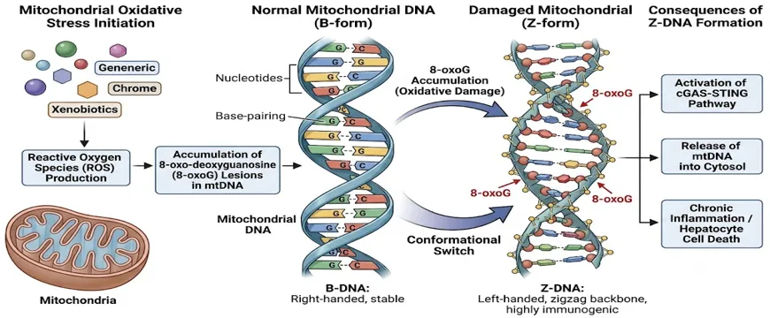
Figure 1: Illustration depicting the molecular pathways of xenobiotic-induced mitochondrial oxidative stress. The diagram highlights the conversion of normal right-handed B-form mitochondrial DNA into the highly immunogenic left-handed Z-form following the accumulation of 8-oxoG lesions.
Valproic acid, a broad-spectrum antiepileptic and mood-stabilizing agent, causes idiosyncratic acute liver failure in a small number of individuals, but with a severe clinical course, is an example of an entirely separate pathway of mitochondrial injury. The hepatotoxicity of valproic acid appears to be closely and mechanistically related to the intrinsic defect in the mitochondrial DNA replication machinery of the individual patient. Usually, valproic acid is extensively metabolized in mitochondria of the liver. In heterozygous or homozygous carriers of POLG gene mutations, encoding the catalytic subunit of DNA polymerase gamma, the liver's ability to compensate for metabolic stress, such as drug treatment, with high mitochondrial DNA copy number is dramatically reduced.[28],[29] Even common polymorphic variants of the POLG gene, like the p.Q1236H substitution, which may be completely asymptomatic in normal physiological conditions, can greatly sensitise hepatocytes to exposure to valproate. The polymerase defect not only causes a failure to replicate the correct mitochondrial genes, but also impairs normal fatty acid metabolism and the growth of cells. The dual hit leads to catastrophic bioenergetic failure, to microvesicular steatosis, and to hepatocyte death in a short space of time; this process often affects children with undiagnosed Alpers-Huttenlocher syndrome.[30],[31]
Moreover, many commonly used antimicrobial drugs, including the principal drug used in the first-line therapy for tuberculosis, isoniazid, cause strong damage to mitochondrial DNA through specific inhibition of single respiratory complexes. The major, highly reactive metabolite of isoniazid, hydrazine, is a powerful and specific inhibitor of mitochondrial complex II. The inactivation of this protein disrupts a normal electron flow through the respiratory chain, leading to a local accumulation of reactive oxygen species that selectively damages the mitochondrial genes that encode subunits of complex I, mainly the NADH dehydrogenase subunits 1 and 5.[10] These genome hot-spots are hot-spots for nonsynonymous mutations that further impair respiratory efficiency, which leads to a vicious cycle of oxidative damage and energetic decline which is clinically expressed as cytolytic hepatitis. Likewise, chemotherapeutic agents like doxorubicin, which are well-known for their cardiotoxicity, are associated with a considerable hepatotoxicity due to mitochondrial DNA intercalation and a tremendous production of oxidative stress that results in the poisoning of the hepatic mitochondrial network. These various modes of action make it clear that the damage to mitochondrial DNA caused by drugs is not a monolithic pathological process, but one that is quite complex and involves a number of potential mechanisms including direct oxidative modification, topological distortion, competitive polymerase inhibition, and the fatal triggering of existing genetic susceptibility.[32]
Table 1: Key Drugs and Mechanisms of Mitochondrial DNA Damage in Hepatotoxicity. The table comprehensively delineates the diverse biochemical pathways through which specific xenobiotics compromise the structural and functional integrity of the mitochondrial genome.
|
Repair / Sensor Protein |
Subcellular Localization |
Primary Biological Function in the Context of Liver Injury |
Consequence of Dysfunction or Inhibition |
|
8-Oxoguanine Glycosylase (OGG1) |
Mitochondrial Matrix |
Recognizes and excises 8-oxoG lesions; prevents transition of B-DNA to Z-DNA |
Accumulation of oxidized bases; profound susceptibility to APAP toxicity |
|
Apurinic/ Apyrimidinic Endonuclease 1 (APE1) |
Mitochondrial Matrix |
Cleaves phosphodiester backbone at abasic sites generated by glycosylases |
Stalled repair processes; increased mtDNA depletion under alkylating stress |
|
DNA Polymerase Gamma (POLG) |
Mitochondrial Matrix |
Sole polymerase for mtDNA replication and gap filling during Base Excision Repair |
Idiosyncratic liver failure upon exposure to drugs like Valproic Acid or NRTIs |
|
Z-DNA Binding Protein 1 (ZBP1) |
Cytosol (Hepatocytes) |
Senses extruded oxidized Z-form mtDNA; activates MAVS-caspase-8 apoptotic cascade |
Evasion of programmed cell death; however, normal activation drives acute liver failure |
|
cGAS-STING Complex |
Cytosol / ER (Non-Parenchymal) |
Senses leaked unmodified or fragmented mtDNA; drives pro-inflammatory cytokine release |
Dampened immune response, but hyperactivation leads to massive sterile inflammation |
CYTOSOLIC SENSING OF DAMAGED MITOCHONDRIAL DNA AND INFLAMMATORY CASCADES:
Mitochondrial DNA mutations can have much more serious pathological effects than just impaired oxidative phosphorylation and the loss of energy currency. Once the mitochondrial network is completely compromised by excessive drug-induced oxidative damage, the structurally altered mitochondria with damaged and fragmented DNA molecules literally spill out from the mitochondrial matrix. This catastrophic translocation is mostly from prolonged opening of the mitochondrial permeability transition pore or through pathological oligomerisation of voltage-dependent anion channels of the outer mitochondrial membrane.[33],[34] This displaced genetic material is a very immunogenic danger signal, once introduced into the previously sterile cytosolic or extracellular environments. Spatial mislocalization of mitochondrial DNA functions as a critical amplifying signaling nexus that connects bioenergetic failure within the cell(s) to the devastating sterile inflammation in surrounding normal tissue, which is an organ-wide manifestation of this mechanism.[35],[36]
The sensing of leaked mitochondrial DNA is primarily and robustly associated with the cyclic GMP-AMP synthase (cGAS) and stimulator of interferon genes (STING) signaling pathway in the non-parenchymal cellular compartment of the liver, which is heavily represented by resident Kupffer cells, dendritic cells, and liver sinusoidal endothelial cells. Dying hepatocytes can release extracellular mitochondrial DNA, which can easily be taken up by these innate immune cells or mitochondrial DNA can leak into the cytosol of the immune cell and directly activate the sensor. Once bound to the misplaced, double-stranded DNA, the cytosolic enzyme cGAS changes its conformation and catalyzes the production of the second messenger cyclic GMP-AMP, which then diffuses and binds to STING that's localized on the membrane of the endoplasmic reticulum. This is an essential binding event that results in the oligomerization and translocation of STING, recruiting and phosphorylating TANK-binding kinase 1. This activation cascade in turn results in the phosphorylation of interferon regulatory factor 3 (IRF3) and nuclear factor kappa B (NF-kB), which triggers massive nuclear transcription of type I interferons and a plethora of pro-inflammatory cytokines including interleukin-6 and tumor necrosis factor α (TNF α) respectively.[37] Moreover, the unmethylated cytosine-phosphate-guanine motifs found in the mitochondrial DNA are excellent triggers of Toll-like receptor 9, which is abundant in the endosomal compartment of infiltrating neutrophils and macrophages, thus contributing to the sterile inflammatory response typical of pathological processes such as severe drug-induced hepatotoxicity.[38],[39]
But, in recent high resolution immunological studies of acute liver failure due to acetaminophen, a completely new picture of the response of the actual parenchymal hepatocytes themselves to oxidized mitochondrial DNA has emerged. In contrast, the non-parenchymal compartment of the liver is rich in cGAS and STING proteins that are expressed at high basal levels and have a well-established function in innate immunity.The non-parenchymal compartment of the liver is lined with cells that express cGAS and STING proteins at high basal levels and have an established role in innate immunity, whereas mature, differentiated hepatocytes express extraordinarily low basal levels of both cGAS and STING proteins, making this canonical inflammatory pathway functionally marginal in the parenchymal compartment. On the contrary, a specialized innate immune sensor, mitochondrial DNA binding protein 1 (ZBP1), which is constitutively expressed in hepatocytes, selectively and promptly recognizes oxidized, left-handed Z-form mitochondrial DNA.[40] ZBP1 is a protein with very unique and highly specialized Z-alpha domains that have a high biochemical affinity only to the zigzag sugar-phosphate backbone of the Z-DNA conformation and not to canonical, undamaged B-DNA. This extreme specificity suggests that the mere presence of mitochondrial DNA in the cytosol is not enough to initiate this cascade, but rather that the DNA must have been structurally altered in a fundamental way, by oxidative modification, and make it pathogenic to the hepatocyte.[41]
ZBP1 binding to oxidized mitochondrial Z-DNA leads to a highly unique apoptotic cascade in the hepatocyte. When ZBP1 detects the topological abnormality, it immediately binds the mitochondrial antiviral-signaling protein (MAVS) and creates a special high-molecular-weight signaling complex. This complex is able to directly activate the initiator caspase-8[42], and mostly lacks a number of classical necroptotic mediators like receptor-interacting serine/threonine-protein kinase-3 which is largely not expressed in functional human hepatocytes. Potent activation of caspase-8 quickly triggers downstream activation of executioner caspases, which leads to irreversible and excessive hepatocyte apoptosis. The fact that the oxidized Z-DNA to ZBP1 axis is a profound discovery that the mitochondrial genome undergoes dramatic changes in structure and that such changes are the ultimate molecular execution order in severe drug-induced liver injury without any involvement of the known immune inflammatory networks is a striking example of the inborn immune network independence of the mitochondrial genome. The final fate of the liver in response to the toxic pharmacological insult, therefore, depends not only on an extruded mitochondrial genetic material but also on its topological conformational state.[43]
MITOCHONDRIAL DNA REPAIR PATHWAYS IN HEPATOCYTES:
The maintenance of hepatocyte viability requires an extremely efficient, rapid and dynamic mitochondrial DNA repair system, given the extremely vulnerable position of the mitochondrial genome to a broad spectrum of pharmaceutical insults. In contrast to the cellular nucleus that has a huge array of redundant DNA repair pathways to preserve genomic integrity, the mitochondrion lacks a complete array of nucleotide excision repair pathways. This evolutionary limitation is the reason the organelle is unable to recognise or remove bulky chemical adducts, large intra-strand cross-links, or cyclobutane pyrimidine dimers formed by ultraviolet radiation. Thus, in the mitochondrion, the Base Excision Repair pathway is the key and most important defense mechanism against oxidative and alkylating damage by drugs. This multi-step conserved biochemical process is specifically designed to rapidly identify, remove and replace individual oxidized, deaminated or alkylated nucleotide bases before they are able to stall the replication fork, cause catastrophic DSBs or induce lethal topological changes.[44],[45]
Specific DNA glycosylases constantly patrol the mitochondrial genome for modified and damaged bases, thus performing the first, rate-limiting step of the mitochondrial Base Excision Repair pathway. In the particular context of drug induced oxidative stress, the enzyme 8-oxoguanine DNA glycosylase (OGG1) is the main molecular sentinel. The recognition of the highly mutagenic lesions, 8-oxo-7,8-dihydroguanine lesions, directly caused by the overproduction of ROS, is specific to the OGG1. Once OGG1 has exactly identified the oxidized purine, it removes the N-glycosidic bond between the damaged base and the sugar-phosphate backbone, leaving a very reactive abasic site (apurinic or apyrimidinic). The rapid and continuous activity of OGG1 can in theory prevent the potentially harmful and thermodynamically favoured transition of B-DNA to Z-DNA when there is a strong toxic stress, provided the amount of OGG1 activity is not overloaded. APE1 (also often called Apex1) is then rapidly recruited to the site of the damage, following this initial glycosylase activity. APE1 opens the phosphodiester backbone right at the 5-prime side of the newly created abasic site, thus allowing the subsequent replacement of the base.[46],[47]
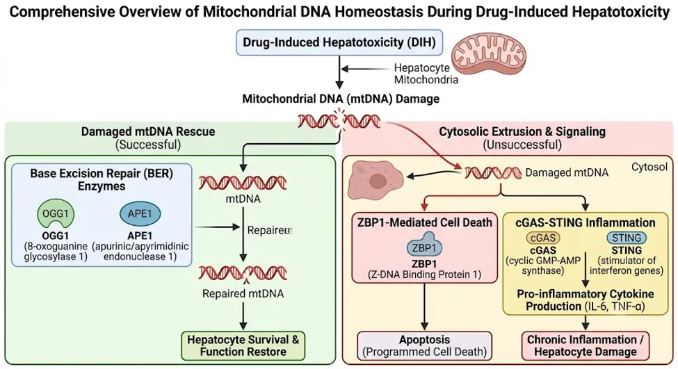
Figure 2: A comprehensive overview of mitochondrial DNA homeostasis during drug-induced hepatotoxicity. The diagram maps the dual outcomes of mitochondrial DNA damage: cytosolic extrusion leading to ZBP1-mediated apoptosis and cGAS-STING inflammation, versus the successful rescue pathway mediated by Base Excision Repair enzymes such as OGG1 and APE1.
The role of these very endonucleases in the protection of liver against xenobiotic and toxic insults has been elegantly shown in highly targeted genetical animal models. For example, the mice that have a haploinsufficient form of the Apex1 gene have very weak abilities to repair mitochondrial DNA. These Apex1 deficient models show a rapid and profound decrease in the levels of mitochondrial DNA and a very high proportion of genetic lesions when they are challenged with high doses of drugs that cause mitochondrial metabolic stress, like alkylating agents or pharmaceutical drugs.[48],[49] Defects in the cell's capacity to process abasic sites directly result in dysfunctional replication forks, collapse of global mitochondrial bioenergetics and a significant increase in hepatocyte death, demonstrating that APE1 is a key, non-redundant survival factor in drug-induced liver injury. The single nucleotide gap is then seamlessly closed by the highly specialized mitochondrial DNA polymerase gamma and the final phosphodiester bond is permanently sealed by the activity of DNA ligase III.[50],[51]
In addition to the enzymes of the Base Excitation Repair, the stability of the whole mitochondrial genome in a very active liver is sustained by a complex network of accessory proteins and structural regulatory factors. Endonuclease G-like 1 (EXOG) and Endonuclease G (ENDOG) perform critical and specific functions in the processing of displaced DNA flaps during long-patch repair and control the degradation of irreparable mitochondrial genomes to stop their pathogenic accumulation. There is a considerable inter-individual genetic variation in these critical repair and degradation systems. Studies of extensive clinical genome-wide association have confirmed that there are strong effects of single-nucleotide polymorphisms in EXOG, ENDOG, FEN1 and POLG genes on human susceptibility to many types of toxic and metabolic liver injuries.[52],[53] In addition, the overall levels of these essential repair proteins are dynamically controlled at the level of their transcription and the import of the proteins into the mitochondria by cell-wide stress responses. The ability of the mitochondrial repair machinery to be rapidly upregulated is regulated by intracellular factors, including the tumor suppressor p53, glycogen synthase kinase 3 beta and circulating systemic interleukins, such as IL-6, which all act to promote cell cycle arrest to give time for mitochondrial DNA to be recovered after acute toxic exposure.[54],[55],[56]
Table 2: Primary DNA Repair Enzymes and Sensors Implicated in Hepatic Mitochondrial Homeostasis. The table summarizes the distinct roles of localized enzymes that definitively dictate the balance between cellular rescue and programmed death following xenobiotic exposure.
|
Repair / Sensor Protein |
Subcellular Localization |
Primary Biological Function in the Context of Liver Injury |
Consequence of Dysfunction or Inhibition |
|
8-Oxoguanine Glycosylase (OGG1) |
Mitochondrial Matrix |
Recognizes and excises 8-oxoG lesions; prevents transition of B-DNA to Z-DNA |
Accumulation of oxidized bases; profound susceptibility to APAP toxicity |
|
Apurinic/ Apyrimidinic Endonuclease 1 (APE1) |
Mitochondrial Matrix |
Cleaves phosphodiester backbone at abasic sites generated by glycosylases |
Stalled repair processes; increased mtDNA depletion under alkylating stress |
|
DNA Polymerase Gamma (POLG) |
Mitochondrial Matrix |
Sole polymerase for mtDNA replication and gap filling during Base Excision Repair |
Idiosyncratic liver failure upon exposure to drugs like Valproic Acid or NRTIs |
|
Z-DNA Binding Protein 1 (ZBP1) |
Cytosol (Hepatocytes) |
Senses extruded oxidized Z-form mtDNA; activates MAVS-caspase-8 apoptotic cascade |
Evasion of programmed cell death; however, normal activation drives acute liver failure |
|
cGAS-STING Complex |
Cytosol / ER (Non-Parenchymal) |
Senses leaked unmodified or fragmented mtDNA; drives pro-inflammatory cytokine release |
Dampened immune response, but hyperactivation leads to massive sterile inflammation |
TRANSLATIONAL IMPLICATIONS AND EMERGING THERAPEUTIC INTERVENTIONS:
The elucidation of mechanisms of mitochondrial damage and the detailed understanding of the tightly coupled cytosolic sensing pathways have rapidly opened up a whole new world of clinical diagnostics and targeted pharmacotherapy in drug-induced liver injury. In clinical diagnostics, highly fragmented mitochondrial DNA in systemic blood circulation is a highly sensitive, specific and early indicator of the extent of actual subclinical liver damage. Being an active, potent, damage associated molecular pattern, rather than a passive product of late stage cell death, circulating cell-free mitochondrial DNA (ccf mtDNA) can supply live and actionable information on the degree of existing sterile inflammation and the clinical course of acute injury. [57] Mitochondrial DNA has been strongly linked with the severity of acute toxic pharmacological damage and chronic metabolic disorders like non-alcoholic fatty liver disease (NAFLD) and severe alcoholic hepatitis (SAH), with increased levels being a hallmark of both conditions. Clinicians could potentially accurately predict which patients initially did not present with symptoms would be most at risk to develop fulminant hepatic failure, based on the constant assessment of these selected biomarker concentrations, and more aggressive medical intervention could be performed much earlier.[58],[59]
In a clinical setting, the management of drug-induced liver injury has been almost exclusively focused on restoring antioxidant defenses in early stages of the chemical insult (therapeutics). The intravenous administration of N-acetylcysteine in a severe acetaminophen overdose is the most important clinical example, and is very effective when administered within the first 8 hours of the critical time before the irreversible secondary cascade of mitochondrial structural destruction sets in. Targeting the fundamental topology and active repair of mitochondrial DNA, however, promises a paradigm-shifting therapeutic approach, which significantly widens that crucial therapeutic window. In recent years, however, there have been several advances in pharmacology that have enabled the identification of novel small molecule agonists that directly and potently activate the enzymatic activity of specific DNA repair enzymes. The highly specific pharmacological activator of the OGG1 glycosylase synthesized compound TH10785 is a giant step forward in acute hepatoprotection. TH10785 actively promotes the physical elimination of 8-oxoguanine lesions from the genetic backbone, thus quickly and efficiently reverting the left-handed Z-DNA pathogenic conformation to the safe, physiological right-handed B-DNA structure.[60],[61]
By inducing a completely new topology, this is sufficient to disrupt the highly specific binding interface that is needed to activate ZBP1, and abruptly and safely terminates the MAVS-caspase-8 apoptotic pathway even when the mitochondria have been severely damaged within the cell. In a series of severe preclinical animal models of lethal acetaminophen toxicity, delayed treatment with TH10785 well outside the accepted therapeutic window of N-acetylcysteine resulted in significantly better survival. The beneficial synergistic biological activities of glutathione augmentation along with boosting DNA lesion repair resulted in almost complete protection from drug-induced acute liver failure when used together in combination therapy.[62] The capability for structural genome integrity was found to be a protective mechanism that has profound implication for the immense translational potential of shifting the clinical paradigm from passive (reactive oxygen species scavenging) to active, targeted maintenance of structural genome integrity.
Moreover, numerous natural bioactive molecules have been found to be very effective in modulating these destructive inflammatory pathways. Emodin is an extensively extracted well characterized anthraquinone derivative from traditional medicinal plants, which has been widely documented in recent literature to attenuate severe hepatotoxicity induced by drugs by actively suppressing the cGAS-STING signaling axis. Emodin successfully inhibits the extruded mitochondrial DNA (mtDNA) mediated secondary inflammatory amplification by significantly downregulating the baseline expression of STING and prominently reducing downstream activation of the NLRP3 inflammasome.[63],[64] Moreover, emodin is hepatoprotective, and it simultaneously upregulates Nrf2-mediated antioxidative stress pathway, which utilize a multi-target approach to the prevention of hepatocyte death, in a highly efficient manner.[64] Additional protection has been recently reported by using advanced pharmacological inhibitors of the mitochondrial permeability transition pore (mPTP) that physically prevent the release of the lethal genome into the cytosol. Together these varied and inventive strategies, from direct agonism of DNA repair to targeted inhibition of nucleic acid sensor function in the cytosol, emphasize that the toxicological consequences of mitochondrial DNA damage are not necessarily lethal, but rather a highly regulated biochemical network that is extremely receptive to targeted pharmacological manipulation.[65]
FUTURE PERSPECTIVES IN DRUG-INDUCED HEPATOTOXICITY:
The in-depth integration of mitochondrial genomic stability into the wider physiological context of DILI is a giant step forward in contemporary mechanistic toxicology. In the past toxicologists looked at the mitochondrion simply as a passive and unfortunate victim of the accumulation of reactive oxygen species, and the final inevitable death of the cell was blamed on the eventual and inevitable failure of ATP production. Today, however, the molecular scene is quite different: it is a highly dynamic and responsive and interactive system in which the actual structure of the mitochondrial genome determines the final fate of the hepatic tissue: survival or death. The stunning conversion of canonical B-DNA to the highly immunogenic Z-DNA structural form provides a clear and definitive proof that severe oxidative stress can directly switch on death receptors with highly specialized function like ZBP1 – fundamentally changing the pathological picture.[66],[67]
In the future, it is important for the scientific and medical community to focus on characterizing inter-individual genetic variation in genes that make up the Base Excision Repair pathway and mitochondrial maintenance genes. The level of life-threatening vulnerability shown by patients carrying even small POLG, APE1 and EXOG polymorphisms indicates that a blanket, population-wide approach to drug safety and dosage is simply not enough. Routine clinical use of these specific pharmacogenomic tests might provide a reliable way to avoid potentially fatal idiosyncratic liver toxicity caused by commonly used drugs such as valproic acid and nucleoside reverse transcriptase inhibitors. Moreover, next generation pharmaceutical drugs should be designed to be structurally non-toxic to mitochondria, or at least be designed as multi-drug therapy that combines repair-activating molecules with high oxidative burden therapeutic drugs.[68],[69]
Summarizing, the exact biochemical mechanisms responsible for mitochondrial DNA damage, for the cytosolic detection of subsequent damage, and for endogenous base excision repair processes represent a completely new and crucial field in hepatology that has been largely unexplored in modern medicine. These highly complex pathways provide a deep and actionable understanding of the real molecular mechanism of toxic liver disease and the mechanisms that mediate the sterile inflammatory signaling that occurs systemically. The transition to the use of advanced therapies that actively repair genomic architecture and safely disrupt pathogenic DNA conformations holds great promise for transforming the clinical care for patients with acute and currently irreversible hepatic failure and will bring a great deal of hope.[70],[71]
REFERENCES

Sonali Sansare, Mitochondrial DNA Damage and Repair: An Under-Explored Frontier in Drug-Induced Hepatotoxicity, Int. J. of Pharm. Sci., 2026, Vol 4, Issue 9, 2805-2820. https://doi.org/10.5281/zenodo.22912821
 10.5281/zenodo.22912821