

We use cookies to ensure our website works properly and to personalise your experience. Cookies policy
Department of Pharmacy, Mahatma Jyotiba Phule Rohilkhand University, Bareilly, Uttar Pradesh, 243006, India.
The aim of study was pharmacological evaluation of wound healing activity of some medicinal plants using animal model. The Fresh leaves of Caesulia axillaris Roxb, Clerodendrum viscosum Ventenat and Allium stracheyi Baker., were collected and washed making dust-free and dried under shade. The dried leaves were rendered into coarse powders and then finally into fine ones. The powders of each plant were weighed and soaked in aqueous, methanolic and hydroalcoholic solvent (1:1), separately for 15 days with frequent stirrings. Wistar albino rats (160-220 g) were used in this study to assess the wound healing potential of leaves extracts of Caesulia axillaris Roxb, Clerodendrum viscosum Ventenat, and Allium stracheyi Baker. The rats were obtained from the Animal House, Department of Pharmacy, Faculty of Engineering and Technology, MJP Rohilkhand University, Bareilly with the ref. no. MJPRU/PY/IAEC/24/64. The Institutional Animal Ethics Committee (IAEC) authorized the protocol for the animal experiment. Ointment base and extract-based ointment was prepared. Wistar albino rats were divided into 08 groups, with 6 animals in each group i.e., group I (control) topically applied the ointment base, group II applied Framycetin sulfate cream (1% w/w), group III applied F1, group IV applied F2, group V applied F3, group VI applied F4, group VII applied F5 and group VIII applied F6. Wound healing activity was evaluated using excision and incision wound models including wound contraction measurements, tensile strength measurement and epithelialization Time. The methanolic (group V) and hydro alcoholic Clerodendrum viscosum Ventenat (group VI) demonstrated the wound contraction as 92.16±0.31 % and 95.24±0.52 %, respectively. Group IV (hydroalcoholic leaves extract of Caesulia axillaris Roxb.) revealed the tensile strength as 198.45±1.20 which was highest amongst all the test groups. In conclusion, in excision and incision wound models, medicinal plants showed the wound healing potential in this order: Caesulia axillaris Roxb > Clerodendrum viscosum Ventenat > Allium stracheyi Baker., when evaluated for wound contraction (%) measurement, Tensile strength measurement and Epithelisation time.
Caesulia axillaris Roxb flourishes in wetland ecosystems i.e., marshes, riverbanks, and other saturated environments. It is essential to the local ecology and well suited to these environments [1][2]. Along the stalk, the leaves are arranged in alternating order. Their lanceolate to elliptic shape makes photosynthesis more efficient. The majority of leaf edges are entire, although they can occasionally show slight serration, which adds to their variety. In addition to providing shelter and food for a variety of species, including pollinators like bees and butterflies, C. axillaris Roxb improves the overall health of the wetland environment by stabilizing the soil and helping to purify the water [3].
C. axillaris Roxb. is rich in a number of bioactive compounds that improve its pharmacological characteristics. The plant's antioxidant, anti-inflammatory, antibacterial, analgesic, and gastro protective qualities are attributed to a variety of components, including phenolic compounds, flavonoids, alkaloids, terpenoids, saponins and essential oils. The potential therapeutic benefits of C. axillaris Roxb are being revealed by ongoing research on these bioactive compounds [4][5]. Limonene and γ-Asarone make up the majority of it. The blossoming shrub Clerodendrum Viscosum Ventenat is known for its ugly foliage. The straight, unbranched stem is 0.5–4 m (1.6–13.1 ft.) tall and has round leaves that are 15 cm (5.9 in) in diameter. The leaves are simple, opposite, elliptic, wide elliptic, ovate, or elongated oval, with a dentate edge, measuring 3.5-20 cm (1.4-7.9 in) in width and 6-25 cm (2.4-9.8 in) in length. Both surfaces are sparsely covered with villous-pubescent hairs. The inflorescence is a terminal cyme with few flowers that is peduncled [6][7].
C. viscosum Ventenat contains a number of bioactive phytochemicals. Because of their different spatial arrangement, the phytochemical profiles of the leaf, root, and flower show different bioactivities in the seed and stem. Flavonoids, terpenoids, steroids and alkaloids are the main bioactive compounds that have been extracted from C. viscosum Ventenat, according to numerous research. Numerous bioactive compounds with notable pharmacological efficacy were found in many parts of C. viscosum Ventenat, according to thorough research [8][9]. Gallic acid, methyl gallate, tannic acid, ellagic acid, reserpine, limonene, α-pinene, β-pinene and ρ-cymene are the main constituents of C. viscosum Ventenat leaves.
Allium stracheyi Baker. is a perennial herb that can grow up to 35 cm in height. The heads are globose, the pedicel is shorter than the flower, and the tepals are dark pink-reddish; the stem is glabrous, leafy at the base, and has thin, linear, flattened leaves [10]. In the Garhwal region, it is known locally as Jamboo, Dhungar, and Pharan. At elevations between 2500 and 3625 meters, this species can be found in Jammu Kashmir, Himachal Pradesh, Uttarakhand, Nepal, and Pakistan [11].
Herbal medicine constitutes a complex system of formulations. Its formulations for medical use comprise several types of secondary metabolites or bioactive substances. Diverse chemical and analytical methodologies facilitate the comprehension of quality control and chemical ingredients in herbal pharmaceuticals. Allium species have been documented to contain a variety of physiologically active chemicals, including phenolic acids, flavonoids, thio-sulfinates, alkaloids, fixed oils, phytosterols, sulfur-containing compounds, and others [12][13]. A. stracheyi Baker. has sulfur-rich compounds having the antioxidant, anti-inflammatory, and antibacterial properties. Compounds high in sulphur have been documented to reduce blood cholesterol [14]. The current study was based on the phytochemical screening, isolation and estimation of total phenolic contents & total flavonoids contents of some medicinal plants i.e., Caesulia axillaris Roxb, Clerodendrum Viscosum Ventenat and Allium stracheyi Baker. The aim of study was pharmacological evaluation of wound healing activity of some medicinal plants using animal model.
MATERIALS AND METHODS
Experimental requirements
Fresh leaves of Caesulia axillaris Roxb, Clerodendrum viscosum Ventenat, Allium stracheyi Baker. methanol, ethanol, distilled water, rotatory evaporator and weighing machine.
Collection, authentication, and preparation of extract
The Fresh leaves of Caesulia axillaris Roxb, Clerodendrum viscosum Ventenat, Allium stracheyi Baker. were collected from UP East region and authenticated by a Botanist Dr. Vijay Kumar Sinhal, Department of Plant Science, MJP Rohilkhand University Bareilly, UP India, with the ref. no. MJPRU/PS/2024/01. The leaves of Caesulia axillaris Roxb, Clerodendrum viscosum Ventenat, Allium stracheyi Baker. were washed to make dust-free and dried under shade. The leaves were rendered into coarse powder and further in fine ones. The powders of each plant were weighed and soaked in aqueous, methanolic and hydroalcoholic solvent (1:1), separately for 15 days with frequent stirrings. The obtained slurry was kept for drying under partial vacuum using a rotatory evaporator [15].
Preparation of rats
Wistar albino rats (160-220 g) were utilized to determine the wound healing potential of leaves extracts of Caesulia axillaris Roxb, Clerodendrum viscosum Ventenat, and Allium stracheyi Baker. The rats were obtained from the Animal House, Department of Pharmacy, Faculty of Engineering and Technology, MJP Rohilkhand University, Bareilly with the ref. no. MJPRU/PY/IAEC/24/64. Rats were kept in plastic cages at ambient temperature and relative humidity which fed with pellet diet and water ad libitum. Institutional Animal Ethics Committee (IAEC) authorized the protocol for the animal experiment.
Preparation of ointment base and extract-based ointment
Six different formulations were prepared using leaves extracts of Caesulia axillaris Roxb, Clerodendrum viscosum Ventenat and Allium stracheyi Baker. The ointment was carried out with individual plant extracts, in which methanolic and hydroalcoholic extracts were separately used. Paraffin wax (1.5 g) was melted using water bath, and further lanolin, cetostearyl alcohol (1.5 g each) and petroleum jelly (24.5 g) were mixed to make a homogenous mixture. Thus, ointment base was prepared which utilized in control (group I). To this base mixture, 1g of leaves extracts of Caesulia axillaris Roxb, Clerodendrum viscosum Ventenat, and Allium stracheyi Baker. (methanolic and hydroalcoholic) were separately added, and thus 6 different ointments (30g each) were made through triturating by using pestle mortar [16].
Table 1. Composition table for ointment base
|
Ingredients |
Quantity (g) |
|
Paraffin wax |
1.5 |
|
Lanolin |
1.5 |
|
Cetostearyl alcohol |
1.5 |
|
Petroleum jelly |
24.5 |
Table 2. Composition table for extract-based ointment
|
Extract/ Ointment base |
Ointment |
|||||
|
|
F1 |
F2 |
F3 |
F4 |
F5 |
F6 |
|
Methanolic Caesulia axillaris Roxb leaves extract |
1.0 g |
|
|
|
|
|
|
Hydroalcoholic Caesulia axillaris Roxb leaves extract |
|
1.0 g |
|
|
|
|
|
Methanolic Clerodendrum viscosum Ventenat leaves extract |
|
|
1.0 g |
|
|
|
|
Hydroalcoholic Clerodendrum viscosum Ventenat leaves extract |
|
|
|
1.0 g |
|
|
|
Methanolic Allium stracheyi Baker. leaves extract |
|
|
|
|
1.0 g |
|
|
Hydroalcoholic Allium stracheyi Baker. leaves extract |
|
|
|
|
|
1.0 g |
|
Ointment base |
q. s. 30 g |
q. s. 30 g |
q. s. 30 g |
q. s. 30 g |
q. s. 30 g |
q. s. 30 g |
Experimental protocol
All the Wistar albino rats kept separately into 08 groups (n=6) as followings:
Table 3. Experimental protocol
|
Group |
Treatment |
|
Group I (Control) |
Received the topical application of ointment base |
|
Group II (Std.) |
Received the topical application of Framycetin sulfate (1% w/w) cream |
|
Group III |
Received the topical application of ointment of Methanolic leaves extract of Caesulia axillaris Roxb (F1) |
|
Group IV |
Received the topical application of ointment of Hydroalcoholic leaves extract of Caesulia axillaris Roxb (F2) |
|
Group V |
Received the topical application of ointment of Methanolic leaves extract of Clerodendrum viscosum Ventenat (F3) |
|
Group VI |
Received the topical application of ointment of Hydroalcoholic leaves extract of Clerodendrum viscosum Ventenat (F4) |
|
Group VII |
Received the topical application of ointment of Methanolic leaves extract of Allium stracheyi Baker. (F5) |
|
Group VIII |
Received the topical application of ointment of Hydroalcoholic leaves extract of Allium stracheyi Baker. (F6) |
Evaluation models of Wound healing activity
Excision wound model
After giving Ketamine anaesthesia (50mg/kg), dorsal part of the animals was shaved. Using a standard ring, an area approx. 500mm2 on dorsal side was marked. Using sharp scissor, the marked area was gently cut to its maximum thickness. Since wound creation day and then on each 5 days interval up to 20th days wounds were determined using 1 mm2 graph paper to confirm the changes in wound [17].
Incision wound model
After anaesthesia, 4 cm paravertebral incisions were made through the whole thickness of the skin which was 1 cm lateral to the midline of vertebral column. Suturing was done to close wounds. Till 15 days, the wound length was measured on every alternate day. The wound recovery strength in incised rats was measured on the 10th wounding day using standard parameters [18].
Evaluation parameters
Wound contraction measurement
On 5, 10, 15, & 20 days in the excision wound model, wound area is traced using a clear sheet of 1 mm2 graph paper. Wound contraction was measured on every 5th day until the wounds were completely healed and the percentage of wound contraction was calculated with below formula [19].
Wound contraction (%) =
wound size (before) - wound size (after) × 100
wound size (before)
Tensile strength measurement
On 10th day, rats were given ketamine anaesthesia; repaired tissues were excised through removing the sutures. A line was marked as 3 mm far from the wound on either side of the incision line. On the line facing each other, 2 Allis forceps were firmly placed. One forceps was made fixed, while the other forceps was attached by a thread to a freely hanging polypropylene graded container. The container was filled with the standard weights, gradually. The weights were gradually pushed to the incision site, pushing the wound edges far apart. On wound opening, the weight was halted and measured [20].
Epithelisation Time
The epithelisation period was determined as the no. of days needed for dead tissue remains of the wound to fall off completely without leaving any residue of wounds [21].
Statistical Analysis
Standard error of the Mean±S.D. will be used to present all results. Statistical analysis was performed with One-way analysis of variance (ANOVA) and the result will be considered statistically significant if (P<0.05).
RESULTS AND DISCUSSION
Evaluation models of Wound healing activity
A. Excision model
Wound contraction measurement
Wound contraction was measured in its percentage. At day 20, group I (control) showed the wound contraction as 65.32±0.11 %. Moreover, group II (std.) exhibited the highest wound contraction as 100±0.32 %. At day 20, the methanolic (group III) and hydroalcoholic leaves extract of Caesulia axillaris Roxb (group IV) exhibited the wound contraction as 96.81±0.11 % and 100±0.23 % respectively. Similarly, methanolic (group V) and hydroalcoholic Clerodendrum viscosum Ventenat (group VI) demonstrated the wound contraction as 92.16±0.31 % and 95.24±0.52 %, respectively. While, wound contraction was found as 93.42±0.16 % and 96.10±0.37 % in methanolic (group VII) and hydroalcoholic Allium stracheyi Baker. (group VIII), respectively.
Table 4. Wound contraction (%) of excision wound model
|
Group |
Wound contraction (%) |
|||
|
Day 5 |
Day 10 |
Day 15 |
Day 20 |
|
|
Group I |
31.64±0.19 |
36.21±0.57 |
62.11±0.43 |
65.32±0.11 |
|
Group II |
59.26±0.43 |
82.43±0.12 |
92.27±0.64 |
100±0.32 |
|
Group III |
54.43±0.14 |
68.14±0.51 |
84.26±0.10 |
96.81±0.11 |
|
Group IV |
56.32±0.11 |
71.29±0.46 |
87.14±0.53 |
100±0.23 |
|
Group V |
52.12±0.34 |
64.12±0.47 |
81.27±0.32 |
92.16±0.31 |
|
Group VI |
55.24±0.64 |
68.37±0.13 |
83.20±0.33 |
95.24±0.52 |
|
Group VII |
51.27±0.43 |
62.14±0.57 |
79.12±0.31 |
93.42±0.16 |
|
Group VIII |
54.39±0.12 |
66.32±0.24 |
81.27±0.40 |
96.10±0.37 |
n=6, P<0.05; values denoted as Mean ± Std Deviation
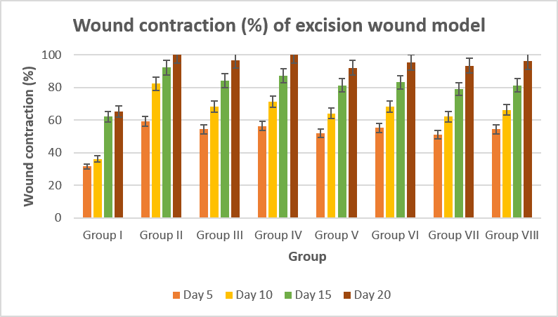
Fig 4. Graphical data of Wound contraction (%) of excision wound model
Thus, it can be observed that wound contraction effect was optimum in hydroalcoholic fractions and less in methanolic fractions. Among all 3 medicinal plant species, hydroalcoholic leaves extract of Caesulia axillaris Roxb (group IV) exhibited the wound contraction as 100±0.23 % which is similar to standard group. It concluded that Caesulia axillaris Roxb has the most effective wound healing potential than other plants (Clerodendrum viscosum Ventenat, Allium stracheyi Baker.) used in the study.
Tensile strength measurement
At 10th day, control (group I) showed the Tensile strength as 163.11±1.54 g and highest tensile strength was observed in group II (standard) as 211.49±1.17 g. Group IV (hydroalcoholic leaves extract of Caesulia axillaris Roxb) revealed the tensile strength as 198.45±1.20 which was highest amongst all the test groups.
Table 5. Tensile strength of excision wound model
|
Group |
Tensile strength (g)) ±S.D. |
|
Group I |
163.11±1.54 |
|
Group II |
211.49±1.17 |
|
Group III |
194.23±1.42 |
|
Group IV |
198.45±1.20 |
|
Group V |
192.16±1.61 |
|
Group VI |
196.23±1.52 |
|
Group VII |
184.20±1.45 |
|
Group VIII |
187.34±1.65 |
n=6, P<0.05; values denoted as Mean ± Std Deviation
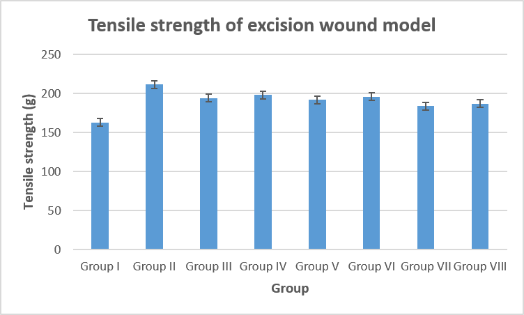
Fig 5. Graphical data of Tensile strength of excision wound model
Epithelisation time
In excision wound model, group I (control) showed the epithelization time as 17.24±0.30 days. The minimum days for epithelisation was recorded for in group II as 8.19±0.51 days.
Caesulia axillaris Roxb showed the decreased epithelization time as 11.57±0.14 days and 10.41±0.23 days, in its methanolic and hydroalcoholic fractions. Moderate wound healing potential was observed in Clerodendrum viscosum Ventenat, however less wound healing potential was found in Allium stracheyi Baker. Thus, Caesulia axillaris Roxb exhibited the highest wound healing potential in epithelisation time of excision wound model.
Table 6. Epithelisation time of excision wound model
|
Group |
Epithelisation time (days)± S.D. |
|
Group I |
17.24±0.30 |
|
Group II |
8.19±0.51 |
|
Group III |
11.57±0.14 |
|
Group IV |
10.41±0.23 |
|
Group V |
13.58±0.17 |
|
Group VI |
12.65±0.24 |
|
Group VII |
14.52±0.20 |
|
Group VIII |
13.46±0.11 |
n=6, P<0.05; values denoted as Mean ± Std Deviation
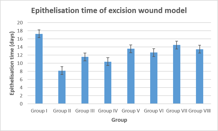
Fig 6. Graphical data of Epithelisation time of excision wound model
B. Incision model
Tensile strength measurement
In incision model, tensile strength was determined to confirm the wound healing activity of herbal extracts. Group III and IV (Caesulia axillaris Roxb) showed the tensile strength as 231.47±1.19 g and 239.41±1.14 g, respectively. In incision wound model, tensile strength observed with higher efficacy than excision wound model. So, among 3 plant species, Caesulia axillaris Roxb showed the highest tensile strength and thus greater wound healing potency.
Table 7. Tensile strength of incision wound model
|
Group |
Tensile strength (g)±S.D. |
|
Group I |
172.18±1.34 |
|
Group II |
258.20±1.16 |
|
Group III |
231.47±1.19 |
|
Group IV |
239.41±1.14 |
|
Group V |
223.52±1.17 |
|
Group VI |
228.61±1.24 |
|
Group VII |
219.32±1.57 |
|
Group VIII |
225.61±1.24 |
n=6, P<0.05; values denoted as Mean ± Std Deviation
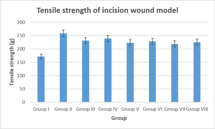
Fig 7. Graphical data of Tensile strength of incision wound model
Many plants and/or their derivatives were used as wound-healing agents by people all over the world, even though there was conflicting evidence on their efficacy and safety. Conventional wound-healing agents are unable to stop all phases of wound healing, which highlights the need for the development of innovative, safe, and effective wound-healing medications [22].
The topical administration of the hydroalcoholic extracts increased the rate of wound reduction in the excision wound model, which may be explained by improved wound healing progression and observable hydration of the wound boundary as a result of tissue regeneration. The percentage closure of all extracts fell between 97.7% to 99.9% on the final day of treatment, compared to 89.9% for the group that received a basic ointment. A decrease in the proportion of the initial wound size is an example of wound contraction that aids in wound closure [23]. Coagulation, inflammation, macrophages, fibroplasias, collagenation, contraction, and epithelization are all directly related to the healing process. Treatment may influence the healing process by mediating at least one stage of the healing process. The major extracellular protein in the granulation tissue of wounds, collagen, is significantly increased in mice treated with WE ointment. Therefore, the beginning of many factors connected to the wound healing process and inflammatory pathways, which may be attributable to the capability of plant materials in GE, is most likely what causes the wound healing activity of the WE ointment [24].
The role of wound contraction is critical because it reduces the size of the wound, increases the amount of extracellular matrix needed to repair the faulty cell, and promotes re-epithelization. The extracts' ability to reduce wounds may be attributed to their mitogenic activity, which increases fibroblast motility and cellular proliferation, inhibition of microbial growth, especially during the inflammatory phase, and subsequent conversion to myofibroblasts during the healing process, which is primarily dermal. One way that plant extracts promote wound healing is by stimulating fibroblasts, which migrate from the wound edge to the site, proliferate, and eventually create collagen, the primary component of extracellular matrix [25]. Restrain the oxidative reactive species developed by phagocytes is important aspect of wound healing [26]. By reducing or delaying the start of cell necrosis and enhancing vascularity, another metabolite known as flavonoids lowers lipid peroxidation. Circulation promotes the viability of collagen fibrils thus preventing cell damage, and promotes DNA synthesis. Due to their antimicrobial and antioxidant properties, which appear to be the cause of wound contraction and an accelerated rate of epithelization, these substances, along with triterpenoids, are also aiding in the healing of wounds. According to a different study, certain flavonoids have astringent properties and inhibit enzymes like phospholipase A2, which are crucial for wound contraction and the pace of epithelization [27].
CONCLUSION
In conclusion, % yield and phytochemical constituents were observed in this order: hydroalcoholic > methanolic > aqueous. Plants species demonstrated as a rich source of phytochemicals including alkaloids, cardiac glycosides, tannins, saponins, coumarins, steroids and flavonoids. They exhibited an excellent quantity of total flavonoids and total phenolic contents. In excision and incision wound models, medicinal plants showed the wound healing potential in this order: Caesulia axillaris Roxb > Clerodendrum viscosum Ventenat > Allium stracheyi Baker., when evaluated for wound contraction (%) measurement, Tensile strength measurement and Epithelisation time.
REFERENCES

Vimal Kumar Singh, Kamal Kishore, Pharmacological Evaluation of Wound Healing Activity of Some Medicinal Plants using Animal Model, Int. J. of Pharm. Sci., 2026, Vol 4, Issue 10, 1093-1103. https://doi.org/10.5281/zenodo.23212481
 10.5281/zenodo.23212481