
View Article

  • Preclinical Models for Drug-Induced Liver Injury with Special Focus on Biomarkers Involved in Pathomechanism

  • Department of Pharmacology, Sarojini Naidu Vanita Pharmacy Maha Vidyalaya (Co- Ed.), Tarnaka, Hyderabad, Telangana, India-500017.

Abstract

Drug-induced liver injury (DILI) remains a major obstacle in drug development and clinical therapy, often causing severe hepatic damage and treatment withdrawal. Experimental animal models are indispensable for elucidating DILI mechanisms, discovering biomarkers, and testing hepatoprotective agents before human trials. This overview synthesizes evidence from peer-reviewed studies on commonly used models-rats, mice, rabbits, guinea pigs, and zebrafish-exposed to hepatotoxins such as paracetamol, carbon tetrachloride, thioacetamide, methotrexate, diclofenac, isoniazid, sodium valproate, gentamicin, and carbamazepine. Dosing regimens, administration routes, exposure durations, and resulting biochemical and histopathological alterations were critically evaluated. Rats and mice predominate due to their reproducibility and physiological similarity to human liver function. Rabbits, guinea pigs, and zebrafish provide complementary advantages for specific toxicological questions, including immune-mediated injury and high-throughput screening. Collectively, these models enable robust investigation of DILI pathogenesis, biomarker validation, and preclinical assessment of safer therapeutics and hepatoprotective strategies.

Keywords

Drug-induced liver injury, Experimental animal models, Hepatotoxicity, Biomarkers and Hepatoprotective agents

Introduction

× Popup Image

The liver is a primary organ involved in the metabolism, detoxification, protein synthesis, bile production.[1] The human liver has remarkable regenerative capacity; however, repeated or persistent injury overwhelms its repair mechanisms.[2] The clinical manifestations range from asymptomatic elevations in liver enzymes to acute hepatitis, cholestasis, chronic liver disease, and acute liver failure.[3]

Damaged hepatocytes release danger-associated molecular patterns (DAMPs), activating Kupffer cells and recruiting inflammatory cells that amplify hepatic injury.[4] It remains one of the principal reasons for discontinuing clinical trials and withdrawing approved drugs from the market.[5]

Clinical evaluation is commonly supported by biochemical markers such as alanine aminotransferase (ALT), aspartate aminotransferase (AST), alkaline phosphatase (ALP), bilirubin, and histopathological examination.[6]

Liver injury is a significant global health issue, leading to both acute and chronic problems. [7] In the past two decades, liver injury causes have changed notably. Viral hepatitis remains a major issue in low- and middle-income countries, while in Western countries, drug-induced liver injury (DILI) is a leading cause of acute liver failure and a common reason for drug withdrawals. Over 1,000 medications, herbs, and supplements have been linked to liver damage. The estimated yearly incidence is 14 to 19 cases per 100,000 people, though the real number is probably higher due to under-reporting. [8]

Most studies show that DILI is more common in older adults due to multiple medications, health conditions, and age-related changes in how drugs are processed. While women might be more prone to severe liver damage from some drugs, overall, there is no clear difference in DILI rates between men and women.[9]

Each year, there are about 310 million new cases worldwide, with nearly 2 million deaths and 63.5 million DALYs. While overall global rates have fallen, deaths and DALYs have risen in High-income North America and Australasia, mainly due to increasing cases of fatty liver disease and alcohol-related liver conditions.[7]

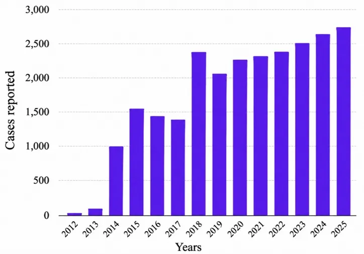
The below figure 1 depicts DILI cases from 2012 to 2025, demonstrating an overall increasing trend in the number of reported cases, with a notable rise after 2017. The rise reflects both an actual increase in certain causes of DILI and a substantial improvement in case detection and reporting, rather than a sudden biological increase in liver injury. Rising use of herbal and dietary supplements, new liver-toxic drugs like cancer immunotherapies, greater physician awareness, expanded DILI registries, improved diagnosis and reporting.
figure

Figure 1: Annual Reported DILI Cases (2012–2025)

In India, data from the Indian Network for Drug-Induced Liver involving 1,288 patients showed that approximately 10% developed acute liver failure, highlighting the substantial disease burden. Morbidity is particularly high among patients receiving anti-tuberculosis drugs, herbal medicines, anticonvulsants, and certain antibiotics.[10]

People affected often face liver dysfunction, extended hospital stays, a lower quality of life, frequent readmissions, and may develop cirrhosis or liver cancer over time. Advanced stages of liver disease can lead to serious issues like portal hypertension, fluid buildup (ascites), brain dysfunction (hepatic encephalopathy), kidney problems, and infections, all of which contribute to heavy healthcare demands and disability.[8]

Early diagnosis, prompt withdrawal of the offending drug, and timely referral for liver transplantation are critical measures to reduce morbidity and mortality.[53] In the Indian INDILI registry, the overall mortality was 12.3%, whereas mortality increased dramatically to 64.8% among patients with DILI-associated acute liver failure (DILI-ALF). Prospective population-based studies have reported lower mortality in the general population because many cases are mild. For example, mortality rates were 5.9% in France, 1% in Iceland, and 0.39% in China, illustrating considerable geographic variation depending on patient characteristics, causative agents, and healthcare systems.[4] The risk of death differs depending on the drug responsible, the extent of liver damage, the patient’s age, and whether any pre-existing liver disease is present. [4]

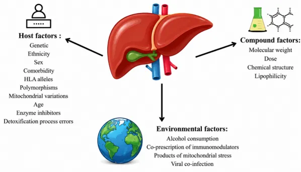
The etiology of DILI is multifactorial and involves interactions between drug characteristics and host susceptibility. Major pathogenic mechanisms include reactive metabolite formation, oxidative stress, mitochondrial dysfunction, immune-mediated hepatotoxicity, disruption of bile acid homeostasis, and genetic predisposition. Recent studies emphasize that idiosyncratic DILI results from a combination of genetic susceptibility, adaptive immune activation, and environmental factors rather than a single pathogenic pathway as shown in figure 2.
figure

Figure 2: Multifactorial Risk Factors Contributing to Drug Induced Liver Injury

Types of DILI:

DILI is categorized based on three factors: Mechanism of injury, the pattern of liver enzyme changes, and the clinical or tissue characteristics observed,shown in the table 1.

Table 1: Classification of DILI Based on Mechanism of Injury, Biochemical Pattern, and Clinical Phenotype

Classification Type Description Key Features Ref
Mechanism of Injury
Intrinsic (Direct) DILI Liver damage caused directly by the drug or its toxic metabolites, usually dose-related Dose-dependent, predictable, rapid onset (hours to days), reproducible in animals, linked to overdoses 11,12
Idiosyncratic DILI Unpredictable liver injury involving immune responses, often linked to genetic factors Dose-independent, unpredictable timing, variable onset, not reproducible in animals, immune/metabolic involvement 12,13,
14,15
Indirect DILI Liver damage caused indirectly through effects on the immune system or liver function Occurs later, related to drug effects (e.g., cancer immunotherapies), not direct liver toxicity 16
Biochemical Pattern
Hepatocellular DILI Damage mainly to liver cells, identified by high ALT levels and R-value ≥ 5 ALT ≥ 3x ULN, R-value ≥ 5, highest risk for acute liver failure 12,16,
Cholestatic DILI Injury affecting bile production or flow, with high ALP levels and R-value ≤ 2 ALP ≥ 2x ULN, R-value ≤ 2, symptoms include jaundice and itching 12,16
Mixed DILI Combination of liver cell and bile flow injury, with intermediate R-value ALT ≥ 3x ULN, ALP ≥ 2x ULN, R-value between 2 and 5 12,16
Clinical Phenotype
Various phenotypes Different liver conditions identified by symptoms and liver tissue analysis Includes acute hepatitis, cholestatic hepatitis, autoimmune hepatitis, steatosis, chronic hepatitis, sinusoidal obstruction syndrome 12,16

*ALT = Alanine aminotransferase

*ALP = Alkaline phosphatase

*ULN=Upper limit of normal

Severity Grades of DILI:

The DILIN grading system is the most commonly adopted method for classifying the severity of liver injury:[17]

  • Grade 1 (Mild): Elevated ALT /ALP levels, while total bilirubin remains below 2.5 mg/dL and the international normalized ratio is less than 1.5.
  • Grade 2 (Moderate): Elevated ALT or ALP accompanied by bilirubin at or above 2.5 mg/dL or an INR of 1.5 or higher, but without the need for hospitalization due to DILI.
  • Grade 3 (Moderate-Severe): Meets Grade 2 criteria and involves hospitalization or an extended hospital stay because of DILI.
  • Grade 4 (Severe): Raised liver enzymes with bilirubin ≥2.5 mg/dL, along with liver complications such as encephalopathy, fluid buildup (ascites), prolonged jaundice, failure of other organs, or serious blood clotting problems.
  • Grade 5 (Fatal): Death caused by DILI or requiring a liver transplant.

Drugs That Cause Liver Injury in Humans: According to the FDA DILI (Drug injured liver injury) rank database and published human hepatotoxicity studies, drugs consistently associated with liver injury include. [18,19,20,21,22,23,24,25,26]

Table 2: Common Drug and Supplement Categories Linked to Liver Injury and Their Characteristics

Drug Class Key Drugs Liver Injury Pattern
NSAIDs Acetaminophen, Diclofenac, Ibuprofen Acetaminophen: dose-dependent: others: idiosyncratic
Antibiotics Amoxicillin–clavulanate, Isoniazid, Rifampicin Common cause of idiosyncratic DILI worldwide
Antiepileptics Valproic acid, Carbamazepine, Phenytoin Mitochondrial dysfunction or immune-mediated
Antitubercular Isoniazid, Rifampicin, Pyrazinamide Frequent hepatotoxicity in combination therapy
Antifungals Ketoconazole, Itraconazole, Fluconazole Cholestatic or mixed; ketoconazole: high risk
Cardiovascular Amiodarone, Statins, Methyldopa Amiodarone: steatosis; statins: mild enzyme rise
Anticancer Methotrexate, Cisplatin, Imatinib Transient enzymes to fibrosis/hepatic failure
Antiretrovirals Nevirapine, Efavirenz, Ritonavir Hypersensitivity, mitochondrial, or immune-mediated
Immunosuppressants Azathioprine, Tacrolimus, Cyclosporine Cholestatic or vascular liver injury

Animal Models For DILI:

Based on the hepatotoxic agent employed, these models are often divided into drug-induced and chemical-induced liver injury models.

Drugs that can induce liver injury:

Many drugs can cause damage to the liver. Some, like acetaminophen, directly harm the liver, while others, such as amoxicillin-clavulanate, isoniazid, and statins, tend to cause unpredictable, immune-related liver injury. Often arise when these drugs are converted into reactive substances or trigger immune responses. [12]

Chemicals frequently reported in experimental hepatotoxicity studies:

  • Bromobenzene
  • Cyclophosphamide
  • Hydrazine
  • Allyl alcohol
  • Cadmium chloride (CdCl₂)
  • Mercuric chloride (HgCl₂)
  • Potassium dichromate (K₂Cr₂O₇)
  • Sodium fluoride
  • Aflatoxin B₁
  • Microcystin-LR

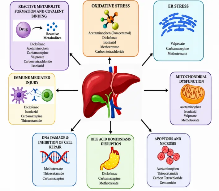
This figure highlights the key ways drugs can cause liver damage, showing the liver at the center to illustrate how various cellular and molecular processes can each, or together, lead to harm. These processes include oxidative stress, the creation of reactive metabolites that bind to liver cells, stress in the endoplasmic reticulum, mitochondrial problems, immune-related damage, DNA harm with faulty repair, disturbances in bile acid balance (cholestasis), and cell death through apoptosis or necrosis. It also points out common drugs like acetaminophen, diclofenac, isoniazid, methotrexate, carbamazepine, valproate, thioacetamide, carbon tetrachloride, and gentamicin, which can trigger liver injury via one or more of these pathways. Overall, the figure shows that drug-induced liver injury is a complex process where overlapping factors combine to cause liver cell dysfunction, inflammation, and damage.
figure

Figure_3 Major Mechanisms of DILI and Representative Hepatotoxic Drugs
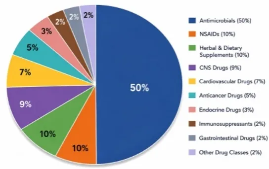
  • The figure shows the estimated share of different drug types linked to DILI, based on combined data from various studies and reviews. Antimicrobials make up the largest group, accounting for about half of the cases. Following them are nonsteroidal anti-inflammatory drugs (NSAIDs) and herbal or dietary supplements, each representing around 10%. Central nervous system drugs account for 9%, cardiovascular drugs 7%, anticancer drugs 5%, endocrine drugs 3%, and immunosuppressants, gastrointestinal drugs, and other drug classes each make up about 2%.

figure

Figure_4 Estimated Percentage Distribution of Drug Classes Causing Drug-Induced Liver Injury.

Paracetamol (Acetaminophen): At normal therapeutic doses, paracetamol is safely metabolized mainly through glucuronidation and sulfation. During an overdose, however, it is converted by CYP2E1, CYP1A2, and CYP3A4 enzymes into the toxic metabolite NAPQI. When glutathione stores become depleted, NAPQI accumulates, triggering oxidative stress, mitochondrial damage, ATP depletion, and ultimately causing centrilobular liver cell injury and death.[27]

This model is quick and reliable, effectively replicating the acute liver damage seen in human overdoses. However, it only reflects acute injury and differs based on species and dosage.

Paracetamol is widely used in animal studies, especially with mice and rats, to mimic human liver injury. Mice receive 200-500 mg/kg and rats 500-1000 mg/kg, orally or by injection, with liver damage appearing within 24 hours. This damage includes liver cell death, oxidative stress, and mitochondrial problems, helping researchers study liver injury and test treatments. [28,27]

Diclofenac: Diclofenac is metabolized into reactive quinone-imine compounds that bind to liver proteins, impair mitochondrial respiration, increase the formation of reactive oxygen species, along with promote programmed cell death and liver cell necrosis.[28]

Diclofenac is used in rats and mice to study both unpredictable and direct liver injury, particularly involving the immune system. The limitation is its low consistency and the need for long-term dosing. Animals are given 10 to 50 mg/kg orally or by injection for 7 to 14 days. This treatment causes a combination of liver cell damage, bile flow problems, and inflammation, helping researchers explore different types of liver injury. [29]

Isoniazid (INH): Isoniazid is broken down into hydrazine and acetyl hydrazine, which cause oxidative stress, disrupt mitochondrial function, reduce glutathione levels, and result in the death of hepatocytes. [30]

This model is relevant clinically for tuberculosis drug toxicity and helps investigate mitochondrial damage. The challenges include variable animal sensitivity and a slow progression of liver injury.

Isoniazid is frequently used rats, mice, and guinea pigs to investigate the unpredictable liver damage associated with tuberculosis therapy. The drug is given orally at a dose of 50–100 mg/kg body weight per day for 14–28 consecutive days.[31]

Methotrexate (MTX): Chronic exposure to methotrexate causes oxidative stress, damages mitochondria, stimulates inflammatory cytokine release, activates hepatic stellate cells, promotes collagen accumulation, and results in liver fibrosis.[32]

Ideal for researching chronic liver fibrosis and oxidative stress, though it requires extended treatment periods and shows less acute liver damage.

Methotrexate is used in animal studies with rats, mice, and rabbits to mimic chronic liver toxicity seen in cancer and autoimmune treatments. Rats typically receive 0.3 to 1 mg/kg by injection once a week for 4 to 8 weeks. This causes liver cell damage, scarring, and oxidative stress, helping researchers understand long-term drug-induced liver injury. [31]

Thioacetamide: Thioacetamide is converted into reactive intermediates thioacetamide-S-oxide and thioacetamide-S,S-dioxide, which generate reactive oxygen species, cause lipid peroxidation, DNA damage, liver cell death, and eventually lead to liver fibrosis. [33] However, it isn’t a therapeutic drug and can cause severe toxicity and high death rates at high doses.

Thioacetamide is frequently used in research involving rats and mice to create models of liver fibrosis and cirrhosis, which mimic chronic liver damage. However, it isn’t a therapeutic drug and can cause severe toxicity and high death rates at high doses.

It is typically given either by injection into the abdomen or orally, usually at doses between 100 and 300 mg per kilogram of body weight, administered two to three times a week for up to eight weeks.[34]

Carbon Tetrachloride (CCl₄): CCl₄ is metabolized by the enzyme CYP2E1 into trichloromethyl radicals, which trigger lipid peroxidation, oxidative stress, damage to cell membranes, and death of liver cells. This process has been well-established in experimental models of CCl₄-induced liver injury.[35]

A well-established, straightforward model for studying fibrosis and cirrhosis with good reproducibility. It’s a chemical toxin rather than a medication, limiting its clinical relevance to drug-induced liver injury.

Carbon tetrachloride is commonly used in animal studies with rats, mice, rabbits, and guinea pigs. Widely used to cchronic liver injury and fibrosis. It causes liver cell damage and oxidative stress. Animals usually receive doses of 0.5 to 1.0 mL/kg by injection or orally, given 2-3 times a week for several weeks. This leads to liver tissue death, inflammation, and scarring, helping researchers study long-term liver damage. [37]

Sodium valproate: It is chemically known as 2-propylpentanoic acid, is a versatile antiepileptic drug commonly used to manage epilepsy, bipolar disorder, and prevent migraines. As a branched-chain fatty acid, VPA undergoes significant metabolism in the liver, which is closely associated with its liver toxicity. Silva et al. identified mitochondrial β-oxidation as the main metabolic route for VPA, with disruption of this process being a key factor in its toxic effects.

This model mimics mitochondrial issues and fatty liver disease (steatosis), but often results in mild liver injury and variable reproducibility across species.

Sodium Valproate is widely used in rat and mouse models to mimic mitochondrial toxicity and liver fat accumulation caused by valproate treatment. Typically administer doses between 200 and 600 mg/kg, either orally or through injection, for a period of 7 to 14 days.[31]

In mice,the treatment consists of administering the compound orally at a dose of 400 mg/kg/day for 21 consecutive days.

Gentamicin: Gentamicin is a broad-spectrum aminoglycoside antibiotic produced by Micromonospora purpurea. It is commonly used to manage severe infections caused by aerobic Gram-negative bacteria and may be administered with β-lactam antibiotics to enhance bactericidal activity against certain Gram-positive organisms.[38]

Useful for exploring oxidative stress and combined liver-kidney toxicity, although it mainly affects the kidneys, with only mild liver injury.

Gentamicin is studied primarily in rats, mice, and rabbits for its toxic effects on the kidneys, but it also causes liver damage. It is widely used to explore liver injury related to oxidative stress. The typical experimental dose ranges from 40 to 100 mg/kg, given intraperitoneally once daily for 7 to 14 days. [31]

Crabamazepine: Carbamazepine is a first-generation anticonvulsant used to treat epilepsy, trigeminal neuralgia, and bipolar disorder. It stabilizes overactive neurons by blocking voltage-gated sodium channels, reducing abnormal brain activity. After oral intake, it’s well absorbed and metabolized in the liver to an active form. Its unique feature is autoinduction, where prolonged use speeds up its own metabolism, shortening its elimination half-life.[39]

It models unpredictable and immune-related liver damage and is clinically relevant. However, liver injury is rare in animals, making this model less consistent.

Carbamazepine is tested on rats and mice to investigate unpredictable liver damage that is often immune-related. The drug is administered at doses of 50 to 100 mg/kg, orally or by injection, for 14 to 28 days.[31]

Table 3: Changes in Biomarkers Commonly Reported in Experimental DILI

Hepatotoxicity Animal model Dose Route Duration
Paracetamol (Acetaminophen) Rats (Wistar) 1000–2000 mg/kg, Oral Or
Intraperitoneal
Single dose or 1–2 days
Mice (C57BL/6 or BALB/c) 300–500 mg/kg Intraperitoneal 6 weeks
Rabbit 2 g/kg Intragastric, Oral 1-3 days
Zebrafish (Wild type embryos) 500–5000 μM Immersion 72–120 hpf (hours post fertilization)
Diclofenac Rats (Wistar) 10–100 mg/kg/day Oral,
Intraperitoneal
7–28 days
Mice (BALB/c) 30–150 mg/kg Intraperitoneal
,oral
3 days
Isoniazid Rats (Wistar)  50–100 mg/kg/day Oral 14–28 days
Mice (C57BL/6) 50–200 mg/kg Oral Acute
Zebrafish 1–10 mM Immersion 72–96 h
Methotrexate Rats (Wistar) 10-20mg/kg /week Intraperitoneal 4–8 weeks
Mice (C57BL/6) 20 mg/kg Intraperitoneal 1 week
Rabbit 20 mg/kg Intraperitoneal Days 1 & 7
Zebrafish 1.5 mM Immersion 6–72 hpf
Thioacetamide (TAA) Rats (Wistar) 100–300 mg/kg Intraperitoneal
2–3 times/week
6–12 weeks
C57BL/6 200 mg/kg Intraperitoneal Single dose
Guinea pig (Hartley) 25–100 mg/kg Intraperitoneal Single dose
Carbon Tetrachloride (CCl₄) Rats (Wistar) 0.5–2 mL/kg (1:1 in olive oil) Intraperitoneal twice weekly for 4–8 weeks
Mouse (C57BL/6) 1mL/kg (1:3 in corn oil) Intraperitoneal Single dose
Rabbit 0.5–1 mL/kg Oral Single dose
Sodium valproate Rats (Wistar) 300–500 mg/kg/day Oral 14–28 days
Guinea pig (Hartley) 20–600 mg/kg Intravenous Acute
Gentamicin Rats (Wistar) 80 mg/kg/day Intraperitoneal 7–8 days
Mice (Swiss albino) 100 mg/kg/day Intraperitoneal 10 days
Carbamazepine Rats (Wistar) 50–100 mg/kg/day Oral 14–28 days
Mice (BALB/c) 50 mg/kg/day Oral 5 days

Biomarkers Of Liver Injury:

Liver injury is commonly assessed by measuring specific biochemical markers in the blood that reflect hepatocellular damage and impaired liver function. Among these:

Physical Parameters

Body Weight: Body weight and liver weight are routinely measured in experimental DILI studies because they reflect the overall health of the animals and the severity of hepatic injury. Drug-induced hepatotoxicity is commonly associated with reduced body weight gain due to decreased food intake, metabolic disturbances, and systemic toxicity.] Most hepatotoxic drugs reduce body weight due to systemic toxicity.[40]

  • Liver weight: Liver weight may either increase because of inflammation, edema, hepatocyte swelling, and fatty degeneration, or decrease in severe injury due to extensive hepatocellular necrosis and tissue loss. Therefore, both absolute liver weight and the liver-to-body weight ratio are considered important indicators of hepatotoxicity alongside biochemical and histopathological parameters. Liver weight commonly changes depending on the degree of inflammation, steatosis, and necrosis; liver-to-body weight ratio is recommended as an indicator of hepatotoxicity. [40]

Liver-Specific Biochemical Parameters

  • Alanine Aminotransferase and Aspartate Aminotransferase:The most widely used enzymes. ALT is firstly seen in the liver, and its elevated levels in serum indicate hepatocyte injury. AST is present in both liver and other tissues like muscle, so it is less specific but still valuable when interpreted along with ALT. [41] It is the most sensitive marker for liver cell damage and is predominantly present in hepatocytes, providing good liver specificity. It can detect liver injury at an early stage, even before symptoms develop, and is recognized by the FDA, EMA, and DILI guidelines for assessing liver toxicity. It isn’t fully specific to the liver, since mild increases can also result from muscle injury or intense exercise. It doesn’t reflect the severity of liver damage or predict the outcome. Additionally, levels may return to normal even when chronic liver injury continues.[42]
  • Alkaline Phosphatase (ALP): ALP is another important enzyme, especially elevated in conditions involving bile flow obstruction or cholestasis. Raised ALP levels suggest damage to bile ducts or impaired bile secretion.[43] Alkaline Phosphatase (ALP) is a useful, non-invasive, and cost-effective biomarker for diagnosing and monitoring liver and bone diseases. It lacks specificity, can be influenced by age and pregnancy, and may give false positives or negatives. ALP alone cannot identify the exact disease cause and requires additional tests.[44]
  • Total bilirubin: Measures the breakdown product of hemoglobin processed by the liver. Increased bilirubin levels in the blood indicate defective liver uptake, conjugation, or excretion, and often correlate with jaundice in liver diseases. Bilirubin is a valuable diagnostic marker for liver diseases, hemolysis, and bile duct obstruction. It helps in early detection of jaundice and liver dysfunction through simple blood tests and is useful for monitoring treatment efficacy. It is non-specific, can be affected by physiological factors like newborn jaundice, may give false readings due to sample hemolysis, and cannot provide a complete diagnosis without other tests. [36]
  • Albumin: A protein synthesized by the liver, serves as an indicator of the liver’s synthetic capacity. Reduced serum albumin points to chronic liver dysfunction and impaired protein production. Albumin is a useful marker for nutritional status and overall health, as well as liver and kidney function. Low albumin levels often point to underlying illness and are associated with worse outcomes in people with chronic conditions and those who are critically ill. Because albumin testing is inexpensive and widely available, it is frequently used in routine evaluations. Thus, albumin results should be interpreted alongside other clinical data.[45]
  • Lactate Dehydrogenase (LDH): LDH is released during tissue damage, including liver injury. Although not liver-specific, elevated LDH levels can indicate hepatocellular damage and necrosis.Lactate Dehydrogenase (LDH) is a broad diagnostic marker elevated in tissue damage, hemolysis, and cancers. It helps indicate disease severity and monitor treatment response. The LDH test is routine, cost-effective, and widely available. Its lack of specificity, potential false elevation due to hemolysis, and rises in benign conditions limit its diagnostic precision. LDH results must be interpreted with other clinical findings.[46]

Oxidative Stress Markers

  • Malondialdehyde (MDA): It plays a mimic role in liver injury , several biomarkers help gauge its extent. Malondialdehyde (MDA) is a lipid peroxidation product; elevated MDA reflects increased oxidative membrane damage within the liver. Malondialdehyde (MDA) is a key biomarker for oxidative stress and lipid peroxidation, useful for early detection of oxidative damage and monitoring disease progression. It can be measured in blood, urine, and tissues. However, MDA has limitations such as lack of specificity, measurement challenges due to assay variability and sample handling, a short half-life that complicates accurate quantification, and the need for careful interpretation within a broader clinical context.[47]
  • Glutathione (GSH): It is a vital intracellular antioxidant that protects liver cells from oxidative damage. Reduced GSH levels in liver tissues or serum indicate depleted antioxidant defenses. Glutathione (GSH) is a vital antioxidant that protects cells from oxidative stress, supports detoxification, and indicates cellular oxidative status. It has therapeutic potential in treating diseases linked to oxidative damage. However, measuring GSH is challenging due to its rapid turnover and variability among tissues and individuals. Interpretation of GSH levels can be complex, and it is usually assessed alongside other markers rather than used alone for diagnosis.[48]
  • Superoxide Dismutase and Catalase: These enzymatic antioxidants that detoxify reactive oxygen species. Their activities often decline in liver injury models, signifying impaired oxidative stress response. SOD and Catalase are vital antioxidant enzymes that protect cells by neutralizing ROS. They serve as indicators of oxidative stress and have therapeutic potential for diseases related to oxidative damage. However, their measurement can vary, and levels are influenced by factors like age and diet. They lack disease specificity, and their regulation is complex, requiring careful interpretation of results.[49]

Inflammatory Markers:

  • Interleukin-1β (IL-1β): The cytokine you’re describing is IL-1β. It is synthesized as an pro-IL-1β and becomes biologically active only after inflammasome-dependent cleavage by caspase-1. [50]
  • TNF-α: TNF-α drives liver injury in acetaminophen overdose by activating NF-κB–dependent inflammatory signaling, inducing hepatocyte death, and recruiting immune cells.[51]
  • IL-6: Interleukin-6 (IL-6) is a key cytokine that coordinates the liver’s response to injury by driving the acute-phase reaction, modulating inflammation, and stimulating hepatocyte regeneration.[52]

Major Biomarkers And Their Responses In Experimental DILI Models:

The table 4 represents the biochemical and physiological alterations observed during drug-induced liver injury (DILI) in animal models. The changes in these biomarkers indicate the extent of liver damage, oxidative stress, and inflammatory responses.

Table 4: Changes in Biomarkers Commonly Reported in Experimental DILI

Biomarker Change in DILI Range in DILI animal studies
Body weight ↓ Decrease 5-20% reduction from baseline
Liver weight ↑ Increase 10-40% increase (relative liver weight)
AST (U/L) ↑ Increase 150–1500 U/L
ALT (U/L) ↑ Increase 200–3000 U/L
ALP (U/L) ↑ Increase 120–450 U/L
Total Bilirubin (mg/dL) ↑ Increase 0.8–4.5 mg/dL
Albumin (g/dL) ↓ Decrease 2.0–3.5 g/dL
LDH (U/L) ↑ Increase 400–3000 U/L
MDA (nmol/mg protein) ↑ Increase 2–8 nmol/mg protein
GSH (μmol/g tissue) ↓ Decrease 20–70% depletion
SOD (U/mg protein) ↓ Decrease 20–60% reduction
Catalase (U/mg protein) ↓ Decrease 20–60% reduction
TNF-α ↑ Increase 2–8 fold increase
IL-1β ↑ Increase 2–6 fold increase
IL-6 ↑ Increase 2–10 fold increase
NF-κB expression ↑ Increase 1.5–5 fold increase

Histopathological Features of Liver Injury:

Liver injury is marked by a range of microscopic changes that reveal the extent and nature of cellular damage along with the body’s inflammatory response. A key feature often observed is:

  • Hepatocyte necrosis: Hepatocyte necrosis which involves the irreversible death of liver cells following exposure to harmful substances. This necrosis disrupts the normal liver structure, impairing its function and integrity.[53]
  • Fatty degeneration: A frequently seen alteration in injured liver tissue is fatty degeneration, also known as steatosis. This condition is characterized by the buildup of fat droplets inside liver cells, typically due to disturbances in lipid processing. Steatosis is often an early and potentially reversible sign of liver distress.[54]
  • Ballooning degeneration: It describes the swelling of hepatocytes accompanied by a pale and rarified cytoplasm. This change reflects cellular injury and stress, commonly linked to steatohepatitis, indicating ongoing liver damage and impaired cell function.[55]
  • Inflammatory infiltration: Inflammation plays a critical role in liver injury, demonstrated by inflammatory cell infiltration. Immune cells such as neutrophils and macrophages migrate into liver tissue, particularly in the parenchymal and portal areas, as part of the body’s defense mechanism. However, this immune response can further exacerbate tissue damage and promote fibrosis.[56]
  • Sinusoidal congestion: Another notable feature is sinusoidal congestion, where the liver’s small blood vessels, or sinusoids, become dilated and filled with excess blood. This condition is often a sign of vascular injury or impaired blood flow within the liver, commonly seen in toxic or ischemic damage.[57]
  • Fibrosis: The accumulation of excessive connective tissue, emerges as the liver attempts to repair ongoing injury. This scarring process, if persistent, can evolve into cirrhosis, significantly reducing liver functionality.[58]
  • Centrilobular necrosis: A particularly characteristic pattern in many toxin-induced injuries, especially with drugs like paracetamol, is centrilobular necrosis. This refers to cell death localized around the central vein area of the liver lobule, where toxin metabolism by cytochrome P450 enzymes is most active, making these hepatocytes highly vulnerable.[59]

Future Perspectives in Liver Injury Research

  • Better translational models: 3D liver organoids, liver-on-a-chip systems, precision-cut liver slices, and iPSC-derived hepatocytes may better replicate human liver responses than traditional animal models.
  • Personalized medicine: Pharmacogenomics, including HLA variants and enzyme polymorphisms, can help identify individuals at higher risk of DILI and guide safer drug selection and dosing.
  • Improved biomarkers: Emerging biomarkers such as miR-122, HMGB1, GLDH, keratin-18, osteopontin, and MCSFR1 may enable earlier and more accurate detection of liver injury than ALT and AST alone.
  • AI-based prediction: Artificial intelligence and multi-omics can improve the prediction, diagnosis, and management of DILI during drug development and clinical care.
  • Reduction in animal use: Organoids, organ-on-a-chip platforms, stem-cell models, and computational toxicology support the 3Rs principles and may reduce reliance on animal testing.

REFERENCES

  1. Suzuki A, Chen M. Epidemiology and risk determinants of drug-induced liver injury: current knowledge and future research needs. Liver Int. 2025;45 (4):e16146. doi:10.1111/liv.16146.
  2. Merat S, Malekzadeh R, Sohrabi MR, Sotoudeh M, Rakhshani N, Sohrabpour AA, et al. Probucol in the treatment of non-alcoholic steatohepatitis: a double-blind randomized controlled study. J Hepatol. 2003;38 (4):414-418. doi:10.1016/S0168-8278 (02)00441-5.
  3. Bell LN, Chalasani N. Epidemiology of idiosyncratic drug-induced liver injury. Semin Liver Dis. 2009;29 (4):337-347. doi:10.1055/s-0029-1240002.
  4. Malhi H, Gores GJ. Cellular and molecular mechanisms of liver injury. Gastroenterology. 2008;134 (6):1641-1654. doi:10.1053/j.gastro.2008.03.002.
  5. Björnsson ES. The epidemiology of newly recognized causes of drug-induced liver injury: an update. Pharmaceuticals (Basel). 2024;17 (4):520. doi:10.3390/ph17040520.
  6. Hassan A, Fontana RJ. The diagnosis and management of idiosyncratic drug-induced liver injury. Liver Int. 2019;39 (1):31-41. doi:10.1111/liv.13931.
  7. Zhang M, Ma L, Jin W, Xia Q. Global landscape of liver disease burden in 1990-2021. iScience. 2026;29 (7):116406. doi:10.1016/j.isci.2026.116406.
  8. Åberg F, Savikko J, Eerola V, Nordin A, Isoniemi H. High prevalence of liver fibrosis and cirrhosis in a nationwide sample of organ donors with liver histology. J Hepatol. 2024;80 (5):e205-e207. doi:10.1016/j.jhep.2023.09.018.
  9. Li M, Wang Y, Lv TT, Liu JM, Kong YY, Jia JD, et al. Mapping the incidence of drug-induced liver injury: a systematic review and meta-analysis. J Dig Dis. 2023;24 (5):332-339. doi:10.1111/1751-2980.13205.
  10. Acharya SK. Acute liver failure: Indian perspective. Clin Liver Dis (Hoboken). 2021;18 (3):143-149. doi:10.1002/cld.1135.
  11. Bigoniya P, Singh C, Shukla A. A comprehensive review of different liver toxicants used in experimental pharmacology. Int J Pharm Sci Drug Res. 2009:124-135.
  12. Hosack T, Damry D, Biswas S. Drug-induced liver injury: a comprehensive review. Ther Adv Gastroenterol. 2023;16:17562848231163410. doi:10.1177/17562848231163410.
  13. Lammert C, Einarsson S, Saha C, Niklasson A, Bjornsson E, Chalasani N. Relationship between daily dose of oral medications and idiosyncratic drug-induced liver injury: search for signals. Hepatology. 2008;47:2003-2009.
  14. Uetrecht J. Prediction of a new drug's potential to cause idiosyncratic reactions. Curr Opin Drug Discov Devel. 2001;4:55-59.
  15. Walgren JL, Mitchell MD, Thompson DC. Role of metabolism in drug-induced idiosyncratic hepatotoxicity. Crit Rev Toxicol. 2005;35:325-361. doi:10.1080/10408440590935620.
  16. Fontana RJ, Liou I, Reuben A, Suzuki A, Fiel MI, Lee W, et al. AASLD practice guidance on drug, herbal, and dietary supplement-induced liver injury. Hepatology. 2023;77 (3):1036-1065. doi:10.1002/hep.32689.
  17. Andrade RJ, Chalasani N, Björnsson ES, Suzuki A, Kullak-Ublick GA, Watkins PB, et al. Drug-induced liver injury. Nat Rev Dis Primers. 2019;5 (1):58. doi:10.1038/s41572-019-0105-0.
  18. Larson AM, Polson J, Fontana RJ, Davern TJ, Lalani E, Hynan LS, et al. Acetaminophen-induced acute liver failure: results of a United States multicenter, prospective study. Hepatology. 2005;42 (6):1364-1372. doi:10.1002/hep.20948.
  19. Aithal GP, Day CP. Nonsteroidal anti-inflammatory drug-induced hepatotoxicity. Clin Liver Dis. 2007;11 (3):563-575. doi:10.1016/j.cld.2007.06.004.
  20. Mitchell JR, Zimmerman HJ, Ishak KG, Thorgeirsson UP, Timbrell JA, Snodgrass WR, et al. Isoniazid liver injury: clinical spectrum, pathology, and probable pathogenesis. Ann Intern Med. 1976;84 (2):181-192. doi:10.7326/0003-4819-84-2-181.
  21. Lucena MI, Molokhia M, Shen Y, Urban TJ, Andrade RJ, Lucena MI, et al. [Reference details incomplete in supplied citation]. Gastroenterology. 2011. doi:10.1053/j.gastro.2011.04.001.
  22. Lawrenson RA, Seaman HE, Sundström A, Williams TJ, Farmer RD. Liver damage associated with minocycline use in acne: a systematic review of the published literature and pharmacovigilance data. Drug Saf. 2000;23 (4):333-349. doi:10.2165/00002018-200023040-00006.
  23. Bellentani S, Marchesini G, Tiribelli C, Masutti F, Miglioli L, Bedogni G. Prevalence of and risk factors for nonalcoholic fatty liver disease: the Dionysos Nutrition and Liver Study. Hepatology. 2005;42 (1):44-52. doi:10.1002/hep.20734.
  24. Whiting-O'Keefe QE, Fye KH, Sack KD. Methotrexate and histologic hepatic abnormalities: a meta-analysis. Am J Med. 1991;90 (6):711-716.
  25. Hoofnagle JH. Categorization of drugs implicated in causing liver injury: critical assessment based on published case reports. Hepatology. 2016;63 (2):590-603. doi:10.1002/hep.28323.
  26. McGill MR, Sharpe MR, Williams CD, Taha M, Curry SC, Jaeschke H. The mechanism underlying acetaminophen-induced hepatotoxicity in humans and mice involves mitochondrial damage and nuclear DNA fragmentation. J Clin Invest. 2012;122 (4):1574-1583. doi:10.1172/JCI59755.
  27. Boelsterli UA. Diclofenac-induced liver injury: a paradigm of idiosyncratic drug toxicity. Toxicol Appl Pharmacol. 2003;192 (3):307-322. doi:10.1016/S0041-008X (03)00368-5.
  28. Hassanpour Z, Shirazi FH, Shokrpoor S, Yarahmadi R, Moghadasi N, Vosoogh M, et al. The effect of subacute co-exposure to carbon tetrachloride and diclofenac on the liver of male Wistar rats. Toxicol Ind Health. 2023;39 (7):364-373. doi:10.1177/07482337231174994.
  29. Li F, Lu J, Cheng J, Wang L, Matsubara T, Csanaky IL, et al. Human PXR modulates hepatotoxicity associated with rifampicin and isoniazid co-therapy. Nat Med. 2013;19 (4):418-420. doi:10.1038/nm.3104.
  30. Ezhilarasan D, Karthikeyan S, Vijayalakshmi P. Preclinical liver toxicity models: advantages, limitations and recommendations. Toxicology. 2024; [volume and article number to verify]. doi:10.1016/j.tox.2024.154020.
  31. Türk E, Güvenç M, Cellat M, Uyar A, Kuzu M, Ağgül AG, et al. Zingerone protects liver and kidney tissues by preventing oxidative stress, inflammation, and apoptosis in methotrexate-treated rats. Drug Chem Toxicol. 2022;45 (3):1054-1065. doi:10.1080/01480545.2020.1804397.
  32. Ahmed KA, Jabbar AAJ, Raouf MMHM, Al-Qaaneh AM, Hassan RR, Salih MI, et al. Phytochemical profiling, acute toxicity, and hepatoprotective effects of Anchusa limbata in thioacetamide-induced liver cirrhosis in rats. Food Sci Nutr. 2024;12 (12):10628-10645. doi:10.1002/fsn3.4544.
  33. Singh S, Nirala SK, Bhadauria M. Comparative role of acetaminophen, carbon tetrachloride and thioacetamide in development of fibrosis in rats. Toxicol Res (Camb). 2024;13 (1):tfad114. doi:10.1093/toxres/tfad114.
  34. Rechnagel RO, Glende EA Jr. Carbon tetrachloride hepatotoxicity: an example of lethal cleavage. CRC Crit Rev Toxicol. 1973;2 (3):263-297. doi:10.3109/10408447309082019.
  35. Jain S, Singla A. Bilirubin: an important diagnostic marker in clinical medicine. Int J Res Med Sci. 2019;7 (8):3002-3008. doi:10.18203/2320-6012.ijrms20193302.
  36. Zhai H, Zhang J, Shang D, Zhu C, Xiang X. The progress to establish optimal animal models for the study of acute-on-chronic liver failure. Front Med (Lausanne). 2023;10:1087274. doi:10.3389/fmed.2023.1087274.
  37. Chaves BJ, Tadi P. Gentamicin. In: StatPearls [Internet]. Treasure Island (FL): StatPearls Publishing; 2026 [updated 2023 Apr 10].
  38. LiverTox: Clinical and Research Information on Drug-Induced Liver Injury [Internet]. Bethesda (MD): National Institute of Diabetes and Digestive and Kidney Diseases; 2012-. Carbamazepine [updated 2017 Jan 15].
  39. Pan Y, Cao M, You D, Qin G, Liu Z. Research progress on the animal models of drug-induced liver injury: current status and further perspectives. Biomed Res Int. 2019;2019:1283824. doi:10.1155/2019/1283824.
  40. Kim WR, Flamm SL, Di Bisceglie AM, Bodenheimer HC Jr. Serum activity of alanine aminotransferase (ALT) as an indicator of health and disease. Hepatology. 2008;47 (4):1363-1370. doi:10.1002/hep.22109.
  41. Church RJ, Watkins PB. The transformation in biomarker detection and management of drug-induced liver injury. Liver Int. 2017;37 (11):1582-1590. doi:10.1111/liv.13441.
  42. Goessling W, et al. Alkaline phosphatase and liver disease. J Hepatol. 2014;60 (5):1101-1108.
  43. Pradhan S, Sahu S. Alkaline phosphatase: a biomarker for diagnosis and monitoring of various diseases. J Clin Diagn Res. 2017;11 (5):BE01-BE04. doi:10.7860/JCDR/2017/25513.9876.
  44. Don BR, Kaysen G. Serum albumin: relationship to inflammation and nutrition. Semin Dial. 2004;17 (6):432-437. doi:10.1046/j.1525-139x.2004.17068.x.
  45. Henry JB. Clinical diagnosis and management by laboratory methods. 22nd ed. St. Louis (MO): Elsevier Saunders; 2012.
  46. Del Rio D, Stewart AJ, Pellegrini N. A review of recent studies on malondialdehyde as toxic molecule and biological marker of oxidative stress. Nutr Metab Cardiovasc Dis. 2005;15 (4):316-328. doi:10.1016/j.numecd.2005.05.003.
  47. Wu G, Fang YZ, Yang S, Lupton JR, Turner ND. Glutathione metabolism and its implications for health. J Nutr. 2004;134 (3):489-492. doi:10.1093/jn/134.3.489.
  48. Birben E, Sahiner UM, Sackesen C, Erzurum S, Kalayci O. Oxidative stress and antioxidant defense. World Allergy Organ J. 2012;5 (1):9-19. doi:10.1097/WOX.0b013e3182439613.
  49. Li Q, Chen F, Wang F. The immunological mechanisms and therapeutic potential in drug-induced liver injury: lessons learned from acetaminophen hepatotoxicity. Cell Biosci. 2022;12 (1):187. doi:10.1186/s13578-022-00921-4.
  50. Jaeschke H, Williams CD, Ramachandran A, Bajt ML. Acetaminophen hepatotoxicity and repair: the role of sterile inflammation and innate immunity. Liver Int. 2012;32 (1):8-20. doi:10.1111/j.1478-3231.2011.02501.x.
  51. Ramachandran A, Jaeschke H. Mechanisms of acetaminophen hepatotoxicity and emerging therapeutic interventions. Redox Biol. 2018;17:275-282. doi:10.1016/j.redox.2018.04.009.
  52. Jaeschke H, McGill MR, Ramachandran A. Oxidant stress, mitochondria, and cell death mechanisms in drug-induced liver injury: lessons learned from acetaminophen hepatotoxicity. Drug Metab Rev. 2012;44 (1):88-106. doi:10.3109/03602532.2011.602688.
  53. Farrell GC, Larter CZ. Nonalcoholic fatty liver disease: from steatosis to cirrhosis. Hepatology. 2006;43 (2 Suppl 1):S99-S112. doi:10.1002/hep.20973.
  54. Brunt EM. Pathology of nonalcoholic fatty liver disease. Nat Rev Gastroenterol Hepatol. 2010;7 (4):195-203. doi:10.1038/nrgastro.2010.21.
  55. Krenkel O, Tacke F. Liver macrophages in tissue homeostasis and disease. Nat Rev Immunol. 2017;17 (5):306-321. doi:10.1038/nri.2017.11.
  56. Schoonhoven R, et al. Sinusoidal congestion in liver injury: pathophysiology and clinical significance. J Hepatol. 2019;70 (3):595-603.
  57. Bataller R, Brenner DA. Liver fibrosis. J Clin Invest. 2005;115 (2):209-218. doi:10.1172/JCI24282.
  58. Lee WM. Acetaminophen (APAP) hepatotoxicity-isn't it time for APAP to go away? J Hepatol. 2017;67 (6):1324-1331. doi:10.1016/j.jhep.2017.07.005.

Reference

  1. Suzuki A, Chen M. Epidemiology and risk determinants of drug-induced liver injury: current knowledge and future research needs. Liver Int. 2025;45 (4):e16146. doi:10.1111/liv.16146.
  2. Merat S, Malekzadeh R, Sohrabi MR, Sotoudeh M, Rakhshani N, Sohrabpour AA, et al. Probucol in the treatment of non-alcoholic steatohepatitis: a double-blind randomized controlled study. J Hepatol. 2003;38 (4):414-418. doi:10.1016/S0168-8278 (02)00441-5.
  3. Bell LN, Chalasani N. Epidemiology of idiosyncratic drug-induced liver injury. Semin Liver Dis. 2009;29 (4):337-347. doi:10.1055/s-0029-1240002.
  4. Malhi H, Gores GJ. Cellular and molecular mechanisms of liver injury. Gastroenterology. 2008;134 (6):1641-1654. doi:10.1053/j.gastro.2008.03.002.
  5. Björnsson ES. The epidemiology of newly recognized causes of drug-induced liver injury: an update. Pharmaceuticals (Basel). 2024;17 (4):520. doi:10.3390/ph17040520.
  6. Hassan A, Fontana RJ. The diagnosis and management of idiosyncratic drug-induced liver injury. Liver Int. 2019;39 (1):31-41. doi:10.1111/liv.13931.
  7. Zhang M, Ma L, Jin W, Xia Q. Global landscape of liver disease burden in 1990-2021. iScience. 2026;29 (7):116406. doi:10.1016/j.isci.2026.116406.
  8. Åberg F, Savikko J, Eerola V, Nordin A, Isoniemi H. High prevalence of liver fibrosis and cirrhosis in a nationwide sample of organ donors with liver histology. J Hepatol. 2024;80 (5):e205-e207. doi:10.1016/j.jhep.2023.09.018.
  9. Li M, Wang Y, Lv TT, Liu JM, Kong YY, Jia JD, et al. Mapping the incidence of drug-induced liver injury: a systematic review and meta-analysis. J Dig Dis. 2023;24 (5):332-339. doi:10.1111/1751-2980.13205.
  10. Acharya SK. Acute liver failure: Indian perspective. Clin Liver Dis (Hoboken). 2021;18 (3):143-149. doi:10.1002/cld.1135.
  11. Bigoniya P, Singh C, Shukla A. A comprehensive review of different liver toxicants used in experimental pharmacology. Int J Pharm Sci Drug Res. 2009:124-135.
  12. Hosack T, Damry D, Biswas S. Drug-induced liver injury: a comprehensive review. Ther Adv Gastroenterol. 2023;16:17562848231163410. doi:10.1177/17562848231163410.
  13. Lammert C, Einarsson S, Saha C, Niklasson A, Bjornsson E, Chalasani N. Relationship between daily dose of oral medications and idiosyncratic drug-induced liver injury: search for signals. Hepatology. 2008;47:2003-2009.
  14. Uetrecht J. Prediction of a new drug's potential to cause idiosyncratic reactions. Curr Opin Drug Discov Devel. 2001;4:55-59.
  15. Walgren JL, Mitchell MD, Thompson DC. Role of metabolism in drug-induced idiosyncratic hepatotoxicity. Crit Rev Toxicol. 2005;35:325-361. doi:10.1080/10408440590935620.
  16. Fontana RJ, Liou I, Reuben A, Suzuki A, Fiel MI, Lee W, et al. AASLD practice guidance on drug, herbal, and dietary supplement-induced liver injury. Hepatology. 2023;77 (3):1036-1065. doi:10.1002/hep.32689.
  17. Andrade RJ, Chalasani N, Björnsson ES, Suzuki A, Kullak-Ublick GA, Watkins PB, et al. Drug-induced liver injury. Nat Rev Dis Primers. 2019;5 (1):58. doi:10.1038/s41572-019-0105-0.
  18. Larson AM, Polson J, Fontana RJ, Davern TJ, Lalani E, Hynan LS, et al. Acetaminophen-induced acute liver failure: results of a United States multicenter, prospective study. Hepatology. 2005;42 (6):1364-1372. doi:10.1002/hep.20948.
  19. Aithal GP, Day CP. Nonsteroidal anti-inflammatory drug-induced hepatotoxicity. Clin Liver Dis. 2007;11 (3):563-575. doi:10.1016/j.cld.2007.06.004.
  20. Mitchell JR, Zimmerman HJ, Ishak KG, Thorgeirsson UP, Timbrell JA, Snodgrass WR, et al. Isoniazid liver injury: clinical spectrum, pathology, and probable pathogenesis. Ann Intern Med. 1976;84 (2):181-192. doi:10.7326/0003-4819-84-2-181.
  21. Lucena MI, Molokhia M, Shen Y, Urban TJ, Andrade RJ, Lucena MI, et al. [Reference details incomplete in supplied citation]. Gastroenterology. 2011. doi:10.1053/j.gastro.2011.04.001.
  22. Lawrenson RA, Seaman HE, Sundström A, Williams TJ, Farmer RD. Liver damage associated with minocycline use in acne: a systematic review of the published literature and pharmacovigilance data. Drug Saf. 2000;23 (4):333-349. doi:10.2165/00002018-200023040-00006.
  23. Bellentani S, Marchesini G, Tiribelli C, Masutti F, Miglioli L, Bedogni G. Prevalence of and risk factors for nonalcoholic fatty liver disease: the Dionysos Nutrition and Liver Study. Hepatology. 2005;42 (1):44-52. doi:10.1002/hep.20734.
  24. Whiting-O'Keefe QE, Fye KH, Sack KD. Methotrexate and histologic hepatic abnormalities: a meta-analysis. Am J Med. 1991;90 (6):711-716.
  25. Hoofnagle JH. Categorization of drugs implicated in causing liver injury: critical assessment based on published case reports. Hepatology. 2016;63 (2):590-603. doi:10.1002/hep.28323.
  26. McGill MR, Sharpe MR, Williams CD, Taha M, Curry SC, Jaeschke H. The mechanism underlying acetaminophen-induced hepatotoxicity in humans and mice involves mitochondrial damage and nuclear DNA fragmentation. J Clin Invest. 2012;122 (4):1574-1583. doi:10.1172/JCI59755.
  27. Boelsterli UA. Diclofenac-induced liver injury: a paradigm of idiosyncratic drug toxicity. Toxicol Appl Pharmacol. 2003;192 (3):307-322. doi:10.1016/S0041-008X (03)00368-5.
  28. Hassanpour Z, Shirazi FH, Shokrpoor S, Yarahmadi R, Moghadasi N, Vosoogh M, et al. The effect of subacute co-exposure to carbon tetrachloride and diclofenac on the liver of male Wistar rats. Toxicol Ind Health. 2023;39 (7):364-373. doi:10.1177/07482337231174994.
  29. Li F, Lu J, Cheng J, Wang L, Matsubara T, Csanaky IL, et al. Human PXR modulates hepatotoxicity associated with rifampicin and isoniazid co-therapy. Nat Med. 2013;19 (4):418-420. doi:10.1038/nm.3104.
  30. Ezhilarasan D, Karthikeyan S, Vijayalakshmi P. Preclinical liver toxicity models: advantages, limitations and recommendations. Toxicology. 2024; [volume and article number to verify]. doi:10.1016/j.tox.2024.154020.
  31. Türk E, Güvenç M, Cellat M, Uyar A, Kuzu M, A?gül AG, et al. Zingerone protects liver and kidney tissues by preventing oxidative stress, inflammation, and apoptosis in methotrexate-treated rats. Drug Chem Toxicol. 2022;45 (3):1054-1065. doi:10.1080/01480545.2020.1804397.
  32. Ahmed KA, Jabbar AAJ, Raouf MMHM, Al-Qaaneh AM, Hassan RR, Salih MI, et al. Phytochemical profiling, acute toxicity, and hepatoprotective effects of Anchusa limbata in thioacetamide-induced liver cirrhosis in rats. Food Sci Nutr. 2024;12 (12):10628-10645. doi:10.1002/fsn3.4544.
  33. Singh S, Nirala SK, Bhadauria M. Comparative role of acetaminophen, carbon tetrachloride and thioacetamide in development of fibrosis in rats. Toxicol Res (Camb). 2024;13 (1):tfad114. doi:10.1093/toxres/tfad114.
  34. Rechnagel RO, Glende EA Jr. Carbon tetrachloride hepatotoxicity: an example of lethal cleavage. CRC Crit Rev Toxicol. 1973;2 (3):263-297. doi:10.3109/10408447309082019.
  35. Jain S, Singla A. Bilirubin: an important diagnostic marker in clinical medicine. Int J Res Med Sci. 2019;7 (8):3002-3008. doi:10.18203/2320-6012.ijrms20193302.
  36. Zhai H, Zhang J, Shang D, Zhu C, Xiang X. The progress to establish optimal animal models for the study of acute-on-chronic liver failure. Front Med (Lausanne). 2023;10:1087274. doi:10.3389/fmed.2023.1087274.
  37. Chaves BJ, Tadi P. Gentamicin. In: StatPearls [Internet]. Treasure Island (FL): StatPearls Publishing; 2026 [updated 2023 Apr 10].
  38. LiverTox: Clinical and Research Information on Drug-Induced Liver Injury [Internet]. Bethesda (MD): National Institute of Diabetes and Digestive and Kidney Diseases; 2012-. Carbamazepine [updated 2017 Jan 15].
  39. Pan Y, Cao M, You D, Qin G, Liu Z. Research progress on the animal models of drug-induced liver injury: current status and further perspectives. Biomed Res Int. 2019;2019:1283824. doi:10.1155/2019/1283824.
  40. Kim WR, Flamm SL, Di Bisceglie AM, Bodenheimer HC Jr. Serum activity of alanine aminotransferase (ALT) as an indicator of health and disease. Hepatology. 2008;47 (4):1363-1370. doi:10.1002/hep.22109.
  41. Church RJ, Watkins PB. The transformation in biomarker detection and management of drug-induced liver injury. Liver Int. 2017;37 (11):1582-1590. doi:10.1111/liv.13441.
  42. Goessling W, et al. Alkaline phosphatase and liver disease. J Hepatol. 2014;60 (5):1101-1108.
  43. Pradhan S, Sahu S. Alkaline phosphatase: a biomarker for diagnosis and monitoring of various diseases. J Clin Diagn Res. 2017;11 (5):BE01-BE04. doi:10.7860/JCDR/2017/25513.9876.
  44. Don BR, Kaysen G. Serum albumin: relationship to inflammation and nutrition. Semin Dial. 2004;17 (6):432-437. doi:10.1046/j.1525-139x.2004.17068.x.
  45. Henry JB. Clinical diagnosis and management by laboratory methods. 22nd ed. St. Louis (MO): Elsevier Saunders; 2012.
  46. Del Rio D, Stewart AJ, Pellegrini N. A review of recent studies on malondialdehyde as toxic molecule and biological marker of oxidative stress. Nutr Metab Cardiovasc Dis. 2005;15 (4):316-328. doi:10.1016/j.numecd.2005.05.003.
  47. Wu G, Fang YZ, Yang S, Lupton JR, Turner ND. Glutathione metabolism and its implications for health. J Nutr. 2004;134 (3):489-492. doi:10.1093/jn/134.3.489.
  48. Birben E, Sahiner UM, Sackesen C, Erzurum S, Kalayci O. Oxidative stress and antioxidant defense. World Allergy Organ J. 2012;5 (1):9-19. doi:10.1097/WOX.0b013e3182439613.
  49. Li Q, Chen F, Wang F. The immunological mechanisms and therapeutic potential in drug-induced liver injury: lessons learned from acetaminophen hepatotoxicity. Cell Biosci. 2022;12 (1):187. doi:10.1186/s13578-022-00921-4.
  50. Jaeschke H, Williams CD, Ramachandran A, Bajt ML. Acetaminophen hepatotoxicity and repair: the role of sterile inflammation and innate immunity. Liver Int. 2012;32 (1):8-20. doi:10.1111/j.1478-3231.2011.02501.x.
  51. Ramachandran A, Jaeschke H. Mechanisms of acetaminophen hepatotoxicity and emerging therapeutic interventions. Redox Biol. 2018;17:275-282. doi:10.1016/j.redox.2018.04.009.
  52. Jaeschke H, McGill MR, Ramachandran A. Oxidant stress, mitochondria, and cell death mechanisms in drug-induced liver injury: lessons learned from acetaminophen hepatotoxicity. Drug Metab Rev. 2012;44 (1):88-106. doi:10.3109/03602532.2011.602688.
  53. Farrell GC, Larter CZ. Nonalcoholic fatty liver disease: from steatosis to cirrhosis. Hepatology. 2006;43 (2 Suppl 1):S99-S112. doi:10.1002/hep.20973.
  54. Brunt EM. Pathology of nonalcoholic fatty liver disease. Nat Rev Gastroenterol Hepatol. 2010;7 (4):195-203. doi:10.1038/nrgastro.2010.21.
  55. Krenkel O, Tacke F. Liver macrophages in tissue homeostasis and disease. Nat Rev Immunol. 2017;17 (5):306-321. doi:10.1038/nri.2017.11.
  56. Schoonhoven R, et al. Sinusoidal congestion in liver injury: pathophysiology and clinical significance. J Hepatol. 2019;70 (3):595-603.
  57. Bataller R, Brenner DA. Liver fibrosis. J Clin Invest. 2005;115 (2):209-218. doi:10.1172/JCI24282.
  58. Lee WM. Acetaminophen (APAP) hepatotoxicity-isn't it time for APAP to go away? J Hepatol. 2017;67 (6):1324-1331. doi:10.1016/j.jhep.2017.07.005.

Photo
Dr. Venu Talla
Corresponding author

Department of Pharmacology, Sarojini Naidu Vanita Pharmacy Maha Vidyalaya (Co- Ed.), Tarnaka, Hyderabad, Telangana, India-500017.

Photo
Kukkali Sneha
Co-author

Department of Pharmacology, Sarojini Naidu Vanita Pharmacy Maha Vidyalaya (Co- Ed.), Tarnaka, Hyderabad, Telangana, India-500017.

Photo
Udutha Bharathi
Co-author

Department of Pharmacology, Sarojini Naidu Vanita Pharmacy Maha Vidyalaya (Co- Ed.), Tarnaka, Hyderabad, Telangana, India-500017.

Kukkali Sneha, Udutha Bharathi, Dr. Venu Talla, Preclinical Models for Drug-Induced Liver Injury with Special Focus on Biomarkers Involved in Pathomechanism, Int. J. of Pharm. Sci., 2026, Vol 4, Issue 11, 2638-2651. https://doi.org/10.5281/zenodo.24301318

More related articles
Design and Characterization of Atenolol Nanostruct...
Manjushree P. R., J. Adlin Jino Nesalin, Ganesh N. S....
Formulation and Development of Mucoadhesive Tablet...
Sathyaraj. A., S. Monesh, K. Lousion, E. Gopinath...
The Skin as a Gateway: Transdermal Drug Delivery A...
Kaviyarasan P., V. Parthasarathi, Dr. S. Thenmozhi...
Related Articles
Natural Deep Eutectic Solvent-Based Enhancement of Naproxen Solubility for Oral ...
Dr. Hardik Koli, Harshada Sutar, Diksha Kognole, Dhananjay Narke, Gayatri Patil, Deeksha Kodole, Dr....
Genipin-Crosslinked Chitosan Hydrogel Scaffolds as Bioactive Delivery Platforms ...
Dr. Koppala R. V. S. Chaitanya, Mahadev Anitha, Hajera Begum, Bommu Vaishnavi...
Anti-Inflammatory Potential of Plants Found in Konkan Region: A Review...
Dr. Ashwini Patil, Shruti Desai, Pranshu Devalekar, Diksha Devalekar, Atharv Devkar, Aishwarya Desai...
Design and Characterization of Atenolol Nanostructured Lipid Carriers for Oral A...
Manjushree P. R., J. Adlin Jino Nesalin, Ganesh N. S....
