

We use cookies to ensure our website works properly and to personalise your experience. Cookies policy
Department of Pharmacology, Sarojini Naidu Vanita Pharmacy Maha Vidyalaya, Tarnaka, Secunderabad, Telangana, India-500017.
Reconstruction of large osseous defects remains a major clinical challenge in orthopaedics and craniofacial surgery because autografts are limited by donor-site morbidity, whereas allografts and synthetic substitutes seldom combine osteoconduction, osteoinduction and tunable degradation. Chitosan, a cationic polysaccharide structurally related to glycosaminoglycans, forms hydrogels and porous scaffolds that support cell adhesion and can carry bioactive cargo, but its weak mechanical strength and rapid, uncontrolled degradation limit its unmodified use as a bone scaffold. Genipin, a naturally derived iridoid crosslinker, covalently bridges chitosan amine groups and has shown markedly lower cytotoxicity than glutaraldehyde, allowing the stability, stiffness, swelling and degradation of the network to be tuned for bone applications. This review integrates bone biology, tissue-engineering principles, the chemistry of genipin crosslinking and the design variables that govern scaffold performance. Physicochemical characterization by FTIR, SEM, XRD, TGA, DSC, swelling and water-uptake studies, porosity determination, in-vitro degradation and compressive testing is critically examined, together with biological readouts and preclinical models.
Bone possesses an intrinsic capacity for regeneration, yet this capacity is overwhelmed when defects result from high-energy trauma, tumour resection, infection or non-union [1]. Autologous bone grafting remains the reference standard, but limited supply and donor-site morbidity restrict its use, while allografts and ceramic or metallic substitutes each carry compromises in biological integration, mechanical compatibility or resorption behaviour [2,3]. These limitations have motivated bone tissue engineering, in which a temporary three-dimensional scaffold guides cells and, ideally, delivers biological signals during repair [4,5].
Among natural polymers, chitosan has attracted sustained interest because it is derived from chitin, is cationic in mildly acidic media, is biodegradable, and resembles glycosaminoglycans of the extracellular matrix [6,7]. Chitosan can be processed into hydrogels, sponges, fibres and printed constructs, and it can host mineral phases and therapeutic molecules [8,9]. However, physically formed chitosan structures tend to swell, dissolve or collapse in physiological conditions, so covalent stabilization is usually required.
Genipin, obtained from the fruit of Gardenia jasminoides after enzymatic hydrolysis of geniposide, reacts with primary amines to form stable intra- and intermolecular bridges [10,11]. Comparative in-vitro data indicate substantially lower cytotoxicity than glutaraldehyde, the traditional chemical crosslinker [12]. Genipin-crosslinked chitosan has therefore been examined as a matrix for bone repair, frequently combined with hydroxyapatite (HA) or β-tricalcium phosphate (β-TCP) [13–15].
Despite a growing literature, reports are scattered across polymer chemistry, materials engineering and bone biology, and methodological heterogeneity makes direct comparison difficult. This review therefore aims to (i) summarize the biological and clinical context, (ii) explain the chemistry and design logic of genipin-crosslinked chitosan scaffolds, (iii) critically explain the analytical techniques used to characterize them, (iv) discuss osteogenic mechanisms and evaluation models, and (v) identify gaps. Throughout, statements referring to published studies are cited, whereas proposed experimental strategies are identified as such; no new experimental data are presented.
2. Bone Biology and Osseous Tissue Regeneration
2.1 Structure and composition of bone
Bone is a hierarchically organized composite of type I collagen, carbonated hydroxyapatite, non-collagenous proteins and water, arranged as dense cortical bone and porous trabecular bone and surrounded by periosteum and endosteum that harbour osteoprogenitor cells [16]. The osteon, a concentric lamellar unit around a Haversian canal, is the basic structural unit of
cortical bone (Figure 1A). This organic–inorganic architecture explains why scaffolds are designed to mimic both a collagen-like polymeric phase and a mineral phase.
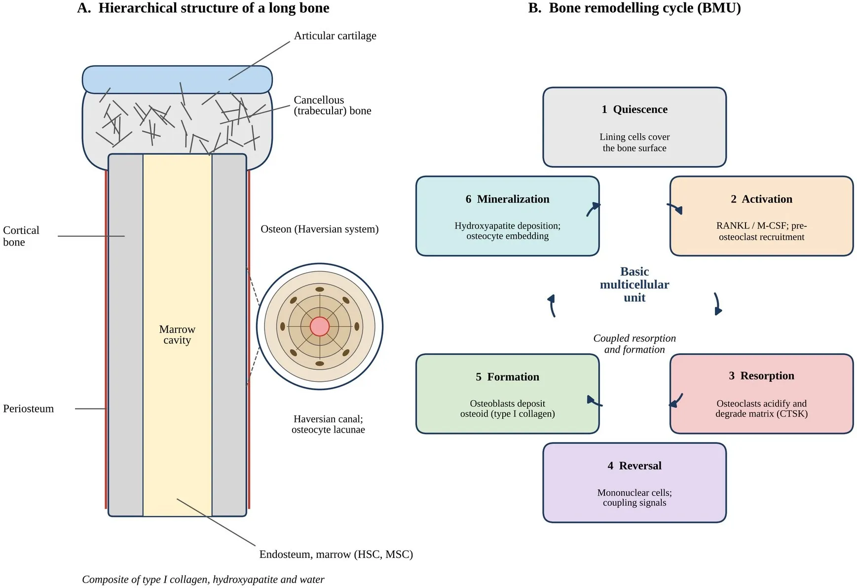
Figure 1. Bone structure and bone remodelling mechanism. (A) Longitudinal schematic of a long bone showing articular cartilage, cancellous and cortical bone, periosteum, endosteum and marrow cavity, with an osteon inset (Haversian canal and osteocyte lacunae). (B) The six phases of the remodelling cycle within a basic multicellular unit (BMU): quiescence, activation, resorption, reversal, formation and mineralization [16,17]. Original conceptual schematic; not to scale.
2.2 Bone cells and remodelling
Bone is continuously renewed through remodelling carried out by BMUs. Activation of osteoclast precursors by RANKL and M-CSF leads to resorption, followed by a reversal phase and then osteoblast-mediated formation of osteoid and its mineralization (Figure 1B) [17]. The balance between RANKL and its decoy receptor OPG, and signals released from the matrix and osteocytes, couple resorption to formation [16,17]. Table 1 summarizes the principal cell types and their relevance for scaffold-based regeneration.
Table 1: Bone Cells, Their Functions and Roles in Bone Regeneration
| Cell type | Principal functions | Key markers / regulators | Relevance to scaffold-guided regeneration |
|---|---|---|---|
| Mesenchymal | Self-renewal; multilineage | Runx2; Osterix; ALP; | Primary target for |
| stem cells | potential; give rise to | periosteal and marrow niches | recruitment, adhesion and |
| (MSCs) and | osteoblasts when committed | [16] | osteogenic differentiation on |
| osteoprogenitors | [16] | scaffolds |
| Cell type | Principal functions | Key markers / regulators | Relevance to scaffold-guided regeneration |
|---|---|---|---|
| Osteoblasts | Synthesize and secrete osteoid (type I collagen); regulate mineralization; produce RANKL and OPG [16,17] | ALP; type I collagen; OPN; OCN; Runx2; Osterix | Readout of osteogenic activity (ALP, matrix deposition, mineralization) |
| Bone-lining cells | Flattened quiescent osteoblast lineage cells covering surfaces; participate in remodelling activation [16] | Surface-resident; regulate local ion exchange | Contribute to surface response and activation of the remodelling cycle |
| Osteocytes | Embedded in lacunae; mechanosensing; orchestrate remodelling via sclerostin and RANKL [16,17] | Sclerostin; DMP1; connexin 43 | Sense scaffold-derived mechanical load; regulate long-term remodelling |
| Osteoclasts | Multinucleated cells that resorb mineralized matrix by acid secretion and proteolysis (CTSK) [16,17] | RANKL/RANK; M-CSF; CTSK; TRAP | Govern scaffold resorption and matrix turnover; coupling to formation |
| Endothelial and hematopoietic cells | Vascularization of the defect; marrow niche; immune modulation [1] | VEGF; CD31; macrophage phenotypes | Vascular ingrowth and inflammatory response determine integration |
Note: Compiled from refs. [1,16,17]. ALP, alkaline phosphatase; OPN, osteopontin; OCN, osteocalcin; CTSK, cathepsin K; TRAP, tartrate-resistant acid phosphatase; DMP1, dentin matrix protein 1.
2.3 Phases of fracture and defect healing
Spontaneous repair proceeds through an inflammatory phase, soft and hard callus formation and remodelling, with osteoprogenitor recruitment, angiogenesis and growth-factor signalling as essential determinants [1]. Scaffolds are intended to support these stages by providing a template for progenitor infiltration and vascular ingrowth, then to degrade as remodelled bone replaces them. Because healing depends on coordinated mechanical, vascular and biochemical conditions, a scaffold cannot be judged by one property alone.
3. Bone Defects and Current Therapeutic Approaches
3.1 Critical-size defects
A critical-size defect is generally understood as one that will not heal spontaneously over the lifetime of the organism or during the study period, but a universal size threshold does not exist and definitions vary with species, anatomical site, soft-tissue environment and fixation [18,19]. This is important for the interpretation of animal studies, where the defect model should be justified rather than assumed to be critical.
3.2 Grafts and bone substitutes
Autografts provide osteogenic cells, osteoinductive factors and an osteoconductive framework, but their supply is limited and harvest can produce pain and morbidity. Allografts avoid harvest but are processed, may have reduced biological activity and carry residual risks, while
ceramics, cements and metals are osteoconductive or load-bearing but generally lack osteoinduction or degrade at rates unmatched to bone formation [2,3,19]. Distraction osteogenesis and induced-membrane techniques can address segmental defects but require prolonged treatment and surgical expertise [19].
3.3 Rationale for polymeric hydrogel scaffolds
Hydrogel-based scaffolds offer high water content, the possibility of minimally invasive delivery, tunable degradation and the capacity to deliver cells or drugs, whereas pure polymers are mechanically weaker than mineralized tissue [3,5]. This motivates composites in which a polymer network is reinforced with calcium phosphates, as in chitosan–HA and chitosan–β-TCP systems [7,9].
4. Fundamentals of Tissue Engineering and Biomaterials
4.1 Scaffold requirements
An ideal bone scaffold is biocompatible, osteoconductive, sufficiently porous with interconnected pores, mechanically adequate for the implantation site, and biodegradable at a rate that permits load transfer to new tissue [4,5,20]. Surface chemistry must permit protein adsorption and cell adhesion, and degradation products must be non-toxic. These requirements are interdependent; for example, higher porosity facilitates ingrowth but typically reduces stiffness [20,21].
4.2 Porosity and architecture
Porosity and pore size influence nutrient diffusion, cell migration, vascularization and bone ingrowth. A minimum pore size of roughly 100 μm is commonly considered necessary for transport and cell penetration, and pores larger than about 300 μm are often recommended for bone ingrowth and capillary formation, although the optimum depends on material and fabrication route [21]. Interconnectivity matters as much as total porosity, and design strategies attempt to balance biological and mechanical needs through controlled architecture [20].
4.3 Hydrogels, composites and bioactive delivery
Hydrogels are crosslinked hydrophilic networks whose properties depend on polymer chemistry, crosslink density and environment [22,23]. Incorporating bioceramics supplies calcium and phosphate ions and increases stiffness, and loading of growth factors, peptides or drugs allows the scaffold to function as a bioactive delivery platform [5,8]. The release behaviour is governed by diffusion, swelling and degradation, which in turn are controlled by the crosslinking approach.
5. Chitosan: Structure, Properties and Biomedical Applications
5.1 Chemical structure and source
Chitosan is a linear copolymer of β-(1→4)-linked D-glucosamine (GlcN) and N-acetyl-D-glucosamine (GlcNAc) units produced by partial deacetylation of chitin; the polymer is conventionally named chitosan when the degree of deacetylation (DD) is high enough to confer solubility in dilute acids [24]. The C-2 primary amine of GlcN, with an apparent pKa near 6.5, is protonated at acidic pH, which explains solubility, polyelectrolyte behaviour and reactivity toward electrophilic crosslinkers (Figure 2A) [24]. Molecular weight, DD and acetyl-group distribution vary with source and processing and strongly affect solubility, viscosity, crystallinity and degradation, making batch characterization essential [24,25].
5.2 Physicochemical and biological properties
Table 2 summarizes key properties relevant to bone scaffolds. Chitosan is biodegradable mainly through lysozyme-mediated hydrolysis of acetylated residues, so the degradation rate depends on DD and crystallinity [6,25]. Structural resemblance to glycosaminoglycans, together with cationic charge, favours interaction with proteins and cells [6,7].
Table 2: Physicochemical and Biomedical Properties of Chitosan
| Property | Description | Implication for bone scaffolds |
|---|---|---|
| Chemical structure | Linear β-(1→4) copolymer of GlcN and GlcNAc; reactive C-2 amine and C-6/C-3 hydroxyl groups [24] | Amine groups are the principal sites for genipin crosslinking and chemical modification |
| Solubility and charge | Soluble in dilute acids (pH below about 6) where amines are protonated; amine pKa near 6.5 [24] | Allows aqueous processing; controls gelation conditions and pH-dependent crosslinking |
| Degree of deacetylation and molecular weight | Vary with source and processing; influence crystallinity, viscosity and solubility [24,25] | Determine crosslinking capacity, mechanical behaviour and degradation; require reporting |
| Biodegradability | Enzymatic degradation (notably lysozyme) of acetylated residues; products are oligosaccharides and glucosamine units [6,25] | Degradation can be tuned via DD and crosslinking; must match bone formation |
| Biocompatibility and toxicity | Generally well tolerated; behaviour depends on purity, DD and molecular weight [25] | Purity and endotoxin control needed for in-vivo use |
| Mechanical properties | Unmodified chitosan structures are mechanically weak and swell extensively [7,14] | Reinforcement by crosslinking and mineral fillers is required for bone applications |
| Biological interactions | Similarity to glycosaminoglycans; supports adhesion, proliferation and osteogenic differentiation of osteoblastic cells in many composite systems [6,7,9] | Basis for use as osteoconductive matrix and delivery carrier |
| Property | Description | Implication for bone scaffolds |
|---|---|---|
| Processability | Forms hydrogels, sponges, films, fibres and printed constructs; hosts HA, β-TCP and bioactive agents [8,9] | Enables diverse fabrication routes for bone scaffolds |
Note: Compiled from refs. [6–9,14,24,25]. GlcN, glucosamine; GlcNAc, N-acetylglucosamine.
5.3 Biomedical applications and limitations
Chitosan has been explored for drug delivery, wound care and orthopaedic tissue engineering, including chitosan–gelatin and chitosan–ceramic composites for bone [7,9,26]. Reviews emphasize that its limitations for bone include low stiffness, high swelling and variable degradation, and that composite design and crosslinking are the principal ways to overcome them [7,9]. These deficiencies define the rationale for a mild but effective covalent crosslinker such as genipin.
6. Genipin as a Natural Crosslinking Agent
6.1 Source and chemistry
Genipin is the aglycone of geniposide, an iridoid glycoside abundant in the fruit of Gardenia jasminoides; it is released by β-glucosidase hydrolysis and has long been used in traditional medicine and as a precursor of natural blue colorants [10,11]. Its structure is a cyclopenta [c]pyran bearing an enol-ether double bond, a methyl ester and a hemiacetal hydroxyl (Figure 2B). These electrophilic sites are responsible for its reaction with primary amino groups of chitosan, collagen, gelatin and other biopolymers [11,27].
6.2 Crosslinking mechanism with chitosan
Two principal reaction routes with amines are described. In the first, the amine attacks C-3 of the dihydropyran ring, opening it and forming a heterocyclic secondary-amine linkage; in the second, the amine substitutes the methyl ester to give an amide [11,27]. Genipin can additionally undergo oligomerization, and oxygen-dependent coupling generates blue-green chromophores that are visible as a colour change and as intrinsic fluorescence of the network (Figure 2C) [11,28]. Reaction pathways, kinetics and the extent of genipin self-polymerization are pH dependent, and small pH changes can markedly alter the structure and properties of the resulting hydrogels [29,30]. Mechanistic assignments differ between studies, which is why crosslinking degree should be measured rather than inferred from reagent ratio.
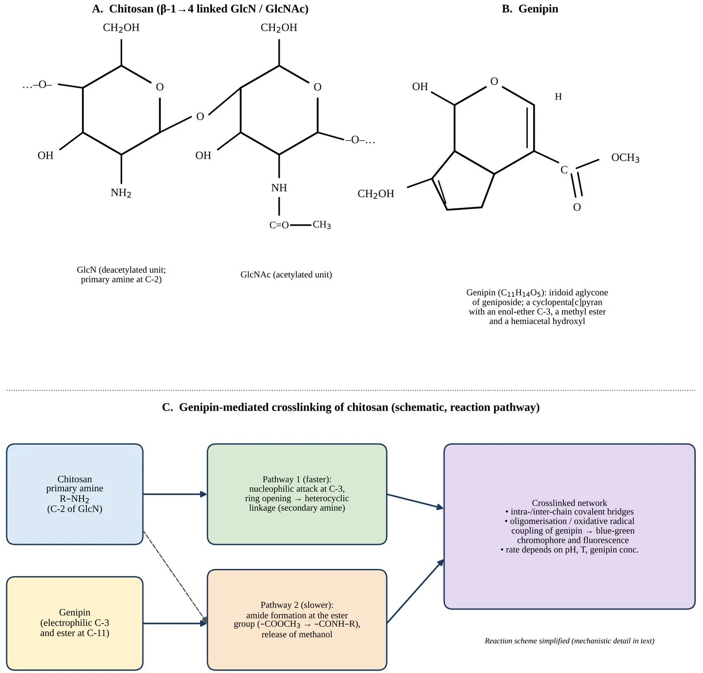
Figure 2. Chemical structures of chitosan and genipin and the proposed genipin-mediated crosslinking mechanism. (A) Chitosan repeating units (GlcN and GlcNAc) linked by β-(1→4) glycosidic bonds; the C-2 primary amine of GlcN is the reactive site. (B) Genipin (C11H14O5). (C) Simplified reaction scheme: nucleophilic attack by chitosan amine at C-3 of genipin leading to ring-opening and heterocyclic linkage, and slower amide formation at the ester group, with formation of a crosslinked, coloured and fluorescent network [11,27,29]. Original schematic redrawn for illustration; atom positions and stereochemistry are simplified.
6.3 Biocompatibility
Comparative in-vitro assays reported markedly lower cytotoxicity for genipin than for glutaraldehyde in fibroblast cultures [12]. In vivo, a genipin-fixed injectable chitosan implant showed acceptable tissue response with degradation governed by crosslinking [31], and genipin crosslinking of chitosan hydrogels did not reduce cell viability but improved cell adhesion on denser networks in a fibroblast model [32]. These observations support, but do not prove, suitability for bone; findings depend on genipin dose, residual unreacted genipin, and cell type, and long-term in-vivo and immunological data in bone sites remain limited [33].
6.4 Comparison with other crosslinking agents
Table 3 compares genipin with glutaraldehyde, EDC/NHS and physical crosslinking. The comparison is qualitative because experimental conditions differ across studies and
quantitative head-to-head data in bone models are scarce; a fuller critical discussion is given in Section 13.
Table 3: Comparison of Genipin and Other Crosslinking Agents for Chitosan-Based Scaffolds
| Feature | Genipin | Glutaraldehyde | EDC/NHS | Physical crosslinking (ionic, e.g., TPP; complexation) |
|---|---|---|---|---|
| Origin | Natural iridoid from Gardenia fruit [10] | Synthetic dialdehyde [34] | Synthetic zero-length carbodiimide system [35] | Polyanions or multivalent ions interacting electrostatically [23] |
| Chemistry with chitosan | Amine attack at C-3 and at the ester group; heterocyclic and amide links [11,27] | Reacts with amines (Schiff-base and more complex oligomeric products) [34] | Forms amide bonds between carboxyl and amine without incorporation; chitosan needs a carboxylated partner [22,35] | Reversible ionic or hydrogen-bond interactions; no covalent bridge [22,23] |
| Cytotoxicity and safety | Substantially lower in-vitro cytotoxicity than glutaraldehyde [12,31] | Cytotoxicity linked to residual or released aldehyde; apoptosis reported [36] | Reagent and by-products are washed out; residual reagent must be removed [35] | Generally mild; ion or polyanion choice must be biocompatible [23] |
| Reaction conditions | Mild, aqueous; slower than glutaraldehyde; depends on pH, temperature, concentration [28,30] | Fast; pH and concentration dependent [34] | Aqueous, pH-sensitive coupling; requires activation [35] | Rapid and spontaneous on mixing; pH and ionic-strength sensitive [23] |
| Effect on mechanics and swelling | Increases modulus and reduces swelling with crosslink density [28,32] | Strong stiffening in collagen systems [34] | Stiffening depends on carboxyl content and conversion [22] | Often weaker, less stable in physiological media [14,23] |
| Special features | Blue colour and fluorescence enable monitoring [11,28] | None relevant; yellowing possible | No colour; zero-length link leaves no spacer [35] | Reversibility can support injectability and self-healing [22] |
| Main limitations | Higher cost; slow kinetics; colour; batch and pH variability [30,33] | Toxicity and calcification concerns limit bone use [36] | Side reactions, hydrolysis of intermediates, limited chitosan compatibility [35] | Instability, burst release, sensitivity to environment [23] |
| Example in bone-related studies | Chitosan–HA fibres, chitosan–β-TCP, chitosan–gelatin, gum arabic/chitosan/HA [13,14,26,37] | Chitosan–gelatin compared with genipin [26] | Mainly collagen or gelatin hybrid systems [22] | TPP-crosslinked chitosan–β-TCP compared with genipin [14] |
Note: Compiled from refs. [10–12,14,22,23,26–28,30,34–36]. Qualitative comparison; EDC, 1-ethyl-3-(3-dimethylaminopropyl)carbodiimide; NHS, N-hydroxysuccinimide; TPP, tripolyphosphate.
7. Genipin-Crosslinked Chitosan Hydrogel Scaffolds
7.1 Network formation and three-dimensional architecture
When chitosan solution is mixed with genipin, covalent bridges between chains convert the solution to a gel whose stiffness, hydrophilicity and swelling depend on the amount of genipin and chitosan [32]. Subsequent freezing and lyophilization, or freeze-gelation, produce porous scaffolds in which ice-crystal templating defines pores and crosslinked walls provide structural integrity (Figure 3) [14,38]. Gao et al. showed that macroporous structure of chitosan hydrogels could be tailored by varying genipin or chitosan amounts, and that crosslinking enhanced storage modulus [32].
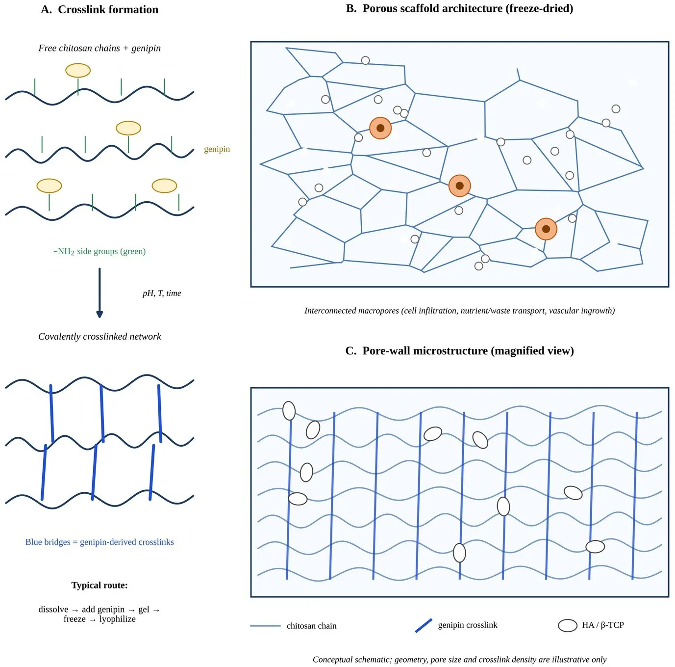
Figure 3. Formation and three-dimensional architecture of genipin-crosslinked chitosan hydrogel scaffolds. (A) Free chitosan chains with amine groups react with genipin to give a covalently crosslinked network. (B) Freeze-dried porous scaffold with interconnected macropores supporting cell infiltration and nutrient transport. (C) Magnified pore wall showing crosslinked chitosan chains and embedded HA or β-TCP particles. Conceptual schematic; geometry and dimensions are illustrative [13,14,32].
7.2 Fabrication platforms
Several routes have been reported. Freeze-gelation has produced genipin-crosslinked chitosan/nano β-TCP scaffolds that were evaluated for porosity, wettability, compressive
strength, mass loss and MSC osteogenesis [14]. Electrospinning has generated genipin-crosslinked chitosan nanofibres containing HA nanoparticles with periosteum-like mechanical properties [13]. Additive manufacturing of HA–chitosan–genipin composites demonstrates the feasibility of computer-designed architectures [15], and injectable chitosan implants crosslinked with genipin illustrate minimally invasive delivery [31]. Hierarchical bilayer scaffolds of chitosan/β-TCP and silk fibroin crosslinked with genipin have been described for osteochondral interfaces [38].
7.3 Composite systems and published osteogenic findings
In electrospun chitosan–HA–genipin fibres, XRD, FTIR and EDS confirmed HA incorporation, and the Young's modulus of composite scaffolds was reported as similar to that of natural periosteum; osteoblast-like cells showed higher ALP activity on composite fibres than on pure chitosan, with a 2.4-fold difference by day 14 [13]. In chitosan/nano β-TCP scaffolds, genipin-crosslinked constructs supported MSC osteogenic differentiation and showed slightly higher mineralization after 21 days than TPP-crosslinked constructs [14]. Chitosan–gelatin scaffolds crosslinked with glutaraldehyde or genipin supported MSC infiltration and increased expression of RUNX2, ALP and osteocalcin, and histology after implantation in mouse femur showed matrix formation with minimal inflammation [26]. A genipin- or ferric-chloride-crosslinked gum arabic/chitosan/nano-HA hydrogel reported compressive strengths in a range comparable to cortical bone and new bone in 6 mm rat calvarial defects, with genipin improving hydrophilicity and porosity [37].
These studies are encouraging but heterogeneous: polymer sources, DD, genipin ratios, mineral content, cell types and animal models differ, and some reports describe qualitative bone formation rather than quantitative morphometry. Direct comparison of absolute values between studies is therefore unreliable, and it remains unclear which composition is optimal [9,33].
8. Design Principles and Formulation Development
8.1 Polymer parameters
Chitosan DD, molecular weight and concentration determine the number of reactive amines, chain entanglement and solution viscosity, and therefore the achievable crosslink density and pore morphology [24,28]. Because commercial chitosan batches vary, DD and molecular weight should be determined and reported; spectroscopic and titrimetric methods are widely used for DD estimation, and XRD-based determination has also been described [39].
8.2 Crosslinker concentration, pH, temperature and time
Genipin concentration, genipin-to-amine ratio, pH, temperature and reaction time govern kinetics and network structure, with higher crosslinking generally raising stiffness and reducing swelling and degradation [28,29,32]. pH is particularly influential because it affects both amine
protonation and the reaction pathway of genipin [29,30]. A fixed crosslinking time may thus produce different degrees of crosslinking at different pH or temperature, so reporting a fixation index or measured degree of crosslinking, not only nominal concentrations, is advisable.
8.3 Bioceramic and bioactive additives
HA and β-TCP supply calcium and phosphate ions and raise stiffness, and they have been paired with genipin-crosslinked chitosan in fibre, freeze-gelled and printed formats [13–15]. Filler content, particle size and dispersion influence viscosity, crosslinking and pore structure and may interact with the crosslinking chemistry. Growth factors, peptides or drugs can be incorporated to create a delivery platform; the release is expected to depend on crosslink density, swelling and degradation, but systematic release studies in genipin-crosslinked bone scaffolds remain limited [8,33].
8.4 Architecture, processing and translation-oriented factors
Freezing rate, polymer concentration and drying conditions control pore size and interconnectivity in lyophilized scaffolds, whereas printing parameters define macroscopic geometry; architecture should be tuned within the biological and mechanical limits discussed in Section 4 [15,20,21]. Practical issues for translation include removal of unreacted genipin, endotoxin control, sterilization compatible with the polymer network, storage stability, and reproducible scale-up. The overall workflow from raw materials to candidate scaffold is summarized in Figure 4; it is a proposed framework linking formulation variables, structure, properties and biological performance.
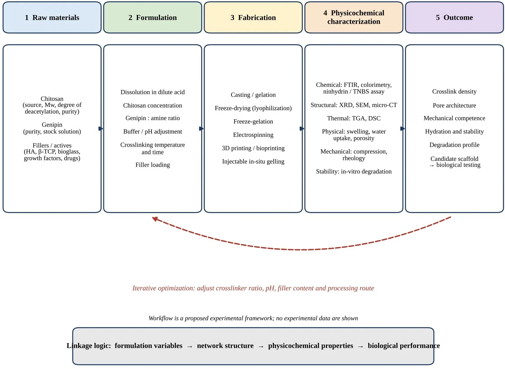
Figure 4: Hydrogel Preparation, Formulation Development and Physicochemical Characterization Workflow. Raw Materials (Chitosan, Genipin, Fillers and Bioactive Agents) Are Formulated, Fabricated by Casting, Freeze-Drying, Electrospinning, 3D Printing or Injectable Gelation, and Characterized by Chemical, Structural, Thermal, Physical, Mechanical and Stability Tests. Results Feed Back into Formulation. Proposed Experimental Framework; No Experimental Data Are Shown [14,28,32].
9. Physicochemical Characterization of Hydrogel Scaffolds
Characterization links formulation to biological behaviour. Each technique below is described by its principle, usual method, parameters measured, interpretation and importance; Table 4 summarizes them. Techniques are described generally; specific values depend strongly on material, sample preparation and protocol, and are not given here unless reported in the cited studies.
9.1 Fourier transform infrared spectroscopy (FTIR)
Principle: absorption of mid-infrared radiation excites molecular vibrations that are characteristic of functional groups. Method: attenuated total reflectance or KBr-pellet spectra of dried scaffolds, with chitosan, genipin and uncrosslinked controls. Parameters: position, intensity and shape of amide I/II, N–H bending, O–H/N–H stretching, C–O–C and, in composites, phosphate bands. Interpretation: changes in amine- and amide-related bands relative to uncrosslinked chitosan indicate amine consumption and new amide or heterocyclic linkages, and phosphate bands confirm mineral incorporation [11,13]. Importance: confirms
chemical modification and filler presence, but overlapping bands make FTIR semi-quantitative, so it is best combined with colorimetric amine assays.
9.2 Scanning electron microscopy (SEM)
Principle: a focused electron beam produces secondary-electron images of surface topography. Method: dry or freeze-dried samples are sputter-coated with a conductive metal; cryo-SEM or environmental SEM preserves hydrated structure. Parameters: pore size distribution, wall thickness, interconnectivity, fibre diameter, mineral dispersion and cell morphology. Interpretation: pore dimensions are measured from many fields by image analysis and should be related to the 100–300 μm benchmarks for bone ingrowth [21]. SEM has been used to describe tailored macroporous chitosan structures and cell adhesion on genipin-crosslinked gels [32]. Importance: direct structural evidence; limitations include drying artefacts, two-dimensional sampling and coating effects.
9.3 X-ray diffraction (XRD)
Principle: constructive diffraction of X-rays by ordered lattices gives reflections at characteristic 2θ angles. Method: powder or flat-sample scans of dried material. Parameters: peak position, width and relative intensity, crystallinity and phase identity. Interpretation: chitosan is semicrystalline, with broad reflections that change with DD and crosslinking, so diffractograms have been used to assess crystallinity and even DD [39,40]. Crosslinking generally disrupts chain packing and may reduce crystallinity, while HA or β-TCP show sharp phase-specific reflections, as used to confirm HA in genipin-crosslinked chitosan fibres [13]. Importance: phase identification and crystallinity; amorphous fractions and small particle sizes limit sensitivity.
9.4 Thermogravimetric analysis (TGA)
Principle: mass is recorded as temperature increases under a controlled atmosphere. Method: a few milligrams heated at constant rate under nitrogen or air. Parameters: onset and peak decomposition temperatures, mass-loss stages and residual mass. Interpretation: an early loss relates to bound water, followed by polymer decomposition; shifts in decomposition temperature are used to infer crosslinking-related stabilization, and residue at high temperature estimates inorganic filler content [40]. Importance: thermal stability, hydration state and filler loading; heating rate and atmosphere strongly affect results and must be reported.
9.5 Differential scanning calorimetry (DSC)
Principle: heat flow to a sample is compared with a reference during a temperature program. Method: sealed or pierced pans heated at constant rate, often with a heat–cool–heat cycle. Parameters: endothermic dehydration, glass transition when detectable, and exothermic or endothermic decomposition events. Interpretation: movement or disappearance of thermal events upon crosslinking suggests restricted chain mobility and altered water binding, though
chitosan transitions are often broad and hard to assign [40]. Importance: complements TGA by showing the energetics of water loss and degradation; interpretation requires caution and orthogonal methods.
9.6 Swelling studies
Principle: hydrophilic networks absorb water until osmotic forces balance elastic retraction of crosslinks. Method: pre-weighed dry samples are immersed in PBS or buffer at 37 °C, removed at time points, blotted and weighed. Parameters: swelling ratio, SR = (Ws − Wd)/Wd, kinetics and equilibrium swelling, often as a function of pH. Interpretation: lower equilibrium swelling indicates higher effective crosslink density; pH-dependent swelling reflects amine protonation [23,32]. Importance: swelling influences pore size, mechanical behaviour, dimensional stability in a defect and release kinetics.
9.7 Porosity determination
Principle: porosity is the void fraction of the scaffold. Methods: liquid displacement (ethanol or hexane), gravimetric calculation from apparent and polymer density, mercury intrusion porosimetry, micro-computed tomography and SEM image analysis. Parameters: total porosity, pore size distribution and interconnectivity. Interpretation: values should be correlated with ingrowth benchmarks and with mechanical results, because high porosity tends to lower compressive modulus [20,21]. Micro-CT provides three-dimensional metrics, provided that resolution and thresholding follow accepted guidance [41]. Porosity of genipin-crosslinked chitosan/β-TCP scaffolds has been evaluated together with pore size and water content [14]. Importance: governs transport, vascularization and mechanics.
9.8 In-vitro degradation
Principle: hydrolytic and enzymatic breakdown reduces mass and mechanical integrity. Method: pre-weighed samples are incubated in PBS with or without lysozyme at 37 °C; at intervals they are rinsed, dried and weighed, and medium pH may be recorded. Parameters: mass remaining (%), pH change, molecular and morphological changes. Interpretation: because chitosan is degraded by lysozyme acting on acetylated residues, enzyme-containing media better reflect in-vivo behaviour, and higher crosslinking is expected to slow degradation [25,31]. Mass loss of genipin- and TPP-crosslinked chitosan/β-TCP scaffolds has been compared [14]. Importance: degradation should be matched with bone formation; in-vitro rates may not predict in-vivo rates.
9.9 Mechanical and compressive testing
Principle: a universal testing machine applies controlled compressive load while recording stress and strain. Method: cylindrical hydrated or dry specimens are compressed at constant crosshead speed; rheometry measures storage and loss moduli of gels. Parameters: compressive modulus (linear region), compressive strength, strain at failure and viscoelastic moduli.
Interpretation: hydration state, specimen geometry and rate must be controlled, since dry and wet values differ greatly; values should be compared with the intended site and not only with cortical bone [20]. Genipin crosslinking has been reported to increase storage modulus of chitosan hydrogels [32], and composites have been reported with compressive strengths in a range compared with cortical bone, although such claims need hydration and site context [37]. Importance: determines handling and load-sharing ability.
9.10 Water uptake studies
Principle: the capacity of the network to absorb and retain water reflects hydrophilicity and pore structure. Method: gravimetric measurement after immersion; water content may be expressed as (Ww − Wd)/Ww, and wettability by contact angle. Parameters: equilibrium water content, uptake rate and contact angle. Interpretation: genipin crosslinking has been reported to alter hydrophilicity and swelling of chitosan hydrogels, and water content and wettability have been measured for genipin–chitosan/β-TCP scaffolds [14,32]. Importance: influences protein adsorption, cell adhesion and nutrient diffusion; it should be distinguished from swelling ratio by stating the normalization basis.
9.11 Quantifying crosslinking degree
Because reagent ratios do not reflect actual conversion, crosslinking degree is quantified by ninhydrin or TNBS assays of residual free amines, with colorimetric or fluorescence monitoring of genipin-derived chromophores as supportive evidence [11,28]. This quantification should accompany every characterization panel so that structure–property relationships can be compared across studies.
Table 4: Physicochemical Characterization Techniques and Their Applications
| Technique | Principle | Parameters measured | Interpretation in genipin–chitosan scaffolds | Limitations / key controls |
|---|---|---|---|---|
| FTIR | Vibrational absorption of IR radiation | Band position and intensity (amide, N–H, O–H, C–O–C, phosphate) |
Amine consumption and new linkages; mineral presence [11,13] | Semi-quantitative; overlapping bands; use uncrosslinked control |
| SEM | Electron-beam surface imaging | Pore size, wall thickness, interconnectivity, fibre diameter, cell morphology | Architecture relative to 100–300 μm benchmarks [21,32] | Drying and coating artefacts; 2D sampling |
| XRD | Diffraction by crystalline lattices | 2θ peak position, width, crystallinity, phase identity | Chitosan crystallinity and mineral phase confirmation [13,39] | Poor for amorphous or low-content phases |
| TGA | Mass change versus temperature | Decomposition temperatures, mass-loss stages, residue | Water content, thermal stability, filler fraction [40] | Depends on heating rate and atmosphere |
| Technique | Principle | Parameters measured | Interpretation in genipin–chitosan scaffolds | Limitations / key controls |
|---|---|---|---|---|
| DSC | Heat flow versus temperature | Endotherms, exotherms, transitions | Water binding and chain mobility changes [40] | Broad, ambiguous transitions in chitosan |
| Swelling | Osmotic water absorption balanced by crosslinks | Swelling ratio, kinetics, pH dependence | Indicates crosslink density and stability [23,32] | Blotting and mass-loss errors; state medium and temperature |
| Porosity | Void fraction of scaffold | Total porosity, pore size, interconnectivity | Relation to ingrowth and mechanics [14,20,21] | Method-dependent; micro-CT resolution [41] |
| In-vitro degradation | Hydrolytic or enzymatic mass loss | Mass remaining, pH, morphology | Stability; lysozyme sensitivity [14,25,31] | In-vitro rates may not predict in vivo |
| Compressive / mechanical testing |
Stress–strain under load; rheometry | Modulus, strength, strain, G′ and G″ | Stiffening by crosslinking; site-appropriate comparison [32,37] | Hydration, geometry and rate must be controlled |
| Water uptake / wettability | Gravimetric water absorption; contact angle | Equilibrium water content, uptake rate, angle | Hydrophilicity changes with crosslinking [14,32] | Normalization basis must be stated |
| Crosslink quantification | Colorimetric assay of residual amines; chromophore monitoring | Degree of crosslinking, fixation index | Actual conversion versus nominal ratio [11,28] | Reagent interference; calibration needed |
Note: Compiled from the cited references; techniques are described generally and the values depend on material and protocol.
10. Biological Evaluation and Osteogenic Potential
10.1 Cytocompatibility and cell–material interaction
Initial testing assesses cytotoxicity using extracts and direct contact with relevant cells, with viability assays such as MTT, live/dead staining and DNA quantification, controls for residual unreacted genipin, and comparison with uncrosslinked and alternative crosslinked materials. Genipin-crosslinked chitosan has been reported not to induce cytotoxicity in fibroblasts, with higher crosslink density improving adhesion and viability in that model [32]; MSC viability and proliferation on genipin–chitosan scaffolds have also been reported [14,26]. Because cell type, scaffold stiffness and surface chemistry interact, results from fibroblasts should not be extrapolated to osteogenic cells without testing.
10.2 Osteogenic differentiation markers
Osteogenic potential is assessed by time-dependent markers. ALP activity and Runx2 expression indicate early commitment; type I collagen and osteopontin reflect matrix production; osteocalcin and mineral deposition indicate maturation. Mineral can be quantified by Alizarin Red S staining with extraction-based quantification [42]. Studies of genipin-crosslinked scaffolds have reported higher ALP, osteonectin and osteocalcin-related responses
and mineralization in composites [13,14,26]. Results should be normalized to cell number or DNA and compared with osteogenic-medium controls, since differentiation media alone induce these markers.
10.3 Bioactivity, angiogenesis and immune response
Apatite-forming ability in simulated body fluid (SBF) is commonly used to screen bioactivity, but its power to predict in-vivo bone bonding is limited and results should be interpreted cautiously [43]. Vascularization and macrophage polarization are increasingly recognized as determinants of repair [1], yet they are inadequately examined for genipin-crosslinked scaffolds; endothelial assays, VEGF measurement and immunophenotyping represent proposed additions.
Table 5: Biological Evaluation Methods, Osteogenic Parameters and Preclinical Animal Models
| Level / method | Parameters measured | Typical system or model | Notes and limitations |
|---|---|---|---|
| Cytocompatibility (extract and direct contact) | Cell viability (MTT, live/dead), morphology, DNA content | Fibroblasts, osteoblast-like cells, MSCs [14,32] | Include residual-genipin control; cell type affects outcome |
| Cell adhesion and proliferation | Attachment, spreading, infiltration into pores, proliferation | MSC, MC3T3-E1 or 7F2 cells on scaffolds [13,26] |
Infiltration needs 3D imaging or sectioning |
| Early osteogenic markers | ALP activity, Runx2 mRNA | MSCs in osteogenic medium [13,26] | Compare with medium-only controls; normalize to DNA |
| Late osteogenic markers and mineralization | Type I collagen, OPN, OCN; Alizarin Red S staining and quantification | MSC cultures for 14–28 days [14,42] | Staining may reflect non-specific calcium deposition; verify by other methods |
| Mechanistic assays | qRT-PCR, Western blot and inhibitors for BMP/Smad, Wnt/β-catenin, MAPK pathways | Cell culture with pathway inhibitors [44–46] | Correlative unless inhibitors or knockdown applied |
| Bioactivity screening | Apatite formation on surface | SBF immersion [43] | Limited predictive value for in-vivo performance |
| Rodent critical-size defect | New bone volume, bridging, histology, micro-CT BV/TV | Rat calvarial defect [37,47]; mouse femur implantation [26] | Defect size and site must be justified; calvaria is non-load-bearing |
| Imaging and histology | Micro-CT metrics; H&E, Masson trichrome; immunohistochemistry | Explants from small-animal studies [37,41] | Standardized scanning and thresholds; blinded scoring |
| Larger-animal and load-bearing models | Biomechanics, long-term degradation, safety, integration | Rabbit, sheep, goat or pig long-bone or condylar defects (proposed next stage) | Costly and ethically demanding; needed before clinical translation |
| Reporting standards | Randomization, blinding, sample-size justification, outcomes | All animal studies [48] | ARRIVE 2.0 recommended for transparent reporting |
Note: Compiled from the cited references; larger-animal models are a proposed progression rather than an established finding for genipin–chitosan scaffolds. BV/TV, bone volume fraction; MC3T3-E1 and 7F2, murine osteoblast-like cell lines; H&E, haematoxylin and eosin.
11. In-Vitro and Preclinical Evaluation Strategies
This section outlines a proposed, literature-informed strategy; it describes how evaluation could be designed and is not a report of experimental results.
11.1 Proposed in-vitro design
A rational in-vitro programme would test a genipin dose series with matched uncrosslinked, glutaraldehyde-crosslinked and TPP- or ionically crosslinked controls, using identical chitosan batches and processing [14,26]. Extract-based and direct-contact tests would be combined, MSCs would be the principal osteogenic cell type, and time points would cover early (ALP, Runx2) and late (mineralization) stages [42]. Leachables, residual genipin and degradation products should be quantified. Pathway involvement would be probed with inhibitors (for example, of BMP or Wnt signalling) rather than inferred from expression alone.
11.2 Proposed preclinical animal strategy
Animal models should be justified by the clinical question. A rat calvarial critical-size defect is a well-described, reproducible screening model for bone-regeneration materials, but it is non-load-bearing [47], and mouse femoral implantation has been used for early biocompatibility of chitosan scaffolds [26]. A staged approach could proceed to larger animals and load-bearing sites after safety and efficacy have been shown. Study design should include randomization, blinding, sample-size justification and transparent reporting following ARRIVE 2.0 [48].
11.3 Outcome assessment
Micro-CT with standardized acquisition and analysis [41] provides bone volume fraction and architecture, histology and immunohistochemistry confirm tissue identity and scaffold–bone interface, and biomechanical testing assesses functional restoration. Scaffold degradation, inflammatory response and systemic safety should be reported alongside efficacy. Figure 5 summarizes the proposed signalling hypothesis and staged evaluation pathway.
12. Mechanistic Insights into Bone Regeneration
This section separates established pathway biology from hypotheses on how genipin-crosslinked chitosan scaffolds may engage these pathways. The strongest evidence concerns the roles of the pathways in osteoblast differentiation in general; evidence that genipin-crosslinked chitosan itself activates a specific pathway in bone cells is, within the literature surveyed for this review, limited and largely indirect.
12.1 Established osteogenic signalling
BMP signalling through Smad1/5/8, together with non-canonical routes such as p38 MAPK, induces the transcription factors Runx2 and Osterix that drive osteoblast differentiation and bone formation [44]. Canonical Wnt/β-catenin signalling promotes osteoblast lineage
commitment and bone mass and is modulated by antagonists such as sclerostin and DKK1 [45]. MAPK cascades, including ERK, p38 and JNK, transduce extracellular cues in osteoblasts and regulate Runx2 activity and differentiation [46]. These pathways are interconnected, with crosstalk among BMP, Wnt and MAPK signalling (Figure 5A).
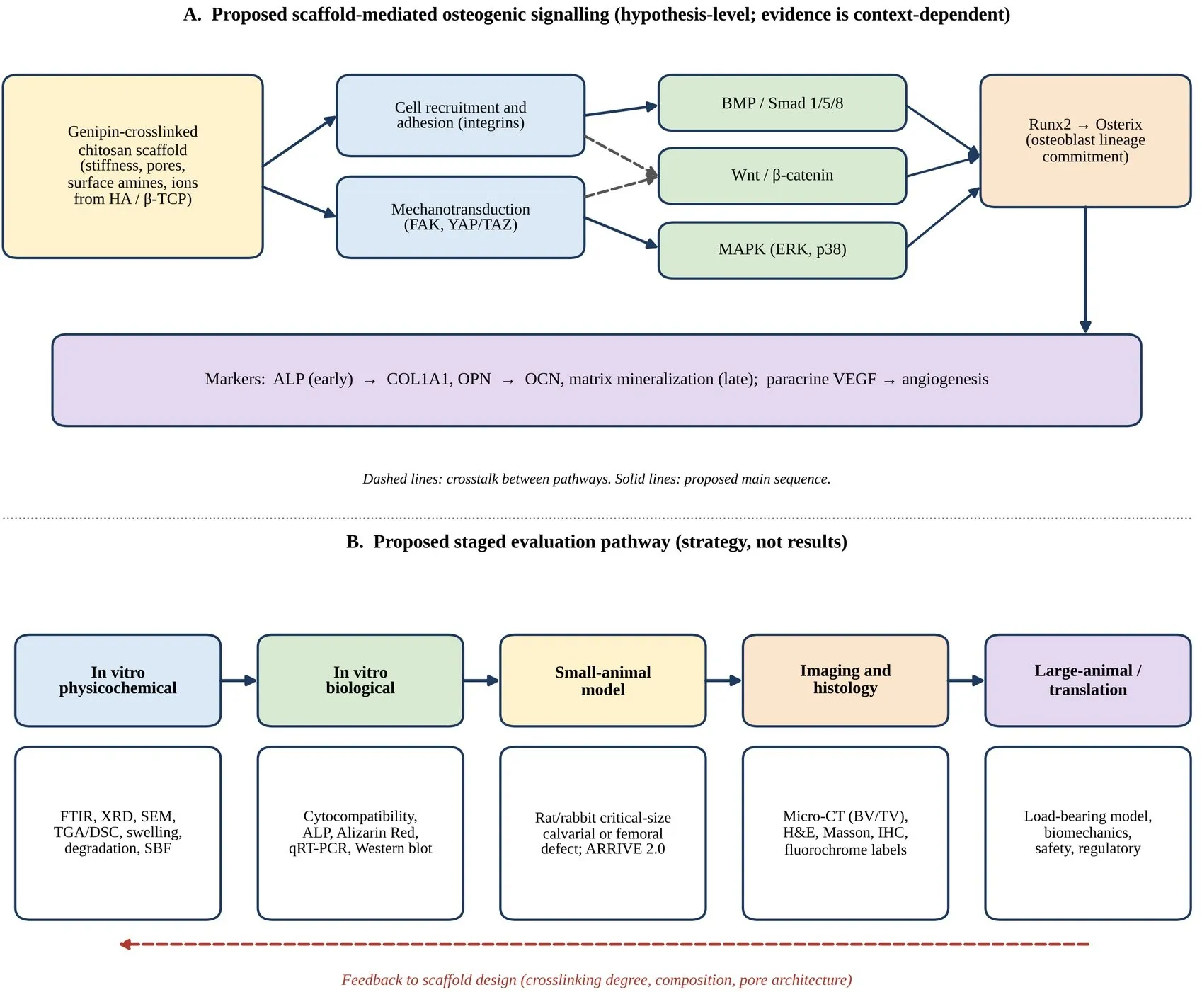
Figure 5: Scaffold-mediated Osteogenic Activity and Preclinical Bone Regeneration Evaluation Pathway.
(A) Proposed hypothesis: scaffold cues (stiffness, pores, surface chemistry, released ions) influence cell adhesion and mechanotransduction, which may converge on BMP/Smad, Wnt/β-catenin and MAPK signalling, Runx2/Osterix activation and osteogenic markers; pathway links for genipin-crosslinked chitosan are not firmly established [44–46,49]. (B) Proposed staged evaluation strategy from in-vitro characterization to large-animal translation [41,47,48]. Conceptual schematic; not experimental data.
12.2 Scaffold-derived cues: stiffness, adhesion and mineral ions
Matrix stiffness directs MSC lineage, with stiffer matrices favouring osteogenic commitment in two-dimensional hydrogel systems [49]. Because genipin crosslinking raises modulus and alters hydrophilicity and cell adhesion of chitosan hydrogels [32], one plausible hypothesis is that crosslinking density tunes integrin-mediated adhesion and mechanotransduction, with downstream engagement of MAPK and other pathways. This is a hypothesis: stiffness effects in three-dimensional porous scaffolds are more complicated, and stiffness is rarely varied independently of porosity or chemistry. Calcium phosphate fillers may add osteogenic cues by ion release or surface effects, and genipin–chitosan composites with HA or β-TCP have shown
increased osteogenic markers compared with polymer alone or alternative crosslinking [13,14]; however, the specific signalling responsible has generally not been dissected.
12.3 Evidence from related compounds
Geniposide, the glycoside precursor of genipin, was reported to alleviate glucocorticoid-induced inhibition of osteogenic differentiation in MC3T3-E1 cells through ERK activation involving the GLP-1 receptor [50]. This supports a biological link between iridoid compounds and MAPK signalling, but it concerns a soluble compound in a specific stress model, not a crosslinked scaffold, and it cannot be extrapolated to scaffold-bound genipin. Clarifying whether leached genipin or genipin-derived fragments are biologically active at concentrations achieved in vivo would be a useful research question.
12.4 Proposed mechanistic experiments
Proposed work would measure phosphorylated Smad1/5/8, nuclear β-catenin and phosphorylated ERK/p38 on scaffolds of defined stiffness and composition, apply pathway inhibitors (such as Noggin, DKK1 or MEK inhibitors) to test necessity, and decouple stiffness from porosity using matched scaffold series. Such experiments would convert correlative marker data into mechanistic evidence.
13. Comparative Analysis of Crosslinking Strategies
Genipin versus glutaraldehyde: the principal advantage of genipin is lower cytotoxicity [12], yet the practical difference in short-term osteogenic cultures can be modest; chitosan–gelatin scaffolds crosslinked with glutaraldehyde or genipin at the same concentration gave similar rheological properties and both supported MSC osteogenic gene expression [26]. This suggests that the safety benefit depends on residual reagent, washing and long-term exposure, and underlines the need for long-term in-vivo comparisons rather than reliance on short assays [34,36].
Genipin versus physical crosslinking: ionically crosslinked chitosan is formed under very mild conditions and may allow injectability, but stability in physiological ionic environments is limited [22,23]. In chitosan/β-TCP freeze-gelled scaffolds, genipin crosslinking was compared with TPP and showed slightly higher mineralization after 21 days [14], while a comparison with ferric chloride in gum arabic/chitosan/nano-HA hydrogels reported compressive strengths in a similar range for both but improved hydrophilicity and porosity with genipin [37]. These results favour covalent genipin networks where mechanical stability is needed, whereas the evidence is insufficient to rank crosslinkers universally.
Genipin versus EDC/NHS: carbodiimide chemistry forms zero-length amide bonds between carboxyl and amine groups without incorporating the reagent [35]; for chitosan, which lacks native carboxyl groups, it is mainly useful in hybrids with carboxylated components, whereas
genipin directly targets chitosan amines [11,22]. Dual or sequential crosslinking, combining physical gelation for handling with genipin for stability, is a promising but largely unvalidated strategy that should be examined with controlled comparisons (Table 3).
14. Recent Advances and Emerging Applications
14.1 Additive manufacturing and hierarchical constructs
Additive manufacturing of HA–chitosan–genipin composites enables patient-specific geometries and controlled macroarchitecture [15], and hierarchical bilayer scaffolds combining chitosan/β-TCP with silk fibroin crosslinked by genipin address osteochondral interfaces [38]. Such constructs raise new characterization requirements, including fidelity of printed geometry and uniformity of crosslinking across thick structures.
14.2 Nanocomposites, fibrous scaffolds and periosteum-like constructs
Electrospun genipin-crosslinked chitosan–HA fibres were designed to mimic periosteum-like mechanical and mineral features for craniofacial repair [13], and gum arabic/chitosan/nano-HA hydrogels incorporate natural HA derived from bone [37]. Reviews note a broad trend toward multifunctional chitosan composites combining bioceramics, polymers and bioactive molecules [9,33].
14.3 Delivery of bioactive molecules and cells
Chitosan hydrogels are established carriers for localized drug delivery [8], and genipin-crosslinked formats offer tunable release through crosslink density. For bone, proposed applications include delivery of osteogenic growth factors, anti-infective agents and cell-laden injectable gels, but systematic release and efficacy data specifically for genipin-crosslinked bone scaffolds remain limited and are an important target for future work [33].
15. Challenges, Limitations and Future Perspectives
15.1 Current limitations
Reproducibility is hindered by variable chitosan DD and molecular weight [24], the strong pH and kinetic dependence of genipin chemistry [30], and inconsistent reporting of actual crosslinking degree. Mechanical performance of most hydrogel composites remains below that of load-bearing cortical bone, and claims of cortical-like strength need hydration and site context [20,37]. Degradation must be matched to bone formation, and in-vitro rates are imperfect predictors [25]. Residual genipin, colour and cost are practical concerns, and long-term in-vivo, immunological and large-animal data are scarce [33]. Studies also differ in cell types, defects and outcomes, preventing quantitative meta-analysis.
15.2 Future perspectives
Priorities include standardized characterization and reporting, with quantified crosslinking degree, hydration-controlled mechanics and defined chitosan specifications; systematic factorial studies of genipin ratio, pH, filler content and architecture to establish structure–property–performance relations; mechanistic work separating stiffness, porosity and chemical effects; and rigorously designed, ARRIVE-compliant animal studies progressing to load-bearing models [41,48]. Dual-crosslinked and ion-doped composite systems, controlled delivery of bioactive molecules and printed patient-specific scaffolds are rational directions, but each should be validated against well-defined controls before clinical translation.
CONCLUSION
Genipin-crosslinked chitosan hydrogel scaffolds combine a biodegradable, amine-rich polysaccharide with a comparatively cytocompatible natural crosslinker, giving tunable stiffness, swelling and degradation and compatibility with bioceramics and bioactive cargo. Published studies indicate that such composites support osteogenic cell responses and, in some models, bone formation, but the evidence is heterogeneous, mostly short-term and rarely benchmarked quantitatively against alternatives. Mechanistic links to BMP/Smad, Wnt/β-catenin and MAPK signalling are plausible but not yet firmly established for these scaffolds. Rigorous characterization, standardized reporting, mechanistic experiments and well-designed preclinical studies are needed to determine whether genipin-crosslinked chitosan can progress from promising biomaterial to clinically useful platform for osseous tissue repair.
LIST OF ABBREVIATIONS
3D Three-dimensional
ALP Alkaline phosphatase
ARRIVE Animal Research: Reporting of In Vivo Experiments
BMP Bone morphogenetic protein
BMU Basic multicellular unit
BV/TV Bone volume fraction (bone volume/total volume)
CD31 Cluster of differentiation 31 COL1A1 Collagen type I alpha 1 chain CTSK Cathepsin K
DD Degree of deacetylation
DKK1 Dickkopf-related protein 1
DMP1 Dentin matrix protein 1
DNA Deoxyribonucleic acid
DSC Differential scanning calorimetry
EDC 1-Ethyl-3-(3-dimethylaminopropyl)carbodiimide
EDS Energy-dispersive X-ray spectroscopy
ERK Extracellular signal-regulated kinase
FAK Focal adhesion kinase
FTIR Fourier transform infrared spectroscopy
GlcN D-Glucosamine
GlcNAc N-Acetyl-D-glucosamine
GLP-1 Glucagon-like peptide-1
HA Hydroxyapatite
H&E Haematoxylin and eosin
HSC Hematopoietic stem cell
IHC Immunohistochemistry
IR Infrared
JNK c-Jun N-terminal kinase
MAPK Mitogen-activated protein kinase
M-CSF Macrophage colony-stimulating factor
MEK Mitogen-activated protein kinase kinase (MAPK/ERK kinase)
micro-CT Micro-computed tomography
MSC Mesenchymal stem cell
MTT 3-(4,5-Dimethylthiazol-2-yl)-2,5-diphenyltetrazolium bromide
NHS N-Hydroxysuccinimide
OCN Osteocalcin
OPG Osteoprotegerin
OPN Osteopontin
PBS Phosphate-buffered saline
qRT-PCR Quantitative reverse transcription polymerase chain reaction
RANK Receptor activator of nuclear factor kappa-B RANKL Receptor activator of nuclear factor kappa-B ligand Runx2 / RUNX2Runt-related transcription factor 2 (protein / gene) SBF Simulated body fluid
SEM Scanning electron microscopy
SR Swelling ratio
β-TCP β-Tricalcium phosphate
TGA Thermogravimetric analysis
TNBS 2,4,6-Trinitrobenzenesulfonic acid
TPP Tripolyphosphate
TRAP Tartrate-resistant acid phosphatase
VEGF Vascular endothelial growth factor
XRD X-ray diffraction
YAP/TAZ Yes-associated protein / transcriptional coactivator with PDZ-binding motif
REFERENCES

Bommu Vaishnavi, Dr. Koppala R. V. S. Chaitanya, Hajera Begum, Mahadev Anitha, Genipin-Crosslinked Chitosan Hydrogel Scaffolds as Bioactive Delivery Platforms for Osseous Tissue Repair: Design Principles, Physicochemical Characterization and Preclinical Evaluation Strategies, Int. J. of Pharm. Sci., 2026, Vol 4, Issue 11, 2617-2637. https://doi.org/10.5281/zenodo.24301229
 10.5281/zenodo.24301229