

We use cookies to ensure our website works properly and to personalise your experience. Cookies policy
East West Education Institute, Talit, Suri Road, P.O-Talit, Dist.-Purba Bardhaman, Wb-713141.
Watermelon (Citrullus lanatus) seeds are rich in bioactive compounds that hold promise for nutritional and therapeutic uses. This study aimed to assess the phytochemical components and antibacterial properties of extracts from watermelon seeds, utilizing solvents with varying polarities. Fresh seeds were gathered, dried in the shade, ground into powder, and then subjected to Soxhlet extraction with petroleum ether, chloroform, and methanol. The resulting extracts were concentrated and analyzed for their extraction yield, phytochemical content, and antimicrobial effectiveness against Escherichia coli using the agar well diffusion technique. Among the three extracts, the methanolic extract yielded the highest percentage at 3.529% w/w, with chloroform and petroleum ether following at 2.032% w/w and 1.417% w/w, respectively. Phytochemical analysis identified alkaloids, carbohydrates, flavonoids, and amino acids in all extracts, while tannins, terpenoids, and glycosides were not detected. Antibacterial activity was evaluated at 500, 1000, and 1500 µg/mL concentrations and compared to the standard drug amoxycillin. Each extract showed antibacterial effects against E. coli that increased with concentration. The petroleum ether extract was the most effective, achieving a 28 mm inhibition zone at 1500 µg/mL, followed by the chloroform extract at 26 mm and the methanolic extract at 25 mm. These findings suggest that watermelon seeds possess phytoconstituents with the potential to inhibit bacterial growth. The study suggests that Citrullus lanatus seeds may serve as a promising natural source of antimicrobial agents and could be further explored for the development of phytopharmaceutical formulations and value-added medicinal products.
Watermelon, a tropical species, flourishes in warm and sunny environments. It requires good drainage and fertile soil with a slightly acidic pH level. Watermelon is propagated from seeds and is frequently grown in the coastal regions, forest areas, and northern savannahs of Ghana [1]. Watermelons, scientifically referred to as Citrullus lanatus, are named for their significant water content (approximately 93%). The "melon" portion of the name pertains to their large, rounded shape and sweet, juicy flesh. The scientific name has its origins in Greek and Latin. "Citrullus" is derived from the Greek term "citrus," which may be a reference to the fruit itself. "Lanatus," from Latin, translates to "woolly," alluding to the seeds [2]. Watermelons (Figure: 1) are multi-purpose fruits with numerous applications. They can be enjoyed as a healthy snack, a delightful dessert, or a refreshing salad [3]. We can also concoct delectable beverages with the watermelon juice. The quality of various watermelon types is mostly determined by their sweetness and sugar content. Watermelons are extremely healthy, thirst-quenching, and low in calories despite their sweetness [4]. It is commonly known that watermelon seeds are extremely nutritious; they are rich in protein, vitamin B, minerals (including magnesium, potassium, phosphorus, salt, zinc, manganese, and copper), fat, and phytochemicals [5]. Often disregarded, watermelon seeds have enormous economic potential, particularly in impoverished nations. These seeds can be used to make a variety of goods, including sauces, flour, and snacks. Both cooking and cosmetics use the oil that is taken from them. As the market for these goods expands, the production of watermelon seeds [6].
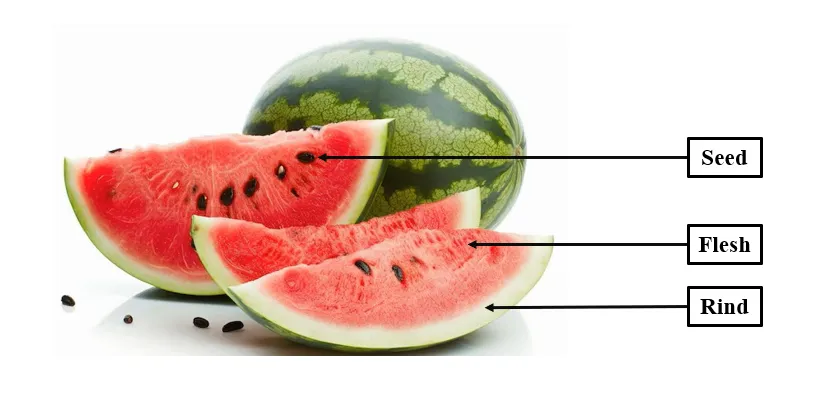
Fig 1: Watermelon Seed
Watermelon (Citrullus lanatus) seeds used for this study were obtained fresh from retailed fruit sellers from Barasat Market, north 24 pargana, West Bengal state.
|
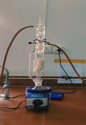
Figure 2: Extraction process |
The extraction process was continued for 6–8 hours. After completion of petroleum ether extraction, the marc (residue) was removed, air-dried to eliminate residual solvent, and subjected to further extraction with chloroform. About 500 mL of chloroform was used, and extraction was continued for 6–8 hours under similar conditions. The residue obtained after chloroform extraction was again dried and subsequently extracted with 500 mL of ethanol using the Soxhlet apparatus for 8–10 hours. Ethanol extraction enabled the isolation of polar compounds such as phenolics, flavonoids, glycosides, tannins, and other bioactive constituents. The obtained petroleum ether, chloroform, and ethanol extracts were separately concentrated under reduced pressure using a rotary evaporator and further dried on a water bath to obtain semisolid masses [9]. The dried extracts were weighed to determine percentage yield and stored in airtight containers at 4°C for subsequent phytochemical and pharmacological studies. The extraction process is shown in figure no 2.
Phytochemical analysis of the extracts were carried qualitatively laboratory techniques. Tests on the presence of alkaloid, flavonoids, glycosides, saponins and tannin were conducted accordingly [10].
Figure 3
Figure 4
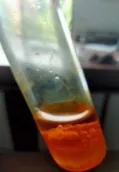
Figure 5
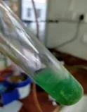
Figure 6
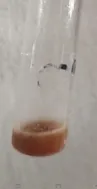
Figure 7

Figure 8
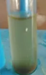
Figure 9
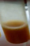
Figure 10
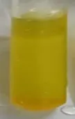
Figure 11
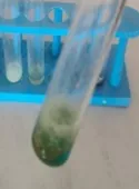
Figure 12
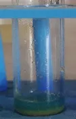
Figure 13
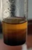
Figure 14
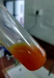
Figure 15
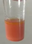
Figure 16

Figure 17
The Antibacterial susceptibility test was performed by agar-well diffusion method. The antimicrobial activity of the Citrullus lanatus seed extracts were tested against 1 microorganisms gram negative bacteria: - Escherichia coli.
Bacterial slants were prepared using Mueller–Hinton agar (MHA) by dissolving the required quantity of medium in distilled water and heating until completely melted. The prepared medium was then dispensed into clean, dry test tubes, which were plugged with cotton and sterilized by autoclaving at 121°C for 15 minutes. After sterilization, the tubes were placed in an inclined position to allow the medium to solidify as a slant, providing a larger surface area for microbial growth. Once solidified, the slants were aseptically inoculated with the desired bacterial culture using a sterile inoculating loop and incubated at 35–37°C for 18–24 hours. The prepared bacterial slants were then stored at refrigerated conditions for further experimental use.
Bacterial culture plates were created using Mueller–Hinton agar (MHA) by mixing the necessary amount of the agar medium with distilled water in a conical flask and heating it until fully dissolved. The flask was then sealed with cotton, covered in aluminum foil, and sterilized through autoclaving at 121°C for 15 minutes. After the sterilization process, the molten agar was allowed to cool to approximately 45–50°C to avoid condensation and contamination. In aseptic conditions, the sterile medium was poured into sterile Petri dishes and permitted to solidify at room temperature. Then solidified the petri dishes. The prepared bacterial plates were then stored at refrigerated conditions for further experimental use.
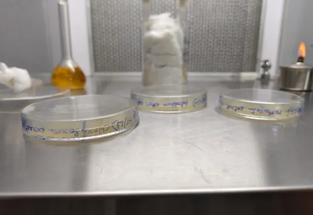
Figure 18: Bacteria plate preparation
In this we have to prepare the Citrullus lanatus seed extracts solution in 3 different concentration (500, 1000, 1500 µg/mL). Citrullus lanatus seed extracts were first prepared as sample solutions by dissolving 10 mg of each dried in 10 mL of dimethyl sulfoxide (DMSO) to obtain a concentration of 1 mg/mL. Then measure 5 mg of extract and dissolved in 10 ml of DMSO solution to obtained the concentration 0.5 mg/mL. Again measure 15 mg of the extract and dissolved in 10 ml of DMSO to obtained the concentration 1.5 mg/ml.
Amoxycillin is used as a standard in this test. Firstly, we have to weight 10 mg of amoxycillin and dissolved in 10 ml of dimethyl sulfoxide (DMSO) to obtain a concentration of 1 mg/mL (1000 µg/mL) (Stock solution). Then take 1 ml from the stock solution and dissolved in 10 ml of dimethyl sulfoxide (DMSO) to obtain a concentration of 100 µg/mL.
The transfer of bacteria onto agar plates was carried out under strict aseptic conditions to prevent contamination. A sterile inoculating loop was first flamed until red hot and allowed to cool. A small amount of the bacterial culture was then picked from a previously prepared slant. The sterile Mueller–Hinton agar (MHA) plates were slightly opened near a flame or inside a laminar airflow chamber, and the bacterial culture was gently transferred onto the surface of the agar. For uniform distribution, the inoculum was spread evenly over the surface using a sterile cotton swab (spread plate method) or streaked using an inoculating loop (streak plate method), depending on the experimental requirement. Care was taken to cover the entire surface to obtain a uniform lawn of bacterial growth, especially for antimicrobial studies [11].
In this process we have to add the sample solution (Citrullus lanatus seed extracts) and the standard solution (Amoxycillin) in to the plates, then the agar plates were allowed to stand for a few minutes to ensure proper absorption of the inoculum into the medium. The inoculated plates were incubated in an incubator at a temperature of 35–37°C for 18–24 hours, depending on the growth requirements of the bacterial species. During incubation, proper environmental conditions such as temperature and humidity were maintained to promote optimal bacterial growth. After the incubation period, the plates were observed for bacterial growth, colony formation, or zones of inhibition in case of antimicrobial activity testing. The antimicrobial activity of the Citrullus lanatus seed extracts was shown in the table no 3.
3.1 Extraction value result: -
In the process of extraction of the Citrullus lanatus seed powder with three solvents (Petroleum ether, Chloroform and ethanol). The extraction yield result shown in the Table no 1. By this table we have to conclude that the ethanolic extract has show better percentage of Yield.
Table no 1: - Extraction value of Citrullus lanatus seed
|
Sr. No. |
Solvent |
Colour |
Consistency |
% Yield w/w |
|
|
Petroleum ether |
White |
Greasy |
1.417 |
|
|
Chloroform |
Yellowish white |
Greasy |
2.032 |
|
|
Ethanol |
Yellowish dark white |
Greasy |
3.529 |
The phytochemical analysis revealed that the seeds of Helianthus annuus extract contain carbohydrates, alkaloids, flavonoids, tannins, saponins, phytosterol, steroids, and fixed oils and fats [Igbinosa et al., 2009]. The diverse phytochemical compounds found in the seed extract, including flavonoids, alkaloids, and saponins, have been documented to exhibit biological activity against microorganisms [Sidambaram et al., 2011]. The phytochemical tests results shown in the table no 2.
Table no 2: - Phytochemical test result of Citrullus lanatus seed
|
Test for |
Methanol |
Chloroform |
Petroleum ether |
|
Alkaloids |
(+) |
(+) |
(+) |
|
Carbohydrate |
(+) |
(+) |
(+) |
|
Flavonoids |
(+) |
(+) |
(+) |
|
Tannins |
(-) |
(-) |
(-) |
|
Terpenoids |
(-) |
(-) |
(-) |
|
Glycosides |
(-) |
(-) |
(-) |
|
Amino acids |
(+) |
(+) |
(+) |
By evaluating the zone of inhibition at various doses, the antibacterial activity of the standard and solvent extracts against Escherichia coli was assessed (Table 3). At a dose of 100 µl, the standard medication demonstrated good antibacterial activity, producing inhibitory zones of 20 mm (CH), 22 mm (PE), and 25 mm (ME). The activity of the chloroform extract increased in a concentration-dependent manner. The inhibitory zone was 12 mm at 500 µg/ml, 18 mm at 1000 µg/ml, and 26 mm at 1500 µg/ml. The chloroform extract's maximum activity was marginally higher than that of the conventional methanolic control. Also, the antibacterial activity of the petroleum ether extract increased with concentration. Out of all the extracts examined, the zones of inhibition were the strongest, measuring 14 mm at 500 µg/ml, 19 mm at 1000 µg/ml, and 28 mm at 1500 µg/ml. With inhibitory zones of 15 mm, 17 mm, and 25 mm at 500, 1000, and 1500 µg/ml, respectively, the methanol extract also showed moderate to high antibacterial activity. The petroleum ether extract at 1500 µg/ml showed the greatest zone of inhibition (28 mm), but all extracts showed dose-dependent antibacterial activity against E. coli.
Table 3: - Antimicrobial activity result
|
Bacteria used in zone of inhibition |
Standard Conc (mg/ml)
|
Chloroform Conc (mg/ml) |
Petroleum ether Conc (mg/ml) |
Methanol Conc(mg/ml) |
|||||||||
|
Dose (µl) |
0.1 |
0.1 |
0.1 |
0.5 |
1 |
1.5 |
0.5 |
1 |
1.5 |
0.5 |
1 |
1.5 |
|
|
Escherichia coli |
20 mm |
22 mm |
25 mm |
12 mm |
18 mm |
26 mm |
14 mm |
19 mm |
28 mm |
15 mm |
17 mm |
25 mm |
|
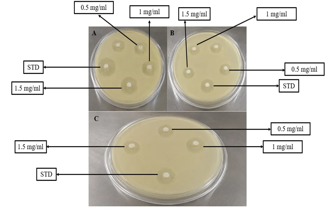
Fig 19: Antimicrobial activity result of Citrullus lanatus seed extracts
CONCLUSION
The current research indicated that the seeds of Citrullus lanatus (watermelon) contain notable phytochemical components and exhibit considerable antibacterial properties, underscoring their potential as a valuable natural source of bioactive substances. Utilizing sequential Soxhlet extraction with petroleum ether, chloroform, and methanol, it was found that the methanolic extract yielded the highest extraction percentage, suggesting a more effective recovery of polar phytochemicals. Qualitative phytochemical analysis confirmed the presence of alkaloids, carbohydrates, flavonoids, and amino acids across all extracts, while tannins, terpenoids, and glycosides were not detected. These phytoconstituents are recognized for contributing to a range of biological activities, including antimicrobial and antioxidant effects, thereby reinforcing the medicinal significance of watermelon seeds. The antibacterial assessment against Escherichia coli revealed concentration-dependent effectiveness for all extracts tested. Among the extracts evaluated, the petroleum ether extract demonstrated the strongest antibacterial properties, achieving a maximum zone of inhibition of 28 mm at a concentration of 1500 µg/mL, closely followed by the chloroform and methanolic extracts. These results suggest that watermelon seeds, which are typically regarded as agricultural waste, can be an effective source of natural antimicrobial substances. In summary, the research emphasizes the potential pharmaceutical and nutraceutical uses of Citrullus lanatus seeds. Nonetheless, further studies focusing on the isolation and characterization of the active compounds, toxicity assessments, and in vivo pharmacological investigations are advised to confirm their safety, effectiveness, and therapeutic potential for upcoming clinical and industrial uses.
REFERENCES
Rezagholizade-Shirvan A, Kalantarmahdavi M, Amiryousefi MR. Evaluation of the effect of basil seed gum, tragacanth gum, pectin, and coating formulation with corn flour on oil absorption and sensory properties of watermelon rind chips. Heliyon. 2023 Jun 1;9(6). doi:10.1016/j.heliyon.2023.e17177.





Mriganka Karmakar, Sanchita Poddar, Debasmita Ghosh, Probal Dhara, Nilay Ganguly,Gargi Seth, `Phytochemical and antibacterial study of Citrullus lanatus Seed extract, Int. J. of Pharm. Sci., 2026, Vol 4, Issue 7, 6085-6096, https://doi.org/10.5281/zenodo.21721700
 10.5281/zenodo.21721700