

We use cookies to ensure our website works properly and to personalise your experience. Cookies policy
1,2 Department Of Pharmaceutical And Medicinal Chemistry, Faculty Of Pharmacy, Niger Delta University, Wilberforce Island, Bayelsa State, Nigeria.
3department Of Haematology And Blood Transfusion Science, College Of Basic Clinical Sciences, Niger Delta University, Wilberforce Island, Bayelsa State, Nigeria.
4department Of Medical Microbiology & Parasitology, Faculty Of Basic Clinical Sciences, Niger Delta University, Wilberforce Island, Bayelsa State, Nigeria
The integration of phytomedicines into modern pharmacotherapy represents a critical frontier in drug discovery, offering diverse bioactive compounds with complex mechanisms of action for disease management. Brillantaisia owariensis is widely utilized in traditional medicines to manage of sickle cell disease, however, scientific validation of this ethnobotanical claim remains scarce. This study evaluated the antisickling potential of Brillantaisia owariensis leaf extracts through phytochemical analyses, alongside antimicrobial assays. The investigation was based on the premise that specific bioactive constituents can inhibit erythrocyte sickling and that certain microbial pathogens contribute to vaso-occlusive crises in sickle cell patients. Qualitative phytochemical screening confirmed the presence of phenolics, cardiac glycosides, saponins, steroids, and gums. Quantitative analysis showed that the plant extract contained phenolics (95.8 ± 0.0015mg/g), flavonoids (122.2 ± 0.0026mg/g) and cardiac glycosides (163.8 ± 0.0031mg/g) for the ethanolic extract and (59.6 ± 0.001mg/g), (43.6 ± 0.002mg/g), and (118 ± 0.0025mg/g) respctively for the n-hexane extract. Both extracts exhibited robust antimicrobial activity, producing zones of inhibition equivalent to 60 –70% of ciprofloxacin control, and exhibited activity against typed bacterial strains (S. aureus NCTC8532, B. subtilis NCTC10400, P. aeruginosa NCTC10662, E. coli NCTC10418) as well as clinical microbial isolates of S. aureus, E. coli, and P. aeruginosa. Statistical analysis showed a significant difference (p < 0.05) between the extracts, establishing the superior activity of the ethanolic extract over the n-hexane extract. These findings provide compelling scientific evidence supporting the traditional use of Brillantaisia owariensis in the management of sickle cell disease and warrants further investigation.
Phytomedicine, known as the science and practice of using plant-derived therapeutics for the prevention and management of diseases, has re-emerged as a vital component of modern healthcare, rooted in centuries of ethnobotanical knowledge and now validated by rigorous pharmacological research. Phytomedicine bridges traditional wisdom and contemporary biomedical science1. In today’s therapeutic landscape, phytomedicine functions as a complementary approach to conventional medicine, It addresses critical gaps in healthcare delivery, particularly in chronic disease management, palliative care, and resource-limited settings where access to synthetic pharmaceuticals may be constrained2. Plant-based formulations often exhibit multi-target mechanisms, acting on several physiological pathways simultaneously, which makes them valuable in complex conditions such as diabetes, hypertension, inflammatory disorders, and antimicrobial resistance etc3,4. Recognized by the World Health Organization as a pillar of primary healthcare for over 80% of populations in developing countries, phytomedicine is no longer viewed as “alternative” but as integrative5. Consequently, it is regarded as an integral component of integrative medicine6.
Brillantaisia owariensis
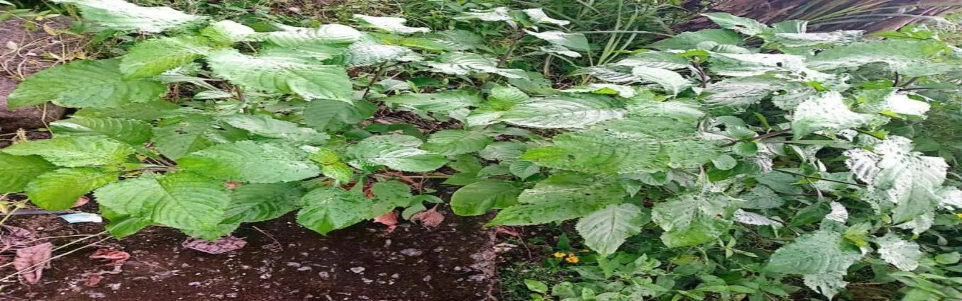
Fig.1 leaves of Brillantaisia owariensis P. Beauv. Family (Acanthaceae).
Other names: giant blue African salvia, Tropical giant salvia
Local names;
Abureni: Ozuzu-oghogho, (Abua/Odual)
Ogbia: Obom-ato , ogbua-emu (Bayelsa)
Brillantaisia owariensis P. Beauv. is a perennial shrub native to tropical Africa, with a distribution spanning Central and West Africa, It typically grows in moist areas, along river banks and in coastal areas reaching heights of up to 2-8 meters. It holds a prominent place in indigenous pharmacopeias, particularly in regions such as the Democratic Republic of Congo, Nigeria, and Angola, where it is traditionally valued as a blood-tonic for managing anaemia and related complications7-9. Phytochemical analyses of B. owariensis have unvailed diverse array of bioactive compounds. Among these are iridolactones including the novel compounds owariensisone B, C and D isolated by Foning10 as well as flavonoids, phenolics, saponins and cadiac glycoside11.
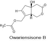
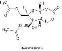
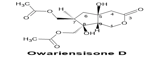
In this context, the present study aims to evaluate the preliminary antisickling properties of Brillantaisia owariensis through phytochemical screening and assessment of its antimicrobial activity. Previous studies have demonstrated that specific phytochemical constituents contribute significantly to antisickling effects. For instance, cardiac glycosides and alkaloids have been reported to regulate ionic balance, particularly through modulation of Na+/K+ pumps, thereby preventing cellular dehydration that promotes hemoglobin S (HbS) polymerization12. Phenolic compounds exhibit potent antioxidant properties, mitigating oxidative stress a key factor in erythrocyte dehydration and subsequent sickling13. Coumarins and anthocyanins possess anti-inflammatory and vasoprotective properties that may reduce the adhesion of sickled erythrocytes to vascular endothelium, thereby decreasing the frequency of vaso-occlusive crises14. Saponins contribute to membrane stabilization by enhancing the structural integrity of red blood cell membranes, reducing osmotic fragility, and preventing premature hemolysis15. Additionally, anthocyanins and flavonoids have been reported to inhibit the polymerization of deoxygenated HbS by increasing oxygen affinity and stabilizing the oxyhemoglobin state16. Complementarily, some phytochemicals have been reported to demonstrate antimicrobial properties these includes; phenolics, known to complex with bacterial cell walls and disrupt microbial membranes; alkaloids, are reported to correlate with the inactivation of microbial adhesions, enzymes, and cell proteins; and terpenoids, which facilitate the penetration of the lipophilic cell wall of microorganisms17,3. Therefore, the presence of these phytochemicals in B. owariensis would serve as a biochemical indicator of its antisickling potential and provide justification for the claim made by locals on its use to manage sickle cell disease complications and anemia.
MATERIAL AND METHOD
Materials
Typed organisms Staphylococcus aureus (NCTC 8532), Bacillus subtilis (NCTC 10400), Pseudomonas aeruginosa (NCTC 10662), and Escherichia coli (NCTC 10418) ). clinicals isolates of S. aureus, E. coli, and P. aeruginosa. Ethanol (BDH Chmicals LTD), n-Hexane (LiChrosolv, Germany), Distilled water, Mercuric (II) Chloride, Potassium iodide (BDH Chemicals LTD England), Hydrochloric acid (LOBA Chemie Mumbai, India), Ferric chloride (BDH Chemical Ltd. England), Ammonia solution (LOBA Chemie Mumbai, India), Benzene (LOBA Chemie Mumbai, India), Sodium hydroxide (LOBA Chemie Mumbai, India), Chloroform(Molychem, India), Sulphuric acid(LOBA Chemie Mumbai, India), Gacial acetic acid (LOBA Chemie Mumbai, India). ( all reagents used in this study were not further purified)
Methodology
Plant Collection and Identification
Fresh leaves of Brillantaisia owariensis were harvested from Otuasega community, Ogbia Local Government Area, Bayelsa State, Nigeria (Latitude 4.92520°N, Longitude 6.40310°E) in August, 2025 and identified by a botanist of the Department of Biological Sciences, Niger Delta University, Dr. Ayo Oyedeji as Brillantaisia owariensis P. beauv. A voucher specimen of the plant was deposited in the university’s herbarium with number (NDUP/0166) for further reference.
Extraction of the plant material
Air-dried and powdered plant material (300 g X 2) was extracted with Ethanol and n-hexane (3L) respectively by maceration process. The extracts were concentrated using a water bath at low temperature (40-50 °C) and dried to a constant weight in a desiccator and the percentage yield determined.
Qualitative phytochemical screening
The freshly prepared plant extracts were qualitatively tested for the presence of phenolics (Ferric chloride test), tannins (Ferric chloride test), phlobatannins (Hydrochloric acid test), flavonoids (Shinoda test), anthocyanins (pH-dependent colour reaction test), terpenoids (Salkowski test), saponons (Frothing test), cardiac glycosides (Keller-Kiliani test), coumarins (sodium hydroxide test), alkaloids (wagners reagent), gums and fixed oils (Precipitation test) using standard colorimetric and precipitation protocols described by Harborne, Trease & Evans and Ismail et al.,18-20 .
Quantitative Phytochemical Determination
Total Phenol Content
Principle: Phenolic compounds react with phosphomolybdic acid in the Folin–Ciocalteu to form a blue-colored molybdenum complex under alkaline condition.
The total phenolic content (TPC) of the extracts was quantified using the Folin–Ciocalteu colorimetric method, based on modified procedures reported by Singleton et al., and Demiray et al.,21-22. A standard calibration curve was established using gallic acid solutions prepared at concentrations between 100 and 1000 µg/mL. For the standards, Folin–Ciocalteu reagent 1.5 mL was added to each solution and allowed to react at room temperature for 5 minutes, then 7.5% (w/v) sodium carbonate solution 1.2 mL was added to neutralize the reaction mixture. The resulting solutions were incubated at ambient temperature, after which absorbance was recorded at 760 nm using a UV–Visible Spectrophotometer. In the case of the test samples, 0.3 mL of each crude extract (5 mg/mL) was subjected to the same procedure. Total Phenolic content was expressed as gallic acid equivalent (mg/g) using the following equation Y = 0.001X + 0.0013, R2 = 0.996 where, y was the absorbance and x was the concentration
Total Flavonoid Content
The total flavonoid content of the extracts were determined using a colorimetric procedure modified from the method described by Zhishen and co-workers23. Where Quercetin was employed as the reference compound, and a series of standard solutions (100, 200, 400, 600, 800, and 1000 µg/mL) were prepared. For each determination, Quercetin solutions 0.3 mL was mixed with 5% sodium nitrite (NaNO₂) solution 0.06 mL and left to react at ambient temperature for 5 minutes. Thereafter, 10% aluminum chloride (AlCl₃) solution 0.06 mL was introduced into the mixture, this was followed by the addition of 0.1 M sodium hydroxide (NaOH) 0.4 mL and finally distilled water 0.5 mL was added to complete the reaction volume. The reaction mixtures were allowed to stand for 30 minutes to ensure full color development, after which the absorbance was measured at a wavelength of 510 nm using a spectrophotometer. For the crude extracts, 0.3 mL of the respective plant extracts (5 mg/mL) solution were treated identically. The flavonoid content was expressed as quercetin equivalent (mg/g) using the following equation Y = 0.001X + 0.015, R2 = 0.999 where, y was the absorbance and x was the concentration
Cardiac Glycoside Content
Cardiac glycoside content was determined using digoxin as the reference standard. A series of digoxin solutions at concentrations of 100, 200, 400, 600, 800, and 1000 µg/mL were prepared. To each of the standard solution was added freshly prepared Baljet’s reagent 10 mL (comprising 1% picric acid and 10% sodium hydroxide) and allowed to stand for 1 hour to ensure complete color development. The reaction mixture was subsequently diluted with distilled water 20 mL and the absorbance was measured at 495 nm using a UV–Visible spectrophotometer. 10 mL of the plant extract (5 mg/mL) was substituted inplace of digoxin standard and similar treatment as above was followed. the same procedure. Cardiac glycoside content was expressed as digoxin equivalent (mg/g) using the following equation Y = 0.001X - 0.002 , R2 = 0.996 where, y was the absorbance and x was the concentration c is the concentration
Anti-microbial Assay
The antimicrobial assay was carried out in the Nigerian Army 16 Division laboratory, Yenagoa, Bayelsa State. The test organisms included typed microbial strains (S. aureus NCTC8532, B. subtilis NCTC1040, P. aeruginosa NCTC10662, and E. coli NCTC10418) and clinical isolate microbial strains (S. aureus, E. coli, and P. aeruginosa). Antimicrobial susceptibility testing was conducted using strict aseptic techniques throughout the procedure. Both typed strains and clinical bacterial isolates were first subcultured in sterile nutrient broth and incubated at 37°C for 24 hours to obtain active growth. The resulting cultures were standardized by adjusting the turbidity with sterile normal saline to match the 0.5 McFarland standard, corresponding to approximately 1.0 × 10⁸ CFU/mL, in accordance with the recommendations of the Clinical and Laboratory Standards Institute (CLSI, 2025). The agar well diffusion technique, as outlined by CLSI24, was employed for the antimicrobial assay. Each experiment was conducted in triplicate. After inoculation and introduction of the test samples into the wells, the plates were left undisturbed at room temperature for 30 minutes to permit adequate diffusion of the extracts into the agar medium. Subsequently, the plates were incubated at 37°C for 24 hours, following the method previously reported by Esimone et al.25. At the end of the incubation period, the diameters of the inhibition zones were carefully measured in millimeters using a transparent, millimeter-calibrated ruler.
Minimum Inhibitory Concentration (MIC):
The minimum inhibitory concentration (MIC) of the extracts was determined in accordance with the methods described by El-Mahmood et al26 and Vinothkumar et al.27, with slight modifications. Briefly, a series of two-fold serial dilutions of each extract was prepared to obtain concentrations of 500, 250, 125, 62.5, 31.25 and 15.625µg/mL (10µL). Each prepared dilution was inoculated with the respective test bacterial strain under aseptic conditions. The inoculated tubes were then incubated at 37°C for 18 hours. Following incubation, the MIC was defined as the lowest concentration of the extract at which no visible turbidity or microbial growth was observed.
Minimum Bactericidal Concentration (MBC):
For the determination of the minimum bactericidal concentration (MBC), equal volumes of broth cultures that showed no visible growth during the MIC assay were selected. These samples were aseptically transferred and streaked onto sterile Mueller–Hinton agar plates in triplicate. The plates were then incubated at 37°C for 18 hours. The MBC was defined as the lowest concentration of the extract at which no bacterial colony formation was observed on the agar surface, in accordance with the procedure described by Mann et al.28.
Statistical analysis
Student’s t-test was used to ascertain the significance of differences between mean values of the two extracts and the level p < 0.05 was considered as the cutoff value or significance. Detailed statistical analyses of the antimicrobial assay data and the susceptibility profiles of all test microorganisms are provided in the Appendix.
RESULTS
The qualitative phytochemical screening of the leaf extract of B. owariensis revealed the prescence of various bioactive compounds as summarized in Table 1. below
Table 1: Qualitative phytochemical screening of the leaf extracts of B. owariensis leaves
|
PHYTOCHEMICALS |
INFERENCE |
|
|
70%-ETHANOL EXTRACT |
|
|
|
Tannins |
++ |
- |
|
Terpenoids |
- |
++ |
|
Coumarins
|
- |
+ |
|
Saponin |
+++ |
- |
|
Alkaloids |
++ |
++ |
|
phlobatannins |
++ |
- |
|
Flavonoids |
+++ |
++ |
|
Cardiac glycosides |
+++ |
++ |
|
Anthocyanins |
+++ |
- |
|
Gums and muscilage |
+++ |
- |
|
Fixed oil |
- |
+++ |
|
Phenolics |
++ |
+ |
KEY; - = Not present + = Present in little quantity
++ = Present in high quantity +++ = Present in large quantity
The quantitative phytochemical analysis of B. owariensis leaf extracts presented in Fig. 2 below revealed varying concentration of cardiac glycoside, phenol and flavonoids present.
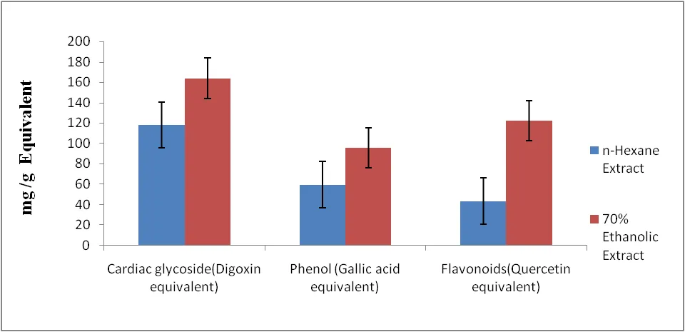
Fig 1: Quantitative phytochemicals screening of the leaf extracts of B. owariensis
The Zone of inbhibition demonstrated by B. owariensis extracts, presented in Table 2. reveals its potential antimicrobial activity against typed and clinical microbial strains.
Table 2. Anti-microbial activity of the leaf extract of B. owariensis
|
Microorganisms |
Mean Zones of Inhibition (mm) and SD |
|||
|
E Extract Mean±SD |
n-Hex Extract Mean±SD |
Cip (5µg/mL) |
Control |
|
|
S. aureus NCTC8532 |
24.5±0.6 |
22.3±0.6 |
35.5±1.2 |
0 |
|
B. subtilis NCTC10400 |
29.7±0.6 |
18.6±0.6 |
35.5±1.2 |
0 |
|
P. aeruginosa NCTC10662 |
15.3±1.2 |
26.3± 1,5 |
35.5±1.2 |
0 |
|
E.coli NCTC10418 |
31.3±0.6 |
30.0±1.0 |
35.5±1.2 |
0 |
|
Clinical Isolates S. aureus E.coli P. aeruginosa |
20.2±0.6 15.3±0.6 23.3±0.6 |
20.2±0.6 15.3±0.6 23.3±0.6
|
R R |
0 0 0 |
Keys E: Ethanolic Extract, n-Hex: n-hexane Extract, CIP: Ciprofloxacin; R: Resistant
The MIC and MBC of the ethanolic extract of B. owariensis presented in Table 3. indicates its potency against various microorganisms.
Table 3. Minimum inhibitory concentration (MIC) and minimum bactericidal concentration (MBC) for the ethanolic extract of Brillantaisia owariensis
|
Microorganisms |
Ethanolic extract (µg/mL) |
|||||||
|
500 |
250 |
125 |
62.5 |
31.25 |
15.625 |
MIC |
MBC |
|
|
S. aureus NCTC8532 |
- |
- |
- |
- |
+ |
+ |
62.5 |
125 |
|
B. subtilis NCTC10400 |
- |
- |
- |
- |
+ |
+ |
62.5 |
125 |
|
P. aeruginosa NCTC10662 |
- |
- |
+ |
+ |
+ |
+ |
62.5 |
125 |
|
E.coli NCTC10418 |
- |
- |
- |
- |
- |
+ |
31.25 |
62.5 |
|
Clinical Isolates S. aureus E.coli P. aeruginosa |
- - - |
- - - |
- - + |
- - + |
+ - + |
+ + + |
62.5 31.25 62.5 |
125 62.5 125 |
Key:
S. aureus = Staphylococcus aureus. B. subtilis = Bacillus subtilis
P. aeruginosa = Pseudomonas aeruginosa. E. coli = Escherichia coli
+ = Microbial growth
-= No microbial growth
The MIC and MBC of the n-Hexane extract of B. owariensis presented in Table 4. indicates its potency against various microorganisms.
Table 4. Minimum inhibitory concentration (MIC) and minimum bactericidal concentration (MBC) of the n-Hexane extract of Brillantaisia owariensis
|
Microorganisms |
n-Hexane (µg/mL) |
|||||||
|
500 |
250 |
125 |
62.5 |
31.25 |
15.625 |
MIC |
MBC |
|
|
S. aureus NCTC8532 |
- |
- |
- |
+ |
+ |
+ |
125 |
125 |
|
B. subtilis NCTC10400 |
- |
- |
- |
+ |
+ |
+ |
125 |
250 |
|
P. aeruginosa NCTC10662 |
- |
- |
- |
+ |
+ |
+ |
125 |
125 |
|
E.coli NCTC10418 |
- |
- |
- |
- |
+ |
+ |
62.5 |
125 |
|
Clinical Isolates S. aureus E.coli P. aeruginosa |
- - - |
- - - |
- - - |
- - + |
+ - + |
+ + + |
62.5 31.25 62.5 |
125 62.5 125 |
Key: S. aureus = Staphylococcus aureus. B. subtilis = Bacillus subtilis
P.aeruginosa = Pseudomonas aeruginosa. E. coli = Escherichia coli
+ = Microbial growth - = No microbial growth
DISCUSSION
The qualitative phytochemical screening result depicted in Table 1, revealed that the plant extracts possess a diverse array of bioactive constituents, including phenolics, saponins, cardiac glycosides, steroids, and gums. These phytochemicals have been widely reported to play significant roles in the management of sickle cell disease (SCD). Specifically, phenolic compounds have been reported to exert their effects through multiple mechanisms, including antioxidant activity, inhibition of hemoglobin S (HbS) polymerization, stabilization of red blood cell (RBC) membranes, anti-inflammatory effects, and enhancement of blood flow. Collectively, these actions contribute to the preservation of erythrocyte integrity, reduction in vaso-occlusive episodes, and prolongation of RBC lifespan as reported by Ochwang’i et al.,13. Saponins have also been reported to reduce osmotic fragility of erythrocytes, exhibit anti-inflammatory properties, and modulate pain pathways via opioid receptors and calcium ion (Ca²⁺) regulation, thereby potentially alleviating chronic pain crises associated with SCD as highlighted by Kouakou et al.,15. Steroidal compounds contribute to erythrocyte membrane stabilization and possess anti-inflammatory effects, which may help reduce pain episodes and prevent cellular damage29. Additionally, gums are believed to form a protective coating on the RBC surface, thereby shielding the cells from mechanical fragility and oxidative stress, ultimately preventing hemolysis as reported by Chikezie,30. the prescence of these phytochemicals suggest that the plant may possess notable anti-inflammatory activity, inhibit hemoglobin S (HbS) polymerization, stabilize red blood cell (RBC) membranes, and enhance blood flow. The quantitative phytochemical analysis result shown in Table 2 further supports the antisickling potential of the plant, as evidenced by the substantial levels of cardiac glycosides (163.4 ± 0.0031mg/g), phenols (98.6 ± 0.0015mg/g), and flavonoids (125.2 ± 0.0026mg/g) in the polar extract and (118 ± 0.00252mg/g), (62.4 ± 0.001mg/g), and (46.6 ± 0.0026 mg/g) respectively for the n-hexane extract. The phytochemical profile is indicative of strong therapeutic potential, particularly as high phenolic and flavonoid contents are known to confer antioxidant protection, stabilize erythrocyte membranes, and reduce hemolysis. Cardiac glycosides may further contribute by modulating ion transport mechanisms, thereby preventing RBC dehydration and delaying HbS polymerization. The combined presence of these compounds suggests that the plant extract may effectively reduce sickling, protect erythrocytes from lysis, and mitigate inflammatory processes as reported by Ochwang’i et al.,13. Furthermore, The antimicrobial evaluation results (Table 2 - 4) demonstrated that the leaf extracts possess broad-spectrum antibacterial activity, the extracts successfully inhibited the growth of several reference typed strains, including Staphylococcus aureus NCTC8532, Bacillus subtilis NCTC10400, Pseudomonas aeruginosa NCTC10662, and Escherichia coli NCTC10418, achieving a zone of inhibition equivalent to 60 -70% of that produced by the reference antibiotic, ciprofloxacin. Crucially, the extracts also exhibited potent efficacy against Ciprofloxacin-resistant clinical isolates of S. aureus, E. coli, and P. aeruginosa. This broad-spectrum anti-microbial potential highlights the therapeutic relevance of B. owariensis, in sickle cell disease management, particurlarly in preventing infection-triggered vaso-occlusive crises. positioning it as a promising candidate for anti-sickling therapy capable of simultaneously managing secondary bacterial infections in sickle cell disease (SCD) patients. Infections are leading cause of morbidity and mortality in SCD patients, often exacerbated by functional asplenia and frequent hospitalization31. By inhibiting key pathogens implicated in osteomyelitis, sepsis, and pulmonary infections, the extracts may mitigate infection-induced inflammation, fever, and hypoxia factors known to precipitate sickling events32. Overall, these findings provide scientific validation for the traditional use of B. owariensis in mitigating SCD complications. The dual presence of bioactive-antisickling phytochemicals and potent antimicrobial properties positions this plant as a promising therapeutic candidate for sickle cell therapy.
CONCLUSION
The findings of this study demonstrated that Brillantaisia owariensis possesses a broad spectrum of bioactive compounds, which includes; phenolics, terpenoids, steroids, alkaloids, coumarins, gums, and lipids. Quantitative analysis further revealed high concentrations of flavonoids, cardiac glycosides, and phenolics, classes of compounds widely associated with antisickling activity. The plant extracts exhibited broad-spectrum antimicrobial activity against pathogens implicated in causing infection triggered crises in SCD, and also efficacious against Ciprofloxacin-resistant microbial strains. these findings provide scientific validation for the traditional use of B. owariensis in the management of sickle cell–related complications and highlights its potential for further pharmacological development.
Ethics approval and consent to participate
Human participants or animal model were not used in the study by any of the authors.
All authors have read and approved the manuscript and grant their formal consent for its publication in the pharmaceutical journal.
All datasets generated or analyzed during the current study are fully included within this published article.
The authors declare that they have no known competing financial interests or personal relationships that could have appeared to influence the work reported in this paper.
This research did not receive any specific grant from funding agencies in the public, commercial or private sector.
Authors’ contributions
Contributor Roles Taxonomy (CRediT)
Ere, Diepreye: Conceptualization, Supervision, Review And Editing, Visualization
Abdulrasheed B. Abdu: Investigation, Formal Analysis.
David T. Tamarabrakemi: Formal Analysis, Methodology
K E. Edoghotu: Investigation, Writing, Data Curation, Software, Visualization.
ACKNOWLEDGEMENTS
The authors gratefully acknowledge Dr. Rauf Raji of the Department of Pharmaceutical and Medicinal Chemistry, Faculty of Pharmacy, NDU, for his valuable laboratory assistance during this investigation.
REFERENCES




Evans Edoghotu*, Diepreye Ere, Torugbene Tamarabrakemi, Abdu Abdulrasheed, Preliminary Screening Of The Antisickling Properties Of Brillantaisia Owariensis Leaf Extracts, Int. J. of Pharm. Sci., 2026, Vol 4, Issue 7, 5189-5201. https://doi.org/10.5281/zenodo.21624127
 10.5281/zenodo.21624127