
View Article

Abstract

Smart drug delivery systems (SDDS) have attracted growing interest in pharmaceutical research because they offer new ways of carrying drugs to, and releasing them at, the intended site in the body. Unlike conventional dosage forms, these systems can be engineered to respond to conditions inside the body or to externally applied triggers, including changes in pH, redox potential, enzyme activity, temperature, light, magnetic fields, ultrasound, and glucose concentration. This review outlines the principles behind SDDS, the main carrier platforms used to build them, their therapeutic applications, and the obstacles that limit wider clinical use. The platforms covered are liposomes, polymeric nanoparticles, exosomes, hydrogels, dendrimers, microneedles, and polymeric micelles; for each, the key properties and drug-release behavior are discussed. Applications in cancer, infections, diabetes, and inflammatory disease are also considered. Despite their advantages, SDDS still face substantial challenges: biological barriers, toxicity to healthy cells, insufficient accumulation at the target site, unstable formulations, difficulty in reproducible manufacturing, and unresolved regulatory questions. If these obstacles can be overcome, SDDS holds considerable promise for improving how drugs are delivered.

Keywords

Smart drug delivery systems, stimuli-responsive nanoplatforms, targeted drug delivery, controlled drug release, nanocarriers, precision medicine, therapeutic applications

Introduction

× Popup Image

Drug delivery covers the approaches, formulations, technologies and systems used to carry a therapeutic agent through the body so that it produces its intended effect safely and efficiently [1]. Delivering a drug to prevent or treat disease is, alongside surgery, radiotherapy, physical therapy and psychotherapy, one of the principal modes of medical treatment [2]. A smart drug delivery system (SDDS) is a delivery system that can sense signals, respond to them, release its drug load and stop releasing it again, largely without outside intervention [1]. The signals that regulate such systems may be external, such as light of different wavelengths, magnetic or electric fields and ultrasound [3], or internal, such as redox state, pH, enzyme activity and the concentration of particular molecules [4]. The central aim of an SDDS is to deliver the right dose to the intended site, at the right moment and in a predetermined manner, with high efficiency and specificity, so that patients are also better able to follow their treatment [5]. Nanocarriers used in SDDS include liposomes, nanospheres, quantum dots (QDs), dendrimers, carbon nanotubes (CNTs) and superparamagnetic iron-oxide nanoparticles [6].

NEED FOR SMART DRUG DELIVERY SYSTEMS:

SDDS are intended to deliver medicines in a controlled and safe way. Carriers such as nanoparticles, liposomes, and polymers can protect the drug, time its release, and direct it to a chosen site, which improves outcomes, lowers side effects, and can reduce the overall cost of treatment. Two early milestones are often cited: Tanaka’s 1978 study of the phase transition of polyacrylamide gels, which advanced research on the volume-phase change of polymer gels [7], and the first reports from the same period of thermosensitive liposomes for drug delivery [8].

A smart system should place more of the drug in the tissue, cell, or organ that needs it; remain intact in body fluids, improve therapeutic performance without added side effects; and allow the drug to be absorbed properly [9]. Drug delivery systems can also address problems such as poor dissolution, degradation by the environment or by enzymes, rapid clearance, toxicity to healthy cells, and difficulty in crossing the body’s barriers [10]. Ideally, the system should not react with the body’s own chemistry, should not provoke an immune response, should stay stable both in storage and in the body, should locate the right cells and keep binding to the correct targets, and should release the drug at the appropriate rate. It should also be cost-effective and biodegradable [11].

ADVANTAGES OF SMART DRUG DELIVERY SYSTEMS:

1. Enhanced therapeutic efficacy: Developing drug delivery approaches that can target specific areas of the body or cell types may improve the efficacy of the treatment. These nanoparticles can detect cancer cells, enabling the tumor to be targeted with higher doses of medication and reducing side effect [12].

2. Controlled drug release: Besides targeting, smart drug delivery strategies can guarantee that the drug is released at defined rates or in response to specific signals. That gives you control over when and how the drug is released [13].

3. Improved adherence: Information technology-based smart medicine delivery systems can be designed to enable the physician to remotely monitor a patient’s dosage [14].

LIMITATIONS OF SMART DRUG DELIVERY SYSTEMS:

1. Complexity of design: The design of pharmaceutical delivery systems requires in-depth knowledge of the behavior of drugs in the body, the progression of disease, and the interaction of pharmaceutical delivery systems with the body [15].

2. Drug loading capacity: Some of the smart drug delivery networks can only load a small amount of drugs. This limited capacity might impair the effectiveness of treatment. The drug release rate with time may also be affected [16].

3. Biocompatibility and toxicity concerns: The success of pharmaceutical delivery systems depends on their biocompatibility. These systems are made of materials that can be toxic to cells, such as nanoparticles, and can cause side effects [15, 17].

STIMULI-RESPONSIVE MECHANISMS IN SMART DRUG DELIVERY SYSTEMS:

1. pH-Responsive Systems:

pH is among the most widely used triggers for controlling drug release [18]. Conventional pH-sensitive carriers exploit the large pH differences between organs, for example between the stomach (about pH 2) and the intestine (about pH 7). One example is Eudragit S100-coated citrus pectin nanoparticles (E-CPNs), which were developed to deliver 5-fluorouracil (5-FU) to the colon [19]. More advanced carriers can detect smaller pH shifts at disease sites such as inflamed, ischaemic and tumour tissue, and in cellular compartments such as endosomes and lysosomes; pH-responsive nanocarriers for solid tumours are a well-known case [20].

Blood and healthy tissue normally have an extracellular pH close to 7.4, but because of heightened glycolytic activity the average extracellular pH of many solid tumours is below 7.0 [21]. This acidity can serve as a specific signal for targeted delivery. Inside cells, endosomes and lysosomes are more acidic than other compartments, typically with a pH of 4.5–5.5 [22]. While pH is a widely used stimulus in smart drug delivery systems, it is often combined with other stimuli, such as temperature and redox conditions, to achieve highly accurate and targeted drug release[23].The pH stimulus can be paired with other factors, including redox and temperature, to enable precise drug release at targeted sites, as demonstrated by poly (2-(diisopropylamino) ethyl methacrylate) (PDPAEMA)[24].

2. Redox-Responsive Systems:

Redox-responsive systems have drawn considerable attention for treating disease and are widely applied in intracellular delivery [25]. Redox potential differs from one tissue microenvironment to another, and these differences can be used to design redox-sensitive carriers. Nanoparticles that respond to glutathione (GSH) are a promising route to targeted delivery [26]. GSH reduction is a well-established redox mechanism in cancer cells: its concentration is about 2–20 μM in blood and normal extracellular matrix but 2–10 mM inside cancer cells, an increase of 100- to 500-fold [27]. In addition, levels of mucosal reactive oxygen species (ROS) in inflamed tissue and colon cancer have been reported to be 10- to 100-fold higher than in normal tissue [28, 29], which makes ROS-responsive systems attractive for regulating localised drug release.

3. Enzyme-Responsive Systems:

Enzymes have emerged as triggers for smart delivery systems in recent years because they act with high substrate specificity and selectivity under mild conditions [30]. Since enzymes such as glycosidases, lipases, phospholipases and proteases take part in almost every biological and metabolic process, they can be exploited to trigger release through biocatalytic action in cancerous or inflamed tissue [31]. A drawback is that the onset of the initial response is difficult to regulate [31].

4. Temperature-Responsive Systems:

Temperature is an especially useful trigger for timing drug release [32]. Conditions such as inflammation and cancer are usually warmer than healthy tissue [33], so nanoparticles can be designed to release drug more effectively inside tumours [34]. Alternatively, external heat sources such as ultrasound or magnetic fields can be used to warm the tumour region and improve release there [35]. Temperature-sensitive nanocarriers are generally designed to retain their drug at normal body temperature (37 °C) and to release it rapidly once the temperature rises above roughly 40–45 °C. A key challenge is to ensure safety without losing sensitivity to small temperature changes [34, 35].

5. Light, Magnetic, and Ultrasound- Responsive Systems:

Light-sensitive carriers can switch drug release on and off, because their nanostructure can open or close when exposed to a single or repeated light irradiation [35]. However, owing to the limited wavelengths that can be used in practice, the penetration depth of light currently restricts non-invasive applications in deep tissue [27].

Magnetic stimuli offer a non-invasive way to control carriers in space and time, guiding them to their targets and releasing the drug under a programmable external magnetic field [35]. Core/shell magnetic nanoparticles (MNPs), for instance, have several distinctive magnetic properties [27]. Their large surface-to-volume ratio provides many sites for biomolecule conjugation, so they can be engineered for functions such as long circulation in the bloodstream, selectivity for diseased tissue, and drug delivery under a localized external magnetic field [36]. When MNPs are encapsulated in colloidal carriers such as micelles, liposomes, or solid nanoparticles, the composite structures can respond to a magnetic field [37], giving multifunctional formulations for both diagnosis and therapy [38].

Ultrasound-sensitive nanocarriers make ultrasound a powerful tool for both locating drug carriers and triggering drug release at chosen sites by adjusting the frequency, duty cycle, and duration of the exposure [3]. This allows carriers to circulate for a time, reach the target, and release their drug where it is needed [39]. Because ultrasound is safe and can reach deep into the body, it is already widely used in hospitals for diagnosis and treatment, and ultrasound-responsive nanocarriers extend its usefulness [3, 39].

6. Other Responsive Systems:

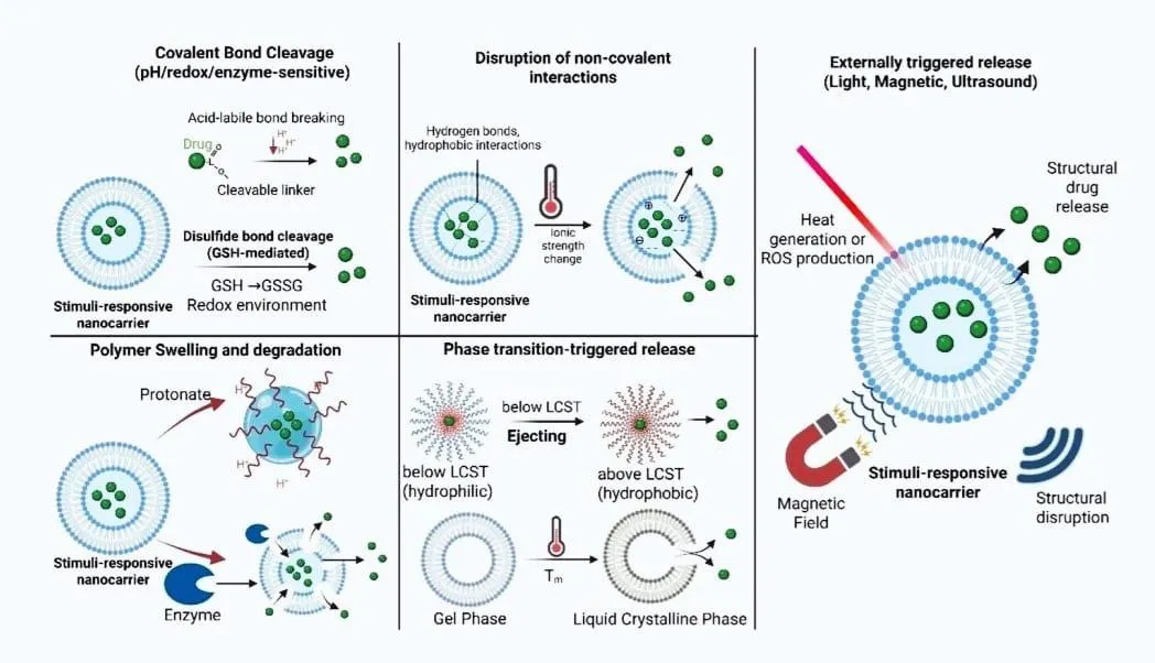
Besides the triggers described above, glucose [40] and electric fields [41] have also been used to control drug release from nanocarriers. Release can be made more precise by combining triggers [42, 43]. The most intensively studied are dual-stimulus systems, such as heat and pH [42, 43], heat and light [44], redox state and pH [45], and ultrasound and magnetic fields [46]. Figure 1 summarises the main mechanisms of stimuli-responsive release from nanocarriers [47].
figure

Figure 1: Mechanisms of Stimuli-Responsive Drug Release from Nanocarriers. Source: [47].

NANOCARRIERS USED IN SMART DRUG DELIVERY SYSTEMS:

1. Liposomes:

Phospholipid systems were first described by Alec Bangham and colleagues in 1965 [48], and closed phospholipid bilayer vesicles were subsequently named liposomes [49]. In 1971, Gregoriadis and co-workers became the first to use liposomes as drug carriers [50]. As preparation methods improved, large unilamellar vesicles (LUVs) could be produced by forcing vesicles through polycarbonate filters. Once liposome size was brought down to 100 nm or less, liposomes came into wide use as drug delivery systems in clinical trials of treatments for cancer, inflammation and fungal infections, as well as in gene therapy [51, 52]. Doxil®, the first nanomedicine delivery system approved by the US Food and Drug Administration (FDA), is based on PEGylated liposomes [53]. Alongside the liposomes already on the market, many other lipid-based nanoparticles are moving from concept toward clinical use, which suggests that liposomal carriers will gain further acceptance in clinical practice (Table 1).

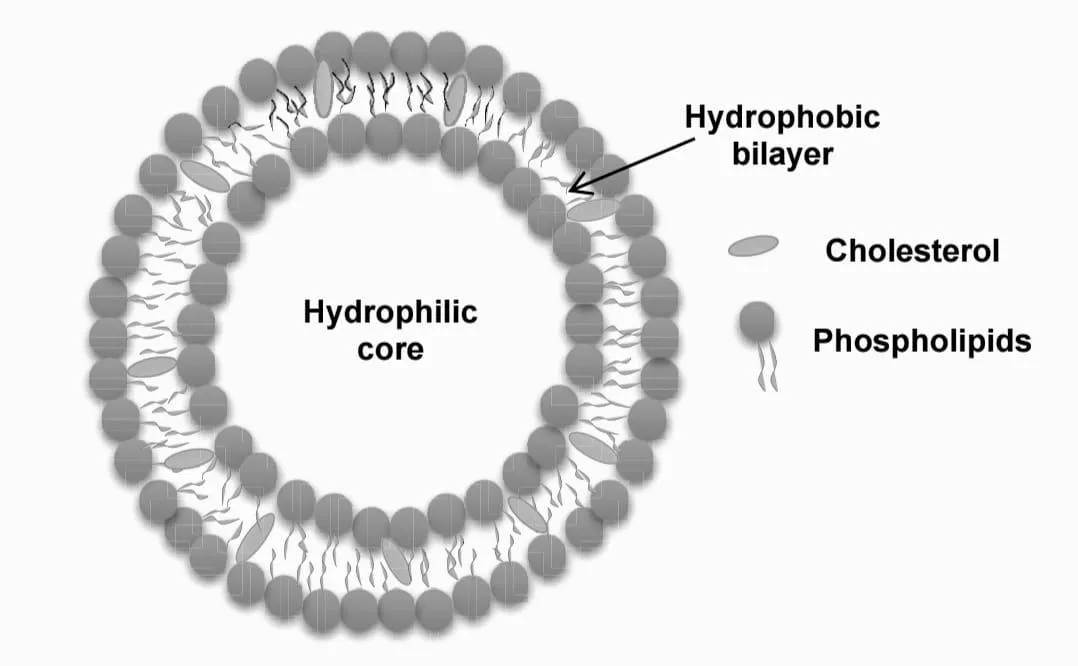
Because of this track record, smart liposomes have become a major focus in nanomedicine. They can be triggered by changes in temperature, pH and enzyme levels, and by ultrasound and light [54, 55]. Although many stimuli can control release from liposomes, temperature is one of the most attractive because of its safety and ease of use [56]. The lipid dipalmitoylphosphatidylcholine (DPPC) has a melting temperature of 41.5 °C, which is the property that allows doxorubicin to be released from ThermoDox® at that temperature [57]. The structure of a liposome is shown in Figure 2.
figure

Figure 2: Schematic Representation of Liposomes Showing the Hydrophilic Core, Hydrophobic Bilayer, Phospholipids and Cholesterol. Source: [58].

Table 1: Examples of Liposomal Pharmaceutical Products and Their Active Ingredients.

Sr. No. Marketed product Active pharmaceutical ingredient References
1 Doxil® Doxorubicin [53, 59]
2 Arikayce® Amikacin [59]
3 Epaxal® Inactivated hepatitis A virus [59]
4 DepoDur® Morphine sulphate [59]
5 AmBisome® Amphotericin B [59, 60]

2. Polymeric Nanoparticles:

Polymers that respond to physical or chemical stimuli are known as smart or stimuli-responsive polymers. Such polymers were used to control the release of active cargo as long as half a century ago, and they have played an important part in the development of nanomedicines. Smart polymeric systems have been reported to respond to a range of stimuli, including ultrasound, pH and magnetic fields [1, 61, 62]. They fall into two groups: single-stimulus responsive polymers and dual- or multi-stimuli-responsive polymers [63].

pH-triggered polymeric delivery systems have advanced considerably in recent years and allow controlled release at tumour sites [64]. A typical example is the pH-sensitive polymeric micelle [65]. In one study, smart micelles were prepared and interfacially crosslinked through hydrazone bonds; these bonds were rapidly cleaved at pH 5.0 and 37 °C [66]. Beyond polyelectrolytes, many self-assembled and other polymeric structures have been developed for controlled delivery, including micelles, polymer gels, polymer nanospheres and dendritic polymers [61]. pH-sensitive polymer systems have also been applied to tumour imaging; for example, the Gao group prepared ultra-pH-sensitive (UPS) fluorescent nanoprobes [67, 68].

Light-responsive polymeric systems can be built by incorporating photosensitive moieties or by triggering a photochemical reaction, and they are useful because their properties change when illuminated [69]. Glucose-responsive polymers, which sense sugar levels, are widely studied for diabetes care and can be used in new medical devices [70]. Some natural polymers can serve as controlled-release biomaterials, and smart release can also be achieved with chitosan blends and cyclodextrin derivatives [71]. Representative marketed nanoparticle-based products are listed in Table 2.

Table 2: Marketed Nanoparticle-Based Drug Delivery Products.

Marketed product Active pharmaceutical ingredient Application References
Doxil® Doxorubicin Cancer therapy [59]
Abraxane® Paclitaxel Cancer therapy [59]
Onpattro® siRNA Hereditary transthyretin amyloidosis [59]
Comirnaty® / Spikevax® mRNA COVID-19 prevention [59, 72]
DepoCyt® Cytarabine Lymphomatous meningitis [59]
Vyxeos® Daunorubicin + cytarabine Acute myeloid leukaemia [59]

3. Exosomes:

Almost every cell type secretes exosomes, a subset of extracellular vesicles with diameters below about 200 nm [73]. They act as messengers between cells and are involved in many physiological and pathological processes [74]. Exosomes can promote tissue regeneration and repair, largely because of their high biosafety, low immunogenicity and ability to carry a range of bioactive compounds to target cells [75, 76]; this property is useful in cardiovascular applications [77]. In addition, exosomes derived from tumour cells tend to accumulate preferentially in tumour tissue [78, 79]. Attaching tumour-targeting ligands such as proteins, peptides or antibodies to exosomes can further increase the specificity of exosome-based delivery [80, 81].

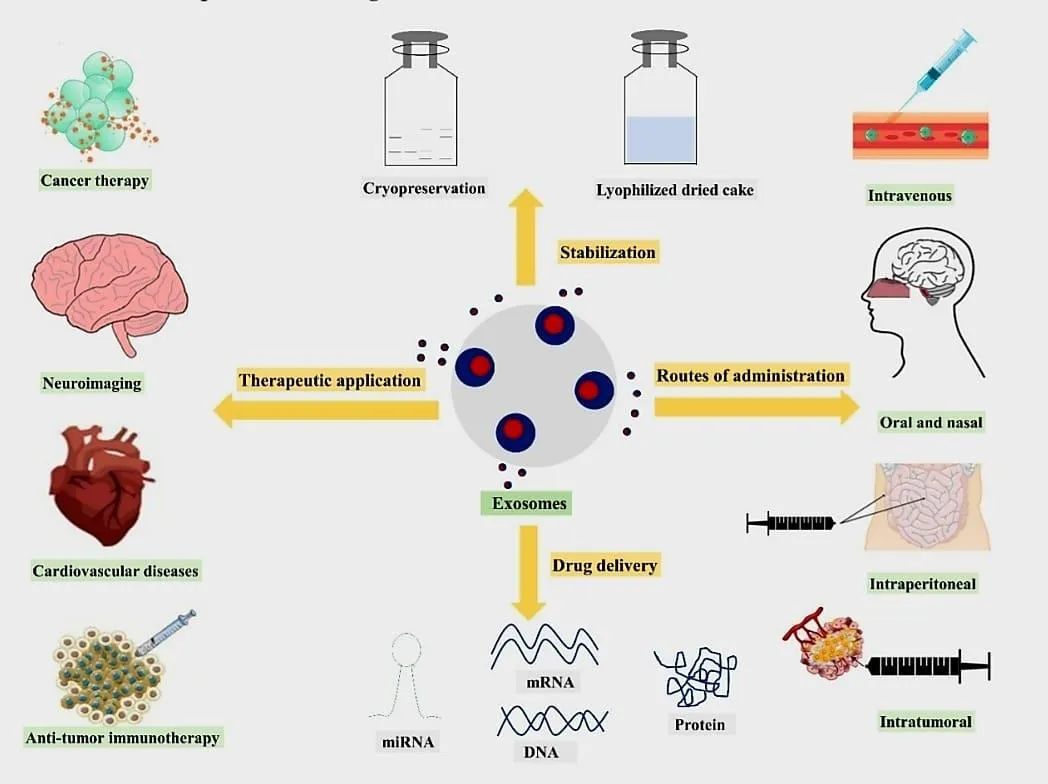
Although exosomes have entered clinical trials, their use as drug carriers may be limited by how much drug they can be loaded with, how well they retain it while circulating in the body, and whether they deliver it to target cells in a functional form [78, 82].Despite clinical trials with exosomes, the ability to load exosomes with enough drug, the ability of exosomes to hold these drugs while moving through the body, and the ability of exosomes to deliver these drugs to the targeted cells in a functional form may limit exosome use in drug delivery applications. Figure 3 gives an overview of exosome-based delivery, and Table 3 summarises representative cargos, loading approaches and reported outcomes.
figure

Figure 3: Overview of Exosome-Based Drug Delivery, Including Stabilisation and Storage, Routes of Administration, Therapeutic Applications, and Delivery of Biological Cargo Such as miRNA, DNA and Proteins. Reproduced from Butreddy Et Al. [77].

Table 3: Exosome-based Drug Delivery Systems: Cargo, Cellular Sources, Loading Approaches and Therapeutic Outcomes.

Exosome cargo Source of exosomes Therapeutic application Drug-loading approach Reported outcome Reference
Signal regulatory protein α Human embryonic kidney 293T cells Cancer Transfection Enhanced phagocytosis of tumour cells [84]
Survivin-T34A Human embryonic kidney 293T cells Pancreatic cancer Transfection Increased apoptosis [85]
Anti-epidermal growth factor receptor Mouse neuroblastoma Epidermoid carcinoma — Target-specific delivery [81]
miRNA Glioblastoma cells Glioblastoma Transfection Diagnostic/prognostic potential [86]
miRNA Human blood endothelial colony-forming cells Ischaemic kidney injury Transfection Improved kidney function [87]
Paclitaxel Prostate cancer cells and LNCaP Autologous prostate cancer Co-incubation Enhanced drug cytotoxicity [88]
Doxorubicin Immature mouse dendritic cells Breast cancer Electroporation Improved tumour drug delivery [89]
Curcumin Glioblastoma-related cell sources Brain tumour/autoimmune conditions Direct mixing Reduced inflammation [90]
Dopamine Kunming mouse brain-derived cells Parkinson’s disease Co-incubation Improved therapeutic effect [91]

4. Hydrogels:

The first hydrogel developed for biological use dates from 1960 [92]. Since then, conventional “dumb” gels have evolved into “smart” gels that respond to specific external stimuli or environmental changes. Smart hydrogels have received considerable attention for drug delivery in recent years because of their responsive behaviour, low invasiveness, ease of administration and ability to release drug in a controlled way over an extended period [93, 94]. Conventional gels may swell or shrink under osmotic pressure, but their responses are generally unpredictable, which leads to inaccurate release and poor control over dose timing [95].

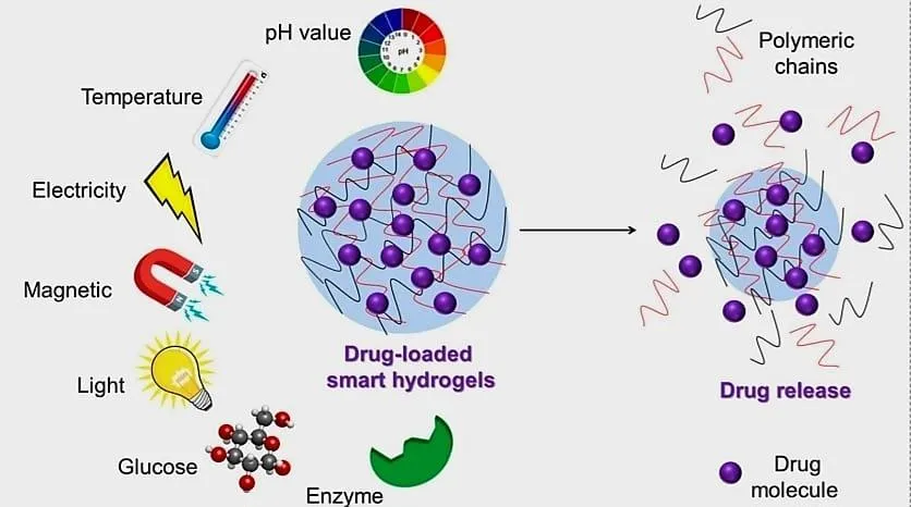
Advances in polymer science and nanotechnology have produced hydrogels that can release drugs at the right time and place [96]. By preventing the drug from accumulating at unwanted sites, smart hydrogels can reduce side effects, lower dosing frequency and maintain stable therapeutic levels after a single injection [97]. They are also easy to prepare and perform well in slow-release devices [95]. The stimuli that can trigger release from smart hydrogels are illustrated in Figure 4, and representative hydrogel types are listed in Table 4.
figure

Figure 4: Stimuli-responsive Smart Hydrogels and Their Drug-Release Mechanisms in Response to External Stimuli, Including pH, Temperature, Electricity, Magnetic Fields, Light, Glucose and Enzymes. Reproduced from Bordbar-Khiabani and Gasik [94].

Table 4: Stimuli-responsive Hydrogels: Examples, Materials, Response Mechanisms and Biomedical Applications.

Type of hydrogel Examples Response / mechanism Application Reference
pH-responsive Chitosan, κ-carrageenan, PEI, PAA, PMAA Swelling or shrinking occurs with changes in pH, allowing drug release Drug delivery, wound healing, sensing [98]
Temperature-responsive Poloxamer, Pluronic, PNIPAM, PEG-based polymers Temperature change causes a polymer phase transition and alters drug release Drug delivery, tissue engineering [99]
Electric-field-responsive PPy nanoparticles, PAA, PEG, chitosan-based hydrogels Electric stimulation changes hydrogel swelling and drug release Drug delivery, wound healing, tissue engineering [100]
Magnetic-field-responsive Alginate, chitosan, magnetic nanoparticles, Fe₃O₄-containing hydrogels A magnetic field produces heating or structural changes that trigger drug release Drug delivery, tissue engineering [101]
Light-responsive Photocleavable polymers, azobenzene-containing hydrogels Light causes structural or chemical changes and enables controlled release Drug delivery, optical delivery, wound healing [102]
Biomolecule-responsive Hyaluronic acid, phenylboronic acid-containing hydrogels Interaction with biomolecules or enzymes changes hydrogel properties and drug release Drug delivery, cell culture, tissue engineering [103]

5. Dendrimers:

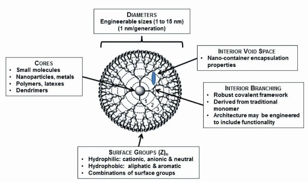
The term "dendrimer" was introduced by D.A. Tomalia and originates from the Greek words “dendron” (tree) and “meros” (part), a common suffix for polymers[104]. Dendrimers are described as highly branched, star-shaped three-dimensional macromolecules with nanometer-scale dimensions. Dendrimers are made up of three parts: (i) The multivalent surface with many reactive sites; (ii) the outer layer just below the multivalent surface providing a unique environment; and (iii) the core, the central part of the dendrimer, is safely shielded by the branches of higher generation dendrimers[105]. The surfaces of higher-generation dendrimers can be decorated with many functional groups, enabling effective host–guest interactions [105].

These synthetic macromolecules are built from branched monomer units and combine a defined structure, uniform size, high biocompatibility, a compact globular shape and a modifiable surface bearing functional groups or ligands, which makes them strong candidates for evaluation as drug carriers [105]. The general architecture is shown in Figure 5, and dendrimer-based products are listed in Table 5.
figure

Figure 5: Schematic Representation of Dendrimer Structure Showing the Central Core, Interior Branching Units, Internal Cavities and Surface Functional Groups. Source: [105].

Table 5: Dendrimer-based Products and Applications.

Dendrimer / product Type of dendrimer Main application Development status References
Priostar® PEHAM/PEA Drug delivery Marketed [106]
Starburst® PAMAM Drug delivery Marketed [104]
Astramol® PPI Drug delivery Marketed [107]
Polylysine Poly-L-lysine Drug delivery Marketed [108]
VivaGel® Poly-L-lysine Prevention of HIV and STDs Clinical trials [108]
Stratus CS® PAMAM Cardiac assay/diagnostic Marketed [109]
SuperFect® PAMAM Transfection agent Marketed [110]
ProfiTec™ PAMAM Transfection agent Marketed [110]
Alert Ticket™ PAMAM Anthrax-detection agent Marketed [111]

6. Microneedles:

The transdermal route is used in many clinical settings to overcome the main limitations of oral drug delivery [10]. Recent work highlights the effectiveness of microneedle (MN) arrays for the transdermal delivery of compounds that cannot cross the skin by passive diffusion or with other external assistance [112]. Microneedles are an effective means of delivering high-molecular-weight molecules, and they also aim to reduce the pain associated with injection by hypodermic needle [112]. They are too small to be seen without magnification, with heights below one millimetre; reported heights are about 50–900 μm, with a diameter of about 1 μm [112].

Microneedles must be mechanically stable and withstand an insertion force of about 10 N [112]. They are intended to support personalised medication at variable dose levels and self-administration [112]. Durability is one of the most important design considerations in fabrication, because the needles must pierce the epidermis without breaking so that drug is delivered efficiently [112]. Representative microneedle products are listed in Table 6.

Table 6: Microneedle-based Drug Delivery Products [112].

Product Type Function Application
Dermaroller® Metallic microneedle Enhances drug absorption Acne, stretch marks, hair loss
MicroHyala® Dissolvable microneedle patch Delivers hyaluronic acid Wrinkle treatment
VaxMat® Dissolvable microneedle patch Delivers macromolecules Proteins, peptides and vaccines
Micro-Trans® Microneedle patch Delivers drugs into the dermis Transdermal drug delivery
Drugmat® Dissolvable microneedle patch Provides rapid drug delivery Transdermal drug delivery
Nanoject® Microneedle array-based device Enables intradermal delivery Drug delivery and diagnostics
Soluvia® Hollow microneedle array Enables accurate intradermal delivery Vaccines
IDflu® / Intanza® Intradermal microneedle injection Delivers vaccine into the skin Influenza vaccination

7. Polymeric Micelles:

Biocompatible polymers have been widely used in pharmaceutical sciences as excipients in the traditional drug formulations and, more recently, in nanomedicines for enhanced therapeutic efficacy of potent drugs[27]. Approximately thirty years ago, micelles created by amphiphilic block copolymers in water were envisioned as vehicles for poorly soluble therapeutic agents that were either covalently bonded to polymer chains or non-covalently integrated within the micelles [27, 35]. Several novel block copolymers have since been proposed for micelle-based delivery systems as promising nanomedicines for humans [61].

To maximize the delivery of medicinal compounds, numerous important developments in polymeric micelles have been made. Such developments have pushed an increasing number of polymeric micelle medication formulations to enter clinical trials for regulatory approval[59]. The design of block copolymers is aimed at effectively encapsulating medicinal chemicals into polymeric micelles by various molecular interactions, which results in the protection of the cargo from the external environment and improvements of the pharmacokinetic (PK) profile [27]. Representative formulations and their clinical status are listed in Table 7.

Table 7: Representative Polymeric Micelle-Based Drug Delivery Systems and Their Clinical Development Status.

Product Drug Polymer Clinical status Reference
Genexol® PM Paclitaxel mPEG-b-PDLLA Approved [59]
Nanoxel® M Docetaxel mPEG-b-PDLLA Approved [59]
NK105 Paclitaxel mPEG-b-modified P(Asp) Phase III completed [59]
NC-6004 Cisplatin PEG-b-P(Glu) Phase III completed [59]
SP1049C Doxorubicin Pluronic® L61/F127 Phase II completed [59]

APPLICATIONS OF SMART DRUG DELIVERY SYSTEMS:

SDDS are being applied across a wide range of diseases. Table 8 summarises representative applications, the role of the delivery system and examples of products or platforms.

Table 8: Applications of Smart Drug Delivery Systems.

Sr. No. Application Role of SDDS Potential benefit Examples Reference
1 Cancer treatment Delivers anticancer drugs in response to specific stimuli, such as heat or changes in the tumour environment Improved drug delivery to tumour tissue and reduced exposure of healthy tissue ThermoDox, Doxil [29, 53]
2 Brain tumour treatment Uses magnetic iron-oxide nanoparticles to generate local heat Localised treatment through magnetic hyperthermia NanoTherm [29]
3 Ovarian cancer treatment Uses polymer-based drug delivery to deliver paclitaxel May improve drug delivery and modify drug distribution in the body Opaxio [29]
4 Fungal infections Delivers amphotericin B through a liposomal formulation Improved drug delivery and reduced toxicity compared with conventional amphotericin B AmBisome [60]
5 Visceral leishmaniasis Uses liposomal encapsulation to deliver amphotericin B Improved delivery of the drug to infected tissues AmBisome [60]
6 Colon-targeted drug delivery Uses pH-responsive nanoparticles to release drugs in response to changes in gastrointestinal pH More localised drug release and reduced premature release Eudragit S100-coated citrus pectin nanoparticles [19, 29]
7 Inflammatory bowel disease Uses stimuli-responsive nanocarriers to deliver therapeutic agents to affected intestinal regions Potentially improves local drug delivery and reduces systemic exposure pH-responsive nanocarriers [28, 113]
8 Diabetes management Uses glucose-responsive systems to regulate insulin release according to glucose levels Potential for glucose-responsive insulin delivery and improved blood glucose control Glucose-responsive nanocarriers [40, 70]
9 Intracellular drug delivery Uses redox-responsive carriers that respond to intracellular reducing conditions, such as elevated glutathione levels Promotes drug release inside cells and may improve therapeutic effectiveness Disulfide-bond-containing nanocarriers [26, 27]
10 Localised cancer therapy Uses temperature-responsive materials to release drugs following local heating Provides better control over the location and timing of drug release Thermosensitive liposomes [8, 57]

CHALLENGES IN SMART DRUG DELIVERY SYSTEMS:

The human body is a complex and constantly changing system with multiple biological barriers to drug delivery. These include rapid clearance by the reticuloendothelial system, uptake of the drug by healthy cells, enzymatic degradation, dilution in the bloodstream, difficulty in penetrating the dense tissue surrounding tumours [114], and barriers such as the blood–brain barrier and cell membranes, which prevent drugs from entering cells [113]. Polyethylene glycol (PEG) is widely used in drug delivery because it prolongs circulation in the bloodstream and helps carriers avoid immediate recognition by the immune system. However, recent studies show that repeated administration of PEGylated products can induce anti-PEG antibodies, which may lead to faster clearance of the drug, reduced therapeutic effect and tissue damage [115].

SDDS must also address the potential toxicity of nanocarriers, the cost of production, the diversity and complexity of cancer types and the lack of clear regulatory guidelines [116]. For antibody–drug conjugates (ADCs), the main concerns are high production cost, limited penetration into solid tumours and the risk of premature drug release [117]. In addition, inconsistencies in the experimental detail and characterisation reported in published studies have made it harder to advance nanotechnology in medicine [118, 119]. Because the available data are limited and heterogeneous, progress in nanomedicine and its translation to clinical use will depend on more consistent, well-characterised studies that can guide industry. Many researchers hold that nanoparticles can be either beneficial or harmful: their advantages are well established, but information on their safety, their interactions with non-specific proteins and their movement and interactions in non-target organs is limited [120].

Although targeting specific sites can reduce side effects and improve treatment, its benefit depends on whether the drug actually reaches the intended location. This is evident for systemically administered siRNA, which often fails to reach its target cell or organ because enzymes rapidly degrade it and because its negative charge is repelled by cell membranes, leading to poor uptake [121]. Lipid-based nanoparticles such as micelles and liposomes are being studied for targeted delivery, but their effectiveness is reduced by phagocytosis by immune cells and filtration by the liver, which can prevent the drug from reaching its target [122]. These nanoparticles may also have toxic effects [120], and, when given orally, they may interact with food and be broken down by bacteria in the digestive tract [123]. Finally, biocompatibility—how well a system is tolerated by the body without causing immune reactions—remains a major challenge, because the body responds differently to biological materials than to synthetic ones [124].

CONCLUSION:

SDDS is a major step forward for health care. SDDS employs carriers such as liposomes, nanoparticles, dendrimers, hydrogels, exosomes, microneedles, and micelles. These vehicles protect drugs so they can be delivered at the right time and targeted to the tissue or cells that require them. SDDS also reacts to other factors like changes in levels, enzyme activity, temperature, light, magnetic fields, and ultrasound. This makes the delivery more accurate and flexible so the medicine can be released when needed. Advantages of SDDS include their ability to effectively deliver drugs and help patients adhere to their treatment regimens. With these benefits, SDDS could revolutionize the treatment of cancer, heart disease, infections, and many other diseases. There are still issues. SDDS designs can be very complex and may contain certain quantities of drug, and hence there are concerns about their interaction with the body and toxicity issues. If scientists are to get past these problems, they have to keep working on multi-functional and stimuli-responsive systems. They also need nanocarriers to be developed. SDDS is the convergence of nanotechnology, biotechnology, and medicine. I believe SDDS can provide effective therapies that are tailored to each patient. New ideas for SDDS may reduce adverse effects and costs. Make patients feel better. It’s a step toward a future of personalized health care.”

REFERENCES

  1. Hrubý M, Filippov SK, Štěpánek P. Smart polymers in drug delivery systems on crossroads: which way deserves following? Eur Polym J. 2015;65:82-97.
  2. Allen TM, Cullis PR. Liposomal drug delivery systems: from concept to clinical applications. Adv Drug Deliv Rev. 2013;65 (1):36-48.
  3. Mullick Chowdhury S, Lee T, Willmann JK. Ultrasound-guided drug delivery in cancer. Ultrasonography. 2017;36:171-84.
  4. Zhang H, Fan T, Chen W, Li Y, Wang B. Recent advances of two-dimensional materials in smart drug delivery nano-systems. Bioact Mater. 2020;5:1071-86.
  5. Singh R, Jadhav K, Vaghasiya K, Ray E, Shukla R, Verma RK. New generation smart drug delivery systems for rheumatoid arthritis. Curr Pharm Des. 2023;29:984-1001.
  6. Adepu S, Ramakrishna S. Controlled drug delivery systems: current status and future directions. Molecules. 2021;26:5905.
  7. Tanaka T. Collapse of gels and the critical endpoint. Phys Rev Lett. 1978;40:820-3.
  8. Yatvin MB, Weinstein JN, Dennis WH, Blumenthal R. Design of liposomes for enhanced local release of drugs by hyperthermia. Science. 1978;202 (4374):1290-3.
  9. Müller RH, Keck CM. Challenges and solutions for the delivery of biotech drugs—a review of drug nanocrystal technology and lipid nanoparticles. J Biotechnol. 2004;113 (1-3):151-70.
  10. Panchagnula R. Transdermal delivery of drugs. Indian J Pharmacol. 1997;29 (3):140-56.
  11. Bertrand N, Wu J, Xu X, Kamaly N, Farokhzad OC. Cancer nanotechnology: the impact of passive and active targeting in the era of modern cancer biology. Adv Drug Deliv Rev. 2014;66:2-25.
  12. Ganta S, Devalapally H, Shahiwala A, Amiji M. A review of stimuli-responsive nanocarriers for drug and gene delivery. J Control Release. 2008;126 (3):187-204.
  13. Jain AK, Swarnakar NK, Das M, Godugu C, Singh RP, Jain S. The effect of the oral administration of polymeric nanoparticles on the efficacy of drugs. Expert Opin Drug Deliv. 2013;10 (3):369-81.
  14. Mei L, Zhang Y, Zheng Y, et al. A multifunctional nanoplatform for imaging, radiotherapy, and the prediction of therapeutic response. Small. 2014;10 (21):4343-54.
  15. Farokhzad OC, Langer R. Impact of nanotechnology on drug delivery. ACS Nano. 2009;3 (1):16-20.
  16. Torchilin VP. Multifunctional nanocarriers. Adv Drug Deliv Rev. 2012;64 (Suppl):302-15.
  17. Attia MF, Anton N, Wallyn J, Omran Z, Vandamme TF. An overview of active and passive targeting strategies to improve the nanocarrier efficiency to tumour sites. J Pharm Pharmacol. 2019;71 (8):1185-98.
  18. Yu P, Yu H, Guo C, Cui Z, Chen X, Yin Q, et al. Reversal of doxorubicin resistance in breast cancer by mitochondria-targeted pH-responsive micelles. Acta Biomater. 2015;14:115-24.
  19. Subudhi MB, Jain A, Jain A, Hurkat P, Shilpi S, Gulbake A, et al. Eudragit S100 coated citrus pectin nanoparticles for colon targeting of 5-fluorouracil. Materials (Basel). 2015;8:832-49.
  20. Stubbs M, McSheehy PM, Griffiths JR, Bashford CL. Causes and consequences of tumour acidity and implications for treatment. Mol Med Today. 2000;6:15-9.
  21. Neri D, Supuran CT. Interfering with pH regulation in tumours as a therapeutic strategy. Nat Rev Drug Discov. 2011;10:767-77.
  22. Lee ES, Oh KT, Kim D, Youn YS, Bae YH. Tumor pH-responsive flower-like micelles of poly (L-lactic acid)-b-poly (ethylene glycol)-b-poly (L-histidine). J Control Release. 2007;123:19-26.
  23. Chen W, Zhong P, Meng F, Cheng R, Deng C, Feijen J, et al. Redox and pH-responsive degradable micelles for dually activated intracellular anticancer drug release. J Control Release. 2013;169:171-9.
  24. Deirram N, Zhang C, Kermaniyan SS, Johnston APR, Such GK. pH-responsive polymer nanoparticles for drug delivery. Macromol Rapid Commun. 2019;40:1800917.
  25. Huo M, Yuan J, Tao L, Wei Y. Redox-responsive polymers for drug delivery: from molecular design to applications. Polym Chem. 2014;5:1519-28.
  26. Wang J, Sun X, Mao W, Sun W, Tang J, Sui M, et al. Tumor redox heterogeneity-responsive prodrug nanocapsules for cancer chemotherapy. Adv Mater. 2013;25:3670-6.
  27. Torchilin VP. Multifunctional, stimuli-sensitive nanoparticulate systems for drug delivery. Nat Rev Drug Discov. 2014;13:813-27.
  28. Wilson DS, Dalmasso G, Wang L, Sitaraman SV, Merlin D, Murthy N. Orally delivered thioketal nanoparticles loaded with TNF-α–siRNA target inflammation and inhibit gene expression in the intestines. Nat Mater. 2010;9:923-8.
  29. Liu D, Yang F, Xiong F, Gu N. The smart drug delivery system and its clinical potential. Theranostics. 2016;6 (9):1306-23. doi:10.7150/thno.14858.
  30. Lock LL, Tang Z, Keith D, Reyes C, Cui H. Enzyme-specific doxorubicin drug beacon as drug-resistant theranostic molecular probes. ACS Macro Lett. 2015;4:552-5.
  31. De La Rica R, Aili D, Stevens MM. Enzyme-responsive nanoparticles for drug release and diagnostics. Adv Drug Deliv Rev. 2012;64:967-78.
  32. Shi Y, Cardoso RM, van Nostrum CF, Hennink WE. Anthracene functionalized thermosensitive and UV-crosslinkable polymeric micelles. Polym Chem. 2015;6:2048-53.
  33. Danhier F, Feron O, Préat V. To exploit the tumor microenvironment: passive and active tumor targeting of nanocarriers for anti-cancer drug delivery. J Control Release. 2010;148:135-46.
  34. Zhao Y, Fan X, Liu D, Wang Z. PEGylated thermo-sensitive poly (amidoamine) dendritic drug delivery systems. Int J Pharm. 2011;409:229-36.
  35. Mura S, Nicolas J, Couvreur P. Stimuli-responsive nanocarriers for drug delivery. Nat Mater. 2013;12:991-1003.
  36. Chen Z, Yin J-J, Zhou Y-T, Zhang Y, Song L, Song M, et al. Dual enzyme-like activities of iron oxide nanoparticles and their implication for diminishing cytotoxicity. ACS Nano. 2012;6:4001-12.
  37. Fang K, Song L, Gu Z, Yang F, Zhang Y, Gu N. Magnetic field activated drug release system based on magnetic PLGA microspheres for chemo-thermal therapy. Colloids Surf B Biointerfaces. 2015;136:712-20.
  38. Hayashi K, Nakamura M, Sakamoto W, Yogo T, Miki H, Ozaki S, et al. Superparamagnetic nanoparticle clusters for cancer theranostics combining magnetic resonance imaging and hyperthermia treatment. Theranostics. 2013;3:366-76.
  39. Paris JL, Cabañas MV, Manzano M, Vallet-Regí M. Polymer-grafted mesoporous silica nanoparticles as ultrasound-responsive drug carriers. ACS Nano. 2015;9:11023-33.
  40. Gu Z, Aimetti AA, Wang Q, Dang TT, Zhang Y, Veiseh O, et al. Injectable nano-network for glucose-mediated insulin delivery. ACS Nano. 2013;7:4194-201.
  41. Curcio M, Spizzirri UG, Cirillo G, Vittorio O, Picci N, Nicoletta FP, et al. On demand delivery of ionic drugs from electro-responsive CNT hybrid films. RSC Adv. 2015;5:44902-11.
  42. Schmaljohann D. Thermo- and pH-responsive polymers in drug delivery. Adv Drug Deliv Rev. 2006;58:1655-70.
  43. Zhang L, Guo R, Yang M, Jiang X, Liu B. Thermo- and pH-dual-responsive nanoparticles for anti-cancer drug delivery. Adv Mater. 2007;19:2988-92.
  44. Jochum FD, Theato P. Thermo- and light-responsive micellation of azobenzene-containing block copolymers. Chem Commun. 2010;46:6717-9.
  45. Pan Y-J, Chen Y-Y, Wang D-R, Wei C, Guo J, Lu D-R, et al. Redox/pH dual stimuli-responsive biodegradable nanohydrogels with varying responses to dithiothreitol and glutathione for controlled drug release. Biomaterials. 2012;33:6570-9.
  46. Cai X, Yang F, Gu N. Applications of magnetic microbubbles for theranostics. Theranostics. 2012;2:103-12.
  47. Pathak P, Chettupalli AK, Bukke SPN, Sharma CB, Thalluri C. Nanotechnology-driven smart drug delivery systems bridge pharmaceutical sciences, biomaterials, and clinical translation. Discov Appl Sci. 2026. doi:10.1007/s42452-026-09530-z.
  48. Bangham A, Standish MM, Watkins J. Diffusion of univalent ions across the lamellae of swollen phospholipids. J Mol Biol. 1965;13:238-52.
  49. Deamer DW. From “Banghasomes” to liposomes: a memoir of Alec Bangham, 1921–2010. FASEB J. 2010;24:1308-10.
  50. Gregoriadis G, Ryman B. Liposomes as carriers of enzymes or drugs: a new approach to the treatment of storage diseases. Biochem J. 1971;124:58P.
  51. Akbarzadeh A, Rezaei-Sadabady R, Davaran S, Joo SW, Zarghami N, Hanifehpour Y, et al. Liposome: classification, preparation, and applications. Nanoscale Res Lett. 2013;8:102.
  52. Torchilin V. Liposomes in drug delivery. In: Fundamentals and Applications of Controlled Release Drug Delivery. Springer; 2012. p. 289-328.
  53. Barenholz Y. Doxil®—the first FDA-approved nano-drug: lessons learned. J Control Release. 2012;160:117-34.
  54. Bibi S, Lattmann E, Mohammed AR, Perrie Y. Trigger release liposome systems: local and remote controlled delivery? J Microencapsul. 2012;29:262-76.
  55. Sawant RR, Torchilin VP. Liposomes as ‘smart’ pharmaceutical nanocarriers. Soft Matter. 2010;6:4026-44.
  56. Kono K, Ozawa T, Yoshida T, Ozaki F, Ishizaka Y, Maruyama K, et al. Highly temperature-sensitive liposomes based on a thermosensitive block copolymer for tumor-specific chemotherapy. Biomaterials. 2010;31:7096-105.
  57. Chen K-J, Chaung E-Y, Wey S-P, Lin K-J, Cheng F, Lin C-C, et al. Hyperthermia-mediated local drug delivery by a bubble-generating liposomal system for tumor-specific chemotherapy. ACS Nano. 2014;8:5105-15.
  58. Nsairat H, Khater D, Sayed U, Odeh F, Al Bawab A, Alshaer W. Liposomes: structure, composition, types, and clinical applications. Heliyon. 2022;8 (5):e09394. doi:10.1016/j.heliyon.2022.e09394.
  59. Desai N, Rana D, Patel M, Bajwa N, Prasad R, Vora LK. Nanoparticle therapeutics in clinical perspective: classification, marketed products, and regulatory landscape. Small. 2025;21:2502315.
  60. Adler-Moore J, Proffitt RT. AmBisome: liposomal formulation, structure, mechanism of action and preclinical experience. J Antimicrob Chemother. 2002;49 (Suppl 1):21-30.
  61. Alvarez-Lorenzo C, Concheiro A. Smart drug delivery systems: from fundamentals to the clinic. Chem Commun. 2014;50:7743-65.
  62. Stumpel JE, Gil ER, Spoelstra AB, Bastiaansen CW, Broer DJ, Schenning AP. Stimuli-responsive materials based on interpenetrating polymer liquid crystal hydrogels. Adv Funct Mater. 2015;25:3314-20.
  63. Cheng R, Meng F, Deng C, Klok H-A, Zhong Z. Dual and multi-stimuli responsive polymeric nanoparticles for programmed site-specific drug delivery. Biomaterials. 2013;34:3647-57.
  64. Liu R, Li D, He B, Xu X, Sheng M, Lai Y, et al. Anti-tumor drug delivery of pH-sensitive poly (ethylene glycol)-poly (L-histidine)-poly (L-lactide) nanoparticles. J Control Release. 2011;152:49-56.
  65. Duan X, Xiao J, Yin Q, Zhang Z, Yu H, Mao S, et al. Smart pH-sensitive and temporal-controlled polymeric micelles for effective combination therapy of doxorubicin and disulfiram. ACS Nano. 2013;7:5858-69.
  66. Shi Y, van Nostrum CF, Hennink WE. Interfacially hydrazone cross-linked thermosensitive polymeric micelles for acid-triggered release of paclitaxel. ACS Biomater Sci Eng. 2015;1:393-404.
  67. Zhou K, Wang Y, Huang X, Luby-Phelps K, Sumer BD, Gao J. Tunable, ultrasensitive pH-responsive nanoparticles targeting specific endocytic organelles in living cells. Angew Chem Int Ed. 2011;50:6109-14.
  68. Wang Y, Zhou K, Huang G, Hensley C, Huang X, Ma X, et al. A nanoparticle-based strategy for the imaging of a broad range of tumours by nonlinear amplification of microenvironment signals. Nat Mater. 2014;13:204-12.
  69. Wang W, Lin J, Cai C, Lin S. Optical properties of amphiphilic copolymer-based self-assemblies. Eur Polym J. 2015;65:112-31.
  70. Kim H, Kang YJ, Kang S, Kim KT. Monosaccharide-responsive release of insulin from polymersomes of polyboroxole block copolymers at neutral pH. J Am Chem Soc. 2012;134:4030-3.
  71. Ryu JH, Hong S, Lee H. Bio-inspired adhesive catechol-conjugated chitosan for biomedical applications: a mini review. Acta Biomater. 2015;27:101-15.
  72. Polack FP, Thomas SJ, Kitchin N, Absalon J, Gurtman A, Lockhart S, et al. Safety and efficacy of the BNT162b2 mRNA Covid-19 vaccine. N Engl J Med. 2020;383:2603-15.
  73. Welsh JA, Goberdhan DCI, O’Driscoll L, Buzas EI, Blenkiron C, Bussolati B, et al. Minimal information for studies of extracellular vesicles (MISEV2023): from basic to advanced approaches. J Extracell Vesicles. 2024;13:e12404.
  74. Kalluri R, LeBleu VS. The biology, function, and biomedical applications of exosomes. Science. 2020;367:eaau6977.
  75. Zhang Y, Liu Y, Liu H, Tang WH. Exosomes: biogenesis, biologic function and clinical potential. Cell Biosci. 2019;9:19.
  76. Tan A, Rajadas J, Seifalian AM. Exosomes as nano-theranostic delivery platforms for gene therapy. Adv Drug Deliv Rev. 2013;65:357-67.
  77. Butreddy A, Kommineni N, Dudhipala N. Exosomes as naturally occurring vehicles for delivery of biopharmaceuticals: insights from drug delivery to clinical perspectives. Nanomaterials (Basel). 2021;11 (6):1481. doi:10.3390/nano11061481.
  78. Johnsen KB, Gudbergsson JM, Skov MN, Pilgaard L, Moos T, Duroux M. A comprehensive overview of exosomes as drug delivery vehicles—endogenous nanocarriers for targeted cancer therapy. Biochim Biophys Acta. 2014;1846:75-87.
  79. Takahashi Y, Nishikawa M, Shinotsuka H, Matsui Y, Ohara S, Imai T, et al. Visualization and in vivo tracking of the exosomes of murine melanoma B16-BL6 cells in mice after intravenous injection. J Biotechnol. 2013;165:77-84.
  80. El-Andaloussi S, Lee Y, Lakhal-Littleton S, Li J, Seow Y, Gardiner C, et al. Exosome-mediated delivery of siRNA in vitro and in vivo. Nat Protoc. 2012;7:2112-26.
  81. Kooijmans SAA, Aleza CG, Roffler SR, van Solinge WW, Vader P, Schiffelers RM. Display of GPI-anchored anti-EGFR nanobodies on extracellular vesicles promotes tumour cell targeting. J Extracell Vesicles. 2016;5:31053.
  82. Antimisiaris SG, Mourtas S, Marazioti A. Exosomes and exosome-inspired vesicles for targeted drug delivery. Pharmaceutics. 2018;10:218.
  83. Jeyaram A, Jay SM. Preservation and storage stability of extracellular vesicles for therapeutic applications. AAPS J. 2017;20:1.
  84. Cho E, Nam G-H, Hong Y, Kim YK, Kim D-H, Yang Y, et al. Comparison of exosomes and ferritin protein nanocages for the delivery of membrane protein therapeutics. J Control Release. 2018;279:326-35.
  85. Aspe JR, Diaz Osterman CJ, Jutzy JMS, Deshields S, Whang S, Wall NR. Enhancement of gemcitabine sensitivity in pancreatic adenocarcinoma by novel exosome-mediated delivery of the survivin-T34A mutant. J Extracell Vesicles. 2014;3.
  86. Skog J, Würdinger T, van Rijn S, Meijer DH, Gainche L, Sena-Esteves M, et al. Glioblastoma microvesicles transport RNA and proteins that promote tumour growth and provide diagnostic biomarkers. Nat Cell Biol. 2008;10:1470-6.
  87. Viñas JL, Burger D, Zimpelmann J, Haneef R, Knoll W, Campbell P, et al. Transfer of microRNA-486-5p from human endothelial colony forming cell-derived exosomes reduces ischemic kidney injury. Kidney Int. 2016;90:1238-50.
  88. Saari H, Lázaro-Ibáñez E, Viitala T, Vuorimaa-Laukkanen E, Siljander P, Yliperttula M. Microvesicle- and exosome-mediated drug delivery enhances the cytotoxicity of paclitaxel in autologous prostate cancer cells. J Control Release. 2015;220:727-37.
  89. Tian Y, Li S, Song J, Ji T, Zhu M, Anderson GJ, et al. A doxorubicin delivery platform using engineered natural membrane vesicle exosomes for targeted tumor therapy. Biomaterials. 2014;35:2383-90.
  90. Sun D, Zhuang X, Xiang X, Liu Y, Zhang S, Liu C, et al. A novel nanoparticle drug delivery system: the anti-inflammatory activity of curcumin is enhanced when encapsulated in exosomes. Mol Ther. 2010;18:1606-14.
  91. Qu M, Lin Q, Huang L, Fu Y, Wang L, He S, et al. Dopamine-loaded blood exosomes targeted to brain for better treatment of Parkinson’s disease. J Control Release. 2018;287:156-66.
  92. Wichterle O, Lim D. Hydrophilic gels for biological use. Nature. 1960;185:117-8.
  93. Andrade F, Roca-Melendres MM, Durán-Lara EF, Rafael D, Schwartz S Jr. Stimuli-responsive hydrogels for cancer treatment: the role of pH, light, ionic strength and magnetic field. Cancers (Basel). 2021;13:1164.
  94. Bordbar-Khiabani A, Gasik M. Smart hydrogels for advanced drug delivery systems. Int J Mol Sci. 2022;23 (7):3665. doi:10.3390/ijms23073665.
  95. Hoare TR, Kohane DS. Hydrogels in drug delivery: progress and challenges. Polymer. 2008;49:1993-2007.
  96. Wang Y, Li J, Tang M, Peng C, Wang G, Wang J, et al. Smart stimuli-responsive hydrogels for drug delivery in periodontitis treatment. Biomed Pharmacother. 2023;162:114688.
  97. Zong S, Wen H, Lv H, Li T, Tang R, Liu L, et al. Intelligent hydrogel with both redox and thermo-response based on cellulose nanofiber for controlled drug delivery. Carbohydr Polym. 2022;278:118943.
  98. Bazban-Shotorbani S, Hasani-Sadrabadi MM, Karkhaneh A, Serpooshan V, Jacob KI, Moshaverinia A, et al. Revisiting structure-property relationship of pH-responsive polymers for drug delivery applications. J Control Release. 2017;253:46-63.
  99. Xian S, Webber MJ. Temperature-responsive supramolecular hydrogels. J Mater Chem B. 2020;8:9197-211.
  100. Agarwala S. Electrically conducting hydrogels for health care: concept, fabrication methods, and applications. Int J Bioprint. 2020;6:273.
  101. Liu Z, Liu J, Cui X, Wang X, Zhang L, Tang P. Recent advances on magnetic sensitive hydrogels in tissue engineering. Front Chem. 2020;8:124.
  102. Li L, Scheiger JM, Levkin PA. Design and applications of photoresponsive hydrogels. Adv Mater. 2019;31:1807333.
  103. Tang W, Chen C. Hydrogel-based colloidal photonic crystal devices for glucose sensing. Polymers (Basel). 2020;12:625.
  104. Tomalia DA, Fréchet JM. Discovery of dendrimers and dendritic polymers: a brief historical perspective. J Polym Sci A Polym Chem. 2002;40:2719-28.
  105. Chauhan AS. Dendrimers for drug delivery. Molecules. 2018;23 (4):938. doi:10.3390/molecules23040938.
  106. Swanson DR, Huang B, Abdelhady HG, Tomalia DA. Unique steric and geometry induced stoichiometries observed in the divergent synthesis of poly (ester-acrylate/amine) (PEA) dendrimers. New J Chem. 2007;31:1368-78.
  107. Tomalia DA, Rookmaker M. Poly (propylene imine) dendrimers. In: Polymer Data Handbook. New York: Oxford University Press; 2009.
  108. Patton DL, Cosgrove Sweeney YT, McCarthy TD, Hillier SL. Preclinical safety and efficacy assessments of dendrimer-based (SPL7013) microbicide gel formulations in a nonhuman primate model. Antimicrob Agents Chemother. 2006;50:1696-700.
  109. Singh P. Dendrimers and their applications in immunoassays and clinical diagnostics. Biotechnol Appl Biochem. 2007;48:1-9.
  110. Liu H, Wang H, Yang W, Cheng Y. Disulfide cross-linked low generation dendrimers with high gene transfection efficacy, low cytotoxicity, and low cost. J Am Chem Soc. 2012;134:17680-7.
  111. Spangler BD, inventor. Biosensors utilizing dendrimer-immobilized ligands and their use thereof. United States patent US 7138121. 2006.
  112. Halder J, Gupta S, Kumari R, Gupta GD, Rai VK. Microneedle array: applications, recent advances, and clinical pertinence in transdermal drug delivery. J Pharm Innov. 2021;16:558-65. doi:10.1007/s12247-020-09460-2.
  113. Sun Y, Davis E. Nanoplatforms for targeted stimuli-responsive drug delivery: a review of platform materials and stimuli-responsive release and targeting mechanisms. Nanomaterials (Basel). 2021;11 (3):746.
  114. Ju C, Mo R, Xue J, Zhang L, Zhao Z, Xue L, et al. Sequential intra-intercellular nanoparticle delivery system for deep tumor penetration. Angew Chem Int Ed. 2014;53:6253-8.
  115. Shi D, Beasock D, Fessler A, et al. To PEGylate or not to PEGylate: immunological properties of nanomedicine’s most popular component, polyethylene glycol and its alternatives. Adv Drug Deliv Rev. 2022;180:114079. doi:10.1016/j.addr.2021.114079.
  116. Shi J, Kantoff PW, Wooster R, Farokhzad OC. Cancer nanomedicine: progress, challenges and opportunities. Nat Rev Cancer. 2017;17:20-37. doi:10.1038/nrc.2016.108.
  117. Lo CF, Chiu TY, Liu YT, Pan PY, Liu KL, Hsu CY, et al. Targeting the phosphatidylserine-immune checkpoint with a small-molecule maytansinoid conjugate. J Med Chem. 2022;65:12802-24. doi:10.1021/acs.jmedchem.2c00631.
  118. Youn YS, Bae YH. Perspectives on the past, present, and future of cancer nanomedicine. Adv Drug Deliv Rev. 2018;130:3-11. doi:10.1016/j.addr.2018.05.008.
  119. Tan YF, Lao LL, Xiong GM, Venkatraman S. Controlled-release nanotherapeutics: state of translation. J Control Release. 2018;284:39-48.
  120. Sharma S, Parveen R, Chatterji BP. Toxicology of nanoparticles in drug delivery. Curr Pathobiol Rep. 2021;9:133-44. doi:10.1007/s40139-021-00227-z.
  121. Pasut G. Grand challenges in nano-based drug delivery. Front Med Technol. 2019;1:1. doi:10.3389/fmedt.2019.00001.
  122. Khalid A, Persano S, Shen H, Zhao Y, Blanco E, Wolfram J, et al. Strategies for improving drug delivery: nanocarriers and microenvironmental priming. Expert Opin Drug Deliv. 2017;14 (7):865-77. doi:10.1080/17425247.2017.1243527.
  123. Zargar SM, Hafshejani DK, Eskandarinia A, Rafienia M, Kharazi AZ. A review of controlled drug delivery systems based on cells and cell membranes. J Med Signals Sens. 2019;9:181-9. doi:10.4103/jmss.JMSS_53_18.
  124. Gautami J. Implantable drug delivery system. Res Rev J Pharm Nanotechnol. 2015;3:24-31.

Reference

  1. Hrubý M, Filippov SK, Št?pánek P. Smart polymers in drug delivery systems on crossroads: which way deserves following? Eur Polym J. 2015;65:82-97.
  2. Allen TM, Cullis PR. Liposomal drug delivery systems: from concept to clinical applications. Adv Drug Deliv Rev. 2013;65 (1):36-48.
  3. Mullick Chowdhury S, Lee T, Willmann JK. Ultrasound-guided drug delivery in cancer. Ultrasonography. 2017;36:171-84.
  4. Zhang H, Fan T, Chen W, Li Y, Wang B. Recent advances of two-dimensional materials in smart drug delivery nano-systems. Bioact Mater. 2020;5:1071-86.
  5. Singh R, Jadhav K, Vaghasiya K, Ray E, Shukla R, Verma RK. New generation smart drug delivery systems for rheumatoid arthritis. Curr Pharm Des. 2023;29:984-1001.
  6. Adepu S, Ramakrishna S. Controlled drug delivery systems: current status and future directions. Molecules. 2021;26:5905.
  7. Tanaka T. Collapse of gels and the critical endpoint. Phys Rev Lett. 1978;40:820-3.
  8. Yatvin MB, Weinstein JN, Dennis WH, Blumenthal R. Design of liposomes for enhanced local release of drugs by hyperthermia. Science. 1978;202 (4374):1290-3.
  9. Müller RH, Keck CM. Challenges and solutions for the delivery of biotech drugs—a review of drug nanocrystal technology and lipid nanoparticles. J Biotechnol. 2004;113 (1-3):151-70.
  10. Panchagnula R. Transdermal delivery of drugs. Indian J Pharmacol. 1997;29 (3):140-56.
  11. Bertrand N, Wu J, Xu X, Kamaly N, Farokhzad OC. Cancer nanotechnology: the impact of passive and active targeting in the era of modern cancer biology. Adv Drug Deliv Rev. 2014;66:2-25.
  12. Ganta S, Devalapally H, Shahiwala A, Amiji M. A review of stimuli-responsive nanocarriers for drug and gene delivery. J Control Release. 2008;126 (3):187-204.
  13. Jain AK, Swarnakar NK, Das M, Godugu C, Singh RP, Jain S. The effect of the oral administration of polymeric nanoparticles on the efficacy of drugs. Expert Opin Drug Deliv. 2013;10 (3):369-81.
  14. Mei L, Zhang Y, Zheng Y, et al. A multifunctional nanoplatform for imaging, radiotherapy, and the prediction of therapeutic response. Small. 2014;10 (21):4343-54.
  15. Farokhzad OC, Langer R. Impact of nanotechnology on drug delivery. ACS Nano. 2009;3 (1):16-20.
  16. Torchilin VP. Multifunctional nanocarriers. Adv Drug Deliv Rev. 2012;64 (Suppl):302-15.
  17. Attia MF, Anton N, Wallyn J, Omran Z, Vandamme TF. An overview of active and passive targeting strategies to improve the nanocarrier efficiency to tumour sites. J Pharm Pharmacol. 2019;71 (8):1185-98.
  18. Yu P, Yu H, Guo C, Cui Z, Chen X, Yin Q, et al. Reversal of doxorubicin resistance in breast cancer by mitochondria-targeted pH-responsive micelles. Acta Biomater. 2015;14:115-24.
  19. Subudhi MB, Jain A, Jain A, Hurkat P, Shilpi S, Gulbake A, et al. Eudragit S100 coated citrus pectin nanoparticles for colon targeting of 5-fluorouracil. Materials (Basel). 2015;8:832-49.
  20. Stubbs M, McSheehy PM, Griffiths JR, Bashford CL. Causes and consequences of tumour acidity and implications for treatment. Mol Med Today. 2000;6:15-9.
  21. Neri D, Supuran CT. Interfering with pH regulation in tumours as a therapeutic strategy. Nat Rev Drug Discov. 2011;10:767-77.
  22. Lee ES, Oh KT, Kim D, Youn YS, Bae YH. Tumor pH-responsive flower-like micelles of poly (L-lactic acid)-b-poly (ethylene glycol)-b-poly (L-histidine). J Control Release. 2007;123:19-26.
  23. Chen W, Zhong P, Meng F, Cheng R, Deng C, Feijen J, et al. Redox and pH-responsive degradable micelles for dually activated intracellular anticancer drug release. J Control Release. 2013;169:171-9.
  24. Deirram N, Zhang C, Kermaniyan SS, Johnston APR, Such GK. pH-responsive polymer nanoparticles for drug delivery. Macromol Rapid Commun. 2019;40:1800917.
  25. Huo M, Yuan J, Tao L, Wei Y. Redox-responsive polymers for drug delivery: from molecular design to applications. Polym Chem. 2014;5:1519-28.
  26. Wang J, Sun X, Mao W, Sun W, Tang J, Sui M, et al. Tumor redox heterogeneity-responsive prodrug nanocapsules for cancer chemotherapy. Adv Mater. 2013;25:3670-6.
  27. Torchilin VP. Multifunctional, stimuli-sensitive nanoparticulate systems for drug delivery. Nat Rev Drug Discov. 2014;13:813-27.
  28. Wilson DS, Dalmasso G, Wang L, Sitaraman SV, Merlin D, Murthy N. Orally delivered thioketal nanoparticles loaded with TNF-α–siRNA target inflammation and inhibit gene expression in the intestines. Nat Mater. 2010;9:923-8.
  29. Liu D, Yang F, Xiong F, Gu N. The smart drug delivery system and its clinical potential. Theranostics. 2016;6 (9):1306-23. doi:10.7150/thno.14858.
  30. Lock LL, Tang Z, Keith D, Reyes C, Cui H. Enzyme-specific doxorubicin drug beacon as drug-resistant theranostic molecular probes. ACS Macro Lett. 2015;4:552-5.
  31. De La Rica R, Aili D, Stevens MM. Enzyme-responsive nanoparticles for drug release and diagnostics. Adv Drug Deliv Rev. 2012;64:967-78.
  32. Shi Y, Cardoso RM, van Nostrum CF, Hennink WE. Anthracene functionalized thermosensitive and UV-crosslinkable polymeric micelles. Polym Chem. 2015;6:2048-53.
  33. Danhier F, Feron O, Préat V. To exploit the tumor microenvironment: passive and active tumor targeting of nanocarriers for anti-cancer drug delivery. J Control Release. 2010;148:135-46.
  34. Zhao Y, Fan X, Liu D, Wang Z. PEGylated thermo-sensitive poly (amidoamine) dendritic drug delivery systems. Int J Pharm. 2011;409:229-36.
  35. Mura S, Nicolas J, Couvreur P. Stimuli-responsive nanocarriers for drug delivery. Nat Mater. 2013;12:991-1003.
  36. Chen Z, Yin J-J, Zhou Y-T, Zhang Y, Song L, Song M, et al. Dual enzyme-like activities of iron oxide nanoparticles and their implication for diminishing cytotoxicity. ACS Nano. 2012;6:4001-12.
  37. Fang K, Song L, Gu Z, Yang F, Zhang Y, Gu N. Magnetic field activated drug release system based on magnetic PLGA microspheres for chemo-thermal therapy. Colloids Surf B Biointerfaces. 2015;136:712-20.
  38. Hayashi K, Nakamura M, Sakamoto W, Yogo T, Miki H, Ozaki S, et al. Superparamagnetic nanoparticle clusters for cancer theranostics combining magnetic resonance imaging and hyperthermia treatment. Theranostics. 2013;3:366-76.
  39. Paris JL, Cabañas MV, Manzano M, Vallet-Regí M. Polymer-grafted mesoporous silica nanoparticles as ultrasound-responsive drug carriers. ACS Nano. 2015;9:11023-33.
  40. Gu Z, Aimetti AA, Wang Q, Dang TT, Zhang Y, Veiseh O, et al. Injectable nano-network for glucose-mediated insulin delivery. ACS Nano. 2013;7:4194-201.
  41. Curcio M, Spizzirri UG, Cirillo G, Vittorio O, Picci N, Nicoletta FP, et al. On demand delivery of ionic drugs from electro-responsive CNT hybrid films. RSC Adv. 2015;5:44902-11.
  42. Schmaljohann D. Thermo- and pH-responsive polymers in drug delivery. Adv Drug Deliv Rev. 2006;58:1655-70.
  43. Zhang L, Guo R, Yang M, Jiang X, Liu B. Thermo- and pH-dual-responsive nanoparticles for anti-cancer drug delivery. Adv Mater. 2007;19:2988-92.
  44. Jochum FD, Theato P. Thermo- and light-responsive micellation of azobenzene-containing block copolymers. Chem Commun. 2010;46:6717-9.
  45. Pan Y-J, Chen Y-Y, Wang D-R, Wei C, Guo J, Lu D-R, et al. Redox/pH dual stimuli-responsive biodegradable nanohydrogels with varying responses to dithiothreitol and glutathione for controlled drug release. Biomaterials. 2012;33:6570-9.
  46. Cai X, Yang F, Gu N. Applications of magnetic microbubbles for theranostics. Theranostics. 2012;2:103-12.
  47. Pathak P, Chettupalli AK, Bukke SPN, Sharma CB, Thalluri C. Nanotechnology-driven smart drug delivery systems bridge pharmaceutical sciences, biomaterials, and clinical translation. Discov Appl Sci. 2026. doi:10.1007/s42452-026-09530-z.
  48. Bangham A, Standish MM, Watkins J. Diffusion of univalent ions across the lamellae of swollen phospholipids. J Mol Biol. 1965;13:238-52.
  49. Deamer DW. From “Banghasomes” to liposomes: a memoir of Alec Bangham, 1921–2010. FASEB J. 2010;24:1308-10.
  50. Gregoriadis G, Ryman B. Liposomes as carriers of enzymes or drugs: a new approach to the treatment of storage diseases. Biochem J. 1971;124:58P.
  51. Akbarzadeh A, Rezaei-Sadabady R, Davaran S, Joo SW, Zarghami N, Hanifehpour Y, et al. Liposome: classification, preparation, and applications. Nanoscale Res Lett. 2013;8:102.
  52. Torchilin V. Liposomes in drug delivery. In: Fundamentals and Applications of Controlled Release Drug Delivery. Springer; 2012. p. 289-328.
  53. Barenholz Y. Doxil®—the first FDA-approved nano-drug: lessons learned. J Control Release. 2012;160:117-34.
  54. Bibi S, Lattmann E, Mohammed AR, Perrie Y. Trigger release liposome systems: local and remote controlled delivery? J Microencapsul. 2012;29:262-76.
  55. Sawant RR, Torchilin VP. Liposomes as ‘smart’ pharmaceutical nanocarriers. Soft Matter. 2010;6:4026-44.
  56. Kono K, Ozawa T, Yoshida T, Ozaki F, Ishizaka Y, Maruyama K, et al. Highly temperature-sensitive liposomes based on a thermosensitive block copolymer for tumor-specific chemotherapy. Biomaterials. 2010;31:7096-105.
  57. Chen K-J, Chaung E-Y, Wey S-P, Lin K-J, Cheng F, Lin C-C, et al. Hyperthermia-mediated local drug delivery by a bubble-generating liposomal system for tumor-specific chemotherapy. ACS Nano. 2014;8:5105-15.
  58. Nsairat H, Khater D, Sayed U, Odeh F, Al Bawab A, Alshaer W. Liposomes: structure, composition, types, and clinical applications. Heliyon. 2022;8 (5):e09394. doi:10.1016/j.heliyon.2022.e09394.
  59. Desai N, Rana D, Patel M, Bajwa N, Prasad R, Vora LK. Nanoparticle therapeutics in clinical perspective: classification, marketed products, and regulatory landscape. Small. 2025;21:2502315.
  60. Adler-Moore J, Proffitt RT. AmBisome: liposomal formulation, structure, mechanism of action and preclinical experience. J Antimicrob Chemother. 2002;49 (Suppl 1):21-30.
  61. Alvarez-Lorenzo C, Concheiro A. Smart drug delivery systems: from fundamentals to the clinic. Chem Commun. 2014;50:7743-65.
  62. Stumpel JE, Gil ER, Spoelstra AB, Bastiaansen CW, Broer DJ, Schenning AP. Stimuli-responsive materials based on interpenetrating polymer liquid crystal hydrogels. Adv Funct Mater. 2015;25:3314-20.
  63. Cheng R, Meng F, Deng C, Klok H-A, Zhong Z. Dual and multi-stimuli responsive polymeric nanoparticles for programmed site-specific drug delivery. Biomaterials. 2013;34:3647-57.
  64. Liu R, Li D, He B, Xu X, Sheng M, Lai Y, et al. Anti-tumor drug delivery of pH-sensitive poly (ethylene glycol)-poly (L-histidine)-poly (L-lactide) nanoparticles. J Control Release. 2011;152:49-56.
  65. Duan X, Xiao J, Yin Q, Zhang Z, Yu H, Mao S, et al. Smart pH-sensitive and temporal-controlled polymeric micelles for effective combination therapy of doxorubicin and disulfiram. ACS Nano. 2013;7:5858-69.
  66. Shi Y, van Nostrum CF, Hennink WE. Interfacially hydrazone cross-linked thermosensitive polymeric micelles for acid-triggered release of paclitaxel. ACS Biomater Sci Eng. 2015;1:393-404.
  67. Zhou K, Wang Y, Huang X, Luby-Phelps K, Sumer BD, Gao J. Tunable, ultrasensitive pH-responsive nanoparticles targeting specific endocytic organelles in living cells. Angew Chem Int Ed. 2011;50:6109-14.
  68. Wang Y, Zhou K, Huang G, Hensley C, Huang X, Ma X, et al. A nanoparticle-based strategy for the imaging of a broad range of tumours by nonlinear amplification of microenvironment signals. Nat Mater. 2014;13:204-12.
  69. Wang W, Lin J, Cai C, Lin S. Optical properties of amphiphilic copolymer-based self-assemblies. Eur Polym J. 2015;65:112-31.
  70. Kim H, Kang YJ, Kang S, Kim KT. Monosaccharide-responsive release of insulin from polymersomes of polyboroxole block copolymers at neutral pH. J Am Chem Soc. 2012;134:4030-3.
  71. Ryu JH, Hong S, Lee H. Bio-inspired adhesive catechol-conjugated chitosan for biomedical applications: a mini review. Acta Biomater. 2015;27:101-15.
  72. Polack FP, Thomas SJ, Kitchin N, Absalon J, Gurtman A, Lockhart S, et al. Safety and efficacy of the BNT162b2 mRNA Covid-19 vaccine. N Engl J Med. 2020;383:2603-15.
  73. Welsh JA, Goberdhan DCI, O’Driscoll L, Buzas EI, Blenkiron C, Bussolati B, et al. Minimal information for studies of extracellular vesicles (MISEV2023): from basic to advanced approaches. J Extracell Vesicles. 2024;13:e12404.
  74. Kalluri R, LeBleu VS. The biology, function, and biomedical applications of exosomes. Science. 2020;367:eaau6977.
  75. Zhang Y, Liu Y, Liu H, Tang WH. Exosomes: biogenesis, biologic function and clinical potential. Cell Biosci. 2019;9:19.
  76. Tan A, Rajadas J, Seifalian AM. Exosomes as nano-theranostic delivery platforms for gene therapy. Adv Drug Deliv Rev. 2013;65:357-67.
  77. Butreddy A, Kommineni N, Dudhipala N. Exosomes as naturally occurring vehicles for delivery of biopharmaceuticals: insights from drug delivery to clinical perspectives. Nanomaterials (Basel). 2021;11 (6):1481. doi:10.3390/nano11061481.
  78. Johnsen KB, Gudbergsson JM, Skov MN, Pilgaard L, Moos T, Duroux M. A comprehensive overview of exosomes as drug delivery vehicles—endogenous nanocarriers for targeted cancer therapy. Biochim Biophys Acta. 2014;1846:75-87.
  79. Takahashi Y, Nishikawa M, Shinotsuka H, Matsui Y, Ohara S, Imai T, et al. Visualization and in vivo tracking of the exosomes of murine melanoma B16-BL6 cells in mice after intravenous injection. J Biotechnol. 2013;165:77-84.
  80. El-Andaloussi S, Lee Y, Lakhal-Littleton S, Li J, Seow Y, Gardiner C, et al. Exosome-mediated delivery of siRNA in vitro and in vivo. Nat Protoc. 2012;7:2112-26.
  81. Kooijmans SAA, Aleza CG, Roffler SR, van Solinge WW, Vader P, Schiffelers RM. Display of GPI-anchored anti-EGFR nanobodies on extracellular vesicles promotes tumour cell targeting. J Extracell Vesicles. 2016;5:31053.
  82. Antimisiaris SG, Mourtas S, Marazioti A. Exosomes and exosome-inspired vesicles for targeted drug delivery. Pharmaceutics. 2018;10:218.
  83. Jeyaram A, Jay SM. Preservation and storage stability of extracellular vesicles for therapeutic applications. AAPS J. 2017;20:1.
  84. Cho E, Nam G-H, Hong Y, Kim YK, Kim D-H, Yang Y, et al. Comparison of exosomes and ferritin protein nanocages for the delivery of membrane protein therapeutics. J Control Release. 2018;279:326-35.
  85. Aspe JR, Diaz Osterman CJ, Jutzy JMS, Deshields S, Whang S, Wall NR. Enhancement of gemcitabine sensitivity in pancreatic adenocarcinoma by novel exosome-mediated delivery of the survivin-T34A mutant. J Extracell Vesicles. 2014;3.
  86. Skog J, Würdinger T, van Rijn S, Meijer DH, Gainche L, Sena-Esteves M, et al. Glioblastoma microvesicles transport RNA and proteins that promote tumour growth and provide diagnostic biomarkers. Nat Cell Biol. 2008;10:1470-6.
  87. Viñas JL, Burger D, Zimpelmann J, Haneef R, Knoll W, Campbell P, et al. Transfer of microRNA-486-5p from human endothelial colony forming cell-derived exosomes reduces ischemic kidney injury. Kidney Int. 2016;90:1238-50.
  88. Saari H, Lázaro-Ibáñez E, Viitala T, Vuorimaa-Laukkanen E, Siljander P, Yliperttula M. Microvesicle- and exosome-mediated drug delivery enhances the cytotoxicity of paclitaxel in autologous prostate cancer cells. J Control Release. 2015;220:727-37.
  89. Tian Y, Li S, Song J, Ji T, Zhu M, Anderson GJ, et al. A doxorubicin delivery platform using engineered natural membrane vesicle exosomes for targeted tumor therapy. Biomaterials. 2014;35:2383-90.
  90. Sun D, Zhuang X, Xiang X, Liu Y, Zhang S, Liu C, et al. A novel nanoparticle drug delivery system: the anti-inflammatory activity of curcumin is enhanced when encapsulated in exosomes. Mol Ther. 2010;18:1606-14.
  91. Qu M, Lin Q, Huang L, Fu Y, Wang L, He S, et al. Dopamine-loaded blood exosomes targeted to brain for better treatment of Parkinson’s disease. J Control Release. 2018;287:156-66.
  92. Wichterle O, Lim D. Hydrophilic gels for biological use. Nature. 1960;185:117-8.
  93. Andrade F, Roca-Melendres MM, Durán-Lara EF, Rafael D, Schwartz S Jr. Stimuli-responsive hydrogels for cancer treatment: the role of pH, light, ionic strength and magnetic field. Cancers (Basel). 2021;13:1164.
  94. Bordbar-Khiabani A, Gasik M. Smart hydrogels for advanced drug delivery systems. Int J Mol Sci. 2022;23 (7):3665. doi:10.3390/ijms23073665.
  95. Hoare TR, Kohane DS. Hydrogels in drug delivery: progress and challenges. Polymer. 2008;49:1993-2007.
  96. Wang Y, Li J, Tang M, Peng C, Wang G, Wang J, et al. Smart stimuli-responsive hydrogels for drug delivery in periodontitis treatment. Biomed Pharmacother. 2023;162:114688.
  97. Zong S, Wen H, Lv H, Li T, Tang R, Liu L, et al. Intelligent hydrogel with both redox and thermo-response based on cellulose nanofiber for controlled drug delivery. Carbohydr Polym. 2022;278:118943.
  98. Bazban-Shotorbani S, Hasani-Sadrabadi MM, Karkhaneh A, Serpooshan V, Jacob KI, Moshaverinia A, et al. Revisiting structure-property relationship of pH-responsive polymers for drug delivery applications. J Control Release. 2017;253:46-63.
  99. Xian S, Webber MJ. Temperature-responsive supramolecular hydrogels. J Mater Chem B. 2020;8:9197-211.
  100. Agarwala S. Electrically conducting hydrogels for health care: concept, fabrication methods, and applications. Int J Bioprint. 2020;6:273.
  101. Liu Z, Liu J, Cui X, Wang X, Zhang L, Tang P. Recent advances on magnetic sensitive hydrogels in tissue engineering. Front Chem. 2020;8:124.
  102. Li L, Scheiger JM, Levkin PA. Design and applications of photoresponsive hydrogels. Adv Mater. 2019;31:1807333.
  103. Tang W, Chen C. Hydrogel-based colloidal photonic crystal devices for glucose sensing. Polymers (Basel). 2020;12:625.
  104. Tomalia DA, Fréchet JM. Discovery of dendrimers and dendritic polymers: a brief historical perspective. J Polym Sci A Polym Chem. 2002;40:2719-28.
  105. Chauhan AS. Dendrimers for drug delivery. Molecules. 2018;23 (4):938. doi:10.3390/molecules23040938.
  106. Swanson DR, Huang B, Abdelhady HG, Tomalia DA. Unique steric and geometry induced stoichiometries observed in the divergent synthesis of poly (ester-acrylate/amine) (PEA) dendrimers. New J Chem. 2007;31:1368-78.
  107. Tomalia DA, Rookmaker M. Poly (propylene imine) dendrimers. In: Polymer Data Handbook. New York: Oxford University Press; 2009.
  108. Patton DL, Cosgrove Sweeney YT, McCarthy TD, Hillier SL. Preclinical safety and efficacy assessments of dendrimer-based (SPL7013) microbicide gel formulations in a nonhuman primate model. Antimicrob Agents Chemother. 2006;50:1696-700.
  109. Singh P. Dendrimers and their applications in immunoassays and clinical diagnostics. Biotechnol Appl Biochem. 2007;48:1-9.
  110. Liu H, Wang H, Yang W, Cheng Y. Disulfide cross-linked low generation dendrimers with high gene transfection efficacy, low cytotoxicity, and low cost. J Am Chem Soc. 2012;134:17680-7.
  111. Spangler BD, inventor. Biosensors utilizing dendrimer-immobilized ligands and their use thereof. United States patent US 7138121. 2006.
  112. Halder J, Gupta S, Kumari R, Gupta GD, Rai VK. Microneedle array: applications, recent advances, and clinical pertinence in transdermal drug delivery. J Pharm Innov. 2021;16:558-65. doi:10.1007/s12247-020-09460-2.
  113. Sun Y, Davis E. Nanoplatforms for targeted stimuli-responsive drug delivery: a review of platform materials and stimuli-responsive release and targeting mechanisms. Nanomaterials (Basel). 2021;11 (3):746.
  114. Ju C, Mo R, Xue J, Zhang L, Zhao Z, Xue L, et al. Sequential intra-intercellular nanoparticle delivery system for deep tumor penetration. Angew Chem Int Ed. 2014;53:6253-8.
  115. Shi D, Beasock D, Fessler A, et al. To PEGylate or not to PEGylate: immunological properties of nanomedicine’s most popular component, polyethylene glycol and its alternatives. Adv Drug Deliv Rev. 2022;180:114079. doi:10.1016/j.addr.2021.114079.
  116. Shi J, Kantoff PW, Wooster R, Farokhzad OC. Cancer nanomedicine: progress, challenges and opportunities. Nat Rev Cancer. 2017;17:20-37. doi:10.1038/nrc.2016.108.
  117. Lo CF, Chiu TY, Liu YT, Pan PY, Liu KL, Hsu CY, et al. Targeting the phosphatidylserine-immune checkpoint with a small-molecule maytansinoid conjugate. J Med Chem. 2022;65:12802-24. doi:10.1021/acs.jmedchem.2c00631.
  118. Youn YS, Bae YH. Perspectives on the past, present, and future of cancer nanomedicine. Adv Drug Deliv Rev. 2018;130:3-11. doi:10.1016/j.addr.2018.05.008.
  119. Tan YF, Lao LL, Xiong GM, Venkatraman S. Controlled-release nanotherapeutics: state of translation. J Control Release. 2018;284:39-48.
  120. Sharma S, Parveen R, Chatterji BP. Toxicology of nanoparticles in drug delivery. Curr Pathobiol Rep. 2021;9:133-44. doi:10.1007/s40139-021-00227-z.
  121. Pasut G. Grand challenges in nano-based drug delivery. Front Med Technol. 2019;1:1. doi:10.3389/fmedt.2019.00001.
  122. Khalid A, Persano S, Shen H, Zhao Y, Blanco E, Wolfram J, et al. Strategies for improving drug delivery: nanocarriers and microenvironmental priming. Expert Opin Drug Deliv. 2017;14 (7):865-77. doi:10.1080/17425247.2017.1243527.
  123. Zargar SM, Hafshejani DK, Eskandarinia A, Rafienia M, Kharazi AZ. A review of controlled drug delivery systems based on cells and cell membranes. J Med Signals Sens. 2019;9:181-9. doi:10.4103/jmss.JMSS_53_18.
  124. Gautami J. Implantable drug delivery system. Res Rev J Pharm Nanotechnol. 2015;3:24-31.

Photo
Sanjivani Khawashi
Corresponding author

P. R. Pote Patil College of Pharmacy, Kathora Road, Amravati – 444604, Maharashtra, India.

Photo
Amit Jaiswal
Co-author

P. R. Pote Patil College of Pharmacy, Kathora Road, Amravati – 444604, Maharashtra, India.

Photo
Arpit Hadole
Co-author

P. R. Pote Patil College of Pharmacy, Kathora Road, Amravati – 444604, Maharashtra, India.

Photo
Janhvi Gawande
Co-author

P. R. Pote Patil College of Pharmacy, Kathora Road, Amravati – 444604, Maharashtra, India.

Photo
Rutika Gandhe
Co-author

P. R. Pote Patil College of Pharmacy, Kathora Road, Amravati – 444604, Maharashtra, India.

Sanjivani Khawashi, Amit Jaiswal, Janhvi Gawande, Rutika Gandhe, Arpit Hadole, Smart Drug Delivery System: An Intelligent Approach Toward Precision Drug Delivery: A Review, Int. J. of Pharm. Sci., 2026, Vol 4, Issue 11, 1-18. https://doi.org/10.5281/zenodo.23876242

More related articles
Synthesis, Characterization, DFT, in Silico ADME, ...
Smriti Raizada, Seema Nagar, Neha Tripathee...
Ocular in Situ Gel-Based Drug Delivery System: Rec...
Pragya Rani, Rozila Rai, Priya Pandey, Pempo Lhamu Bhutia, Debdat...
Probiotics: Therapeutic Applications and Drug Delivery Approaches...
Vedika Pawar, Sanskruti Pawar, Bhagyashree Potdar, Shruti Patil, Trunali Ghude...
Acmella Oleracea in Herbal Cosmeceuticals: A Comprehensive Review...
Aakanksha Bhawal, Shrutika Patil, Sahil Mishra, Prince Yadav, Janhavi Patil, Roshani Shah...
More related articles
Ocular in Situ Gel-Based Drug Delivery System: Recent Development...
Pragya Rani, Rozila Rai, Priya Pandey, Pempo Lhamu Bhutia, Debdatta Sarkar, Pragya Baghel...
Ocular in Situ Gel-Based Drug Delivery System: Recent Development...
Pragya Rani, Rozila Rai, Priya Pandey, Pempo Lhamu Bhutia, Debdatta Sarkar, Pragya Baghel...
