
We use cookies to ensure our website works properly and to personalise your experience. Cookies policy
Shree Mahavir Education Society’s Mahavir Institute of Pharmacy, Nashik
Therapeutic agents are frequently distributed non-specifically in cancer therapy, which might result in lower effectiveness and undesirable side effects. This presents substantial obstacles. As a prospective method for precise and regulated drug delivery, stimulus-responsive intelligent nanocarriers have been created thanks to recent advancements in nanotechnology. These smart nanosystems are designed to release drugs selectively at tumor sites by reacting to particular internal or external triggers, such as light irradiation, raised redox potential, and acidic pH conditions. This targeted delivery increases drug accumulation in malignant tissues while limiting damage to healthy cells. These nanocarriers, besides their therapeutic functions, can also be equipped with imaging agents. This allows for a theranostic approach, enabling the concurrent diagnosis, monitoring, and treatment of cancer. pH-responsive, redox-responsive, and light-responsive nanocarriers have demonstrated outstanding potential in enhancing treatment results and overcoming the disadvantages of traditional chemotherapy among the many systems. This review emphasizes the increasing role of stimulus-responsive nanocarriers in the creation of safer, more effective, and customized cancer management techniques by highlighting their operating principles, therapeutic advantages, and developing applications.
Cancer is a broad term for a group of diseases in which abnormal cells grow and divide uncontrollably. Normally, cells in the body grow, divide, and die in a regulated manner. In cancer, these control mechanisms fail, causing cells to multiply excessively and form tumors or spread to other parts of the body.1Cancer can develop in almost any tissue or organ of the body. Each type of cancer has its own characteristics and behaviour. The disease begins when a cell escapes the normal controls that regulate cell division and starts growing independently. The term "cancer" was first described by Hippocrates about 2,300 years ago. He used the Greek word "Karkinoma" (or Karkinos), which was later translated into the Latin word "Cancer."
Around 10 million people are diagnosed with cancer every year, and more than 6 million deaths occur annually worldwide. Cancer is common in all countries, but rates are higher in developed countries due to smoking and unhealthy lifestyles.
Definition: “Cancer refers to any one of a large number of diseases characterized by the development of abnormal cells that divide uncontrollably and have the ability to infiltrate and destroy normal body tissue”. cancer is caused by the changes to DNA. most of the cancer causing dna changes occurs in section of DNA called genes. these changes are also called genetic changes.
Some Genes Involved in Human Cancer
Proto-oncogenes are normal genes that produce proteins responsible for stimulating cell growth and division. When these genes undergo mutations, they become oncogenes. Oncogenes produce overactive proteins that continuously signal cells to divide, leading to excessive and uncontrolled cell proliferation.12
Tumor suppressor genes are genes that produce proteins that slow down or stop cell division. They act as the body's natural brakes on cell growth. If these genes are mutated, the proteins may become inactive, causing the loss of control over cell division and allowing cells to grow uncontrollably.3
Causes of Cancer
Causes of Cancer: A Macromolecular Investigation
Cancer is a multifactorial disease characterized by uncontrolled cell proliferation resulting from cumulative alterations in cellular macromolecules, including DNA, RNA, proteins, lipids, and carbohydrates. These molecular abnormalities disrupt normal cellular homeostasis, leading to malignant transformation, tumor progression, and metastasis.
DNA Damage and Genetic Mutations
Genomic instability is considered a hallmark of cancer development. Exposure to carcinogenic agents such as tobacco smoke, ultraviolet radiation, ionizing radiation, environmental pollutants, and chemical mutagens can induce DNA damage. These alterations include point mutations, deletions, insertions, chromosomal rearrangements, and gene amplifications. Such mutations may activate oncogenes while simultaneously inactivating tumor suppressor genes, resulting in uncontrolled cellular proliferation and impaired DNA repair mechanisms.
Proto-oncogenes such as RAS, MYC, and HER2 regulate normal cellular growth and differentiation. However, their mutation or overexpression converts them into oncogenes that continuously stimulate cell division. Conversely, tumor suppressor genes including TP53, RB1, BRCA1, and BRCA2 normally inhibit cell proliferation and maintain genomic integrity. Loss of their function promotes malignant transformation.
Epigenetic Alterations
In addition to genetic mutations, epigenetic modifications play a critical role in carcinogenesis. These alterations regulate gene expression without changing the DNA sequence. Major epigenetic mechanisms include DNA methylation, histone modification, and chromatin remodeling. Aberrant DNA methylation can silence tumor suppressor genes, whereas abnormal histone modifications may activate oncogenic pathways, thereby facilitating tumor initiation and progression.
RNA Dysregulation
Various RNA molecules contribute significantly to cancer development. Dysregulated expression of messenger RNA (mRNA), microRNA (miRNA), and long non-coding RNA (lncRNA) affects multiple cellular processes, including proliferation, apoptosis, angiogenesis, and metastasis. Overexpression of oncogenic miRNAs and suppression of tumor-suppressive miRNAs promote cancer progression and therapeutic resistance.
Protein Alterations and Abnormal Signaling Pathways
Genetic and epigenetic abnormalities frequently result in the production of dysfunctional proteins. Overexpression of growth factor receptors such as EGFR, HER2, and VEGFR activates signaling pathways that promote cellular proliferation, survival, and angiogenesis. Similarly, dysregulation of apoptotic proteins including Bcl-2, Bax, and Caspases enables cancer cells to evade programmed cell death, thereby supporting tumor growth and survival.
Oxidative Stress and Reactive Oxygen Species (ROS)
Reactive oxygen species (ROS) are generated during normal cellular metabolism however, excessive ROS production contributes significantly to carcinogenesis. Elevated ROS levels induce DNA strand breaks, protein oxidation, and lipid peroxidation, resulting in genomic instability and cellular dysfunction. Persistent oxidative stress accelerates mutation accumulation and promotes tumor progression.
Lipid Alterations and Membrane Dysfunction
Cancer cells exhibit profound alterations in lipid metabolism to support rapid growth and proliferation. Increased lipid synthesis and lipid peroxidation modify membrane structure and function, affecting cellular signaling pathways associated with proliferation, invasion, and metastasis. These metabolic adaptations facilitate tumor survival within hostile microenvironments.
Chronic Inflammation and Tumor Promotion
Chronic inflammation is strongly associated with cancer development. Inflammatory mediators such as tumor necrosis factor-alpha (TNF-α), interleukin-6 (IL-6), and nuclear factor-kappa B (NF-κB) promote DNA damage, angiogenesis, and cellular proliferation. Persistent inflammatory signaling creates a favorable environment for tumor initiation and progression.
Tumor Microenvironment
The tumor microenvironment consists of cancer cells, stromal cells, immune cells, extracellular matrix components, and signaling molecules. Distinct characteristics of the tumor microenvironment include acidic pH, elevated glutathione (GSH) levels, hypoxia, and increased ROS production. These unique physiological conditions contribute to tumor progression and provide the scientific basis for the development of pH-responsive, redox-responsive, and ROS-responsive smart nanocarriers for targeted cancer therapy and theranostic applications.5,6,7
Types of Cancer
Carcinoma: Originates in the skin or epithelial tissues lining internal organs and includes breast, lung, prostate, and colorectal cancers.
Sarcoma: Develops in connective tissues such as bone, muscle, fat, cartilage, and blood vessels.
Leukemia: A cancer of blood-forming tissues that affects blood cells and bone marrow.
Lymphoma: Arises in the lymphatic system, which is part of the body's immune system.
Central Nervous System (CNS) Cancers: Occur in the brain or spinal cord and include tumors such as gliomas and meningiomas.
Multiple Myeloma: A cancer of plasma cells found in the bone marrow.
Melanoma: Develops from melanocytes, the pigment-producing cells of the skin.
Germ Cell Tumors: Originate from reproductive cells that develop into sperm or eggs.
Neuroendocrine Tumors: Arise from hormone-producing neuroendocrine cells located throughout the body.8,9,10,11
Mechanism of Cancer
Cancer develops when normal cells acquire genetic mutations that interfere with the normal regulation of cell growth, division, and survival. These mutations can activate oncogenes, which promote cell proliferation, and inactivate tumor suppressor genes, which normally control cell growth and repair damaged DNA. As a result, cells begin to divide uncontrollably and evade normal regulatory mechanisms. The abnormal cells continue to proliferate, resist programmed cell death (apoptosis), and accumulate to form a mass of tissue known as a tumor. With further progression, cancer cells gain the ability to invade surrounding tissues and spread to distant organs through the bloodstream or lymphatic system. This process, known as metastasis, is responsible for the progression of cancer and the development of secondary tumors in other parts of the body.
Conventional therapy
Conventional chemotherapy is one of the most widely used approaches for cancer treatment. In this method, anticancer drugs are administered in their free form and circulate throughout the body via the bloodstream. Although these drugs can kill cancer cells, they also affect healthy tissues because of their non-specific distribution. As a result, patients often experience severe side effects such as nausea, hair loss, fatigue, bone marrow suppression, and organ toxicity. Furthermore, many chemotherapeutic drugs have poor solubility, limited stability, rapid clearance from the body, and low accumulation at the tumor site, which can reduce treatment effectiveness.
The persistent rise in cancer-related deaths, coupled with the inherent risks and severe side effects of existing treatments, highlights the urgent need for more effective and personalized options.
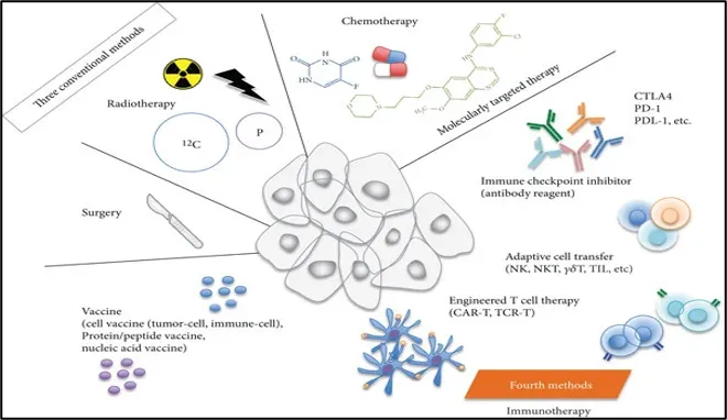
Fig 1: Conventional therapies in cancer
Nanotechnology (nanocarriers)in cancer
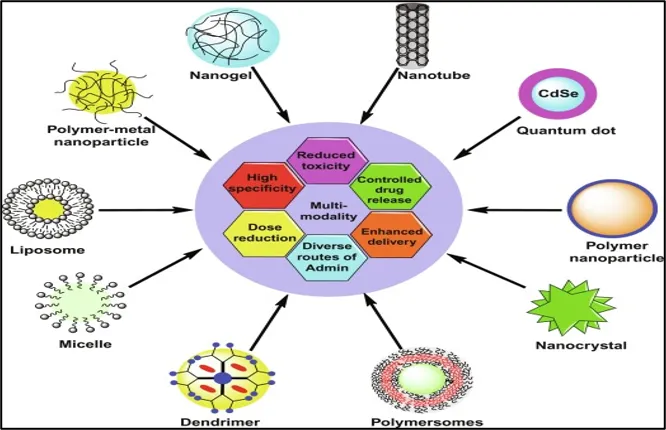
Nanotechnology is the application of materials and devices at the nanoscale (1–100 nm) for diagnosis, treatment, and prevention of diseases. In cancer therapy, nanotechnology has emerged as a promising approach to overcome the limitations of conventional chemotherapy by improving drug delivery, enhancing therapeutic efficacy, and reducing systemic toxicity. The development of nanotechnology is based on the usage of small molecular structures and particles as tools for delivering drugs. Nano-carriers such as liposomes, micelles, dendritic macromolecules, quantum dots, and carbon nanotubes have been widely used in cancer treatment.12 (e.g., bovine serum albumin (BSA) , gelatin , zein, polycaprolactone (PCL, polylactic acid (PLA, chitosan (CS, ultrahigh-molecular-weight polyethylene (UHMWPE) have been widely used in stimulus-responsive nanosearch systems due to their good biological characteristics.13
Advantages of Nanocarriers in Cancer Therapy
Stimulus-Responsive Smart Nanocarriers
Stimulus-responsive smart nanocarriers are advanced drug delivery systems that release therapeutic agents in response to specific internal or external stimuli. These nanocarriers improve targeted drug delivery, reduce side effects, and enhance treatment efficacy by releasing drugs only at the desired site.
Types of Stimuli in Stimulus-Responsive Smart Nanocarriers
1. Internal Stimuli (Endogenous Stimuli)
These stimuli originate from the tumor microenvironment or inside cancer cells.
2. External Stimuli (Exogenous Stimuli)
These stimuli are applied from outside the body to control drug release.
Design Principle
Drug loading
Drug loading refers to the incorporation of therapeutic agents into nanocarriers. An ideal nanocarrier should have high drug-loading capacity, protect the drug from degradation, and maintain stability during circulation. Efficient drug loading ensures adequate drug delivery to the tumor site while minimizing systemic toxicity.
Targeting ligand
Targeting ligands are molecules attached to the surface of nanocarriers that recognize and bind specific receptors overexpressed on cancer cells. Common ligands include folic acid, antibodies, peptides, and aptamers. These ligands enhance selective uptake by tumor cells and improve therapeutic efficacy while reducing off-target effects.
Controlled released mechanism
Controlled release mechanisms enable nanocarriers to release drugs at the desired site and time. In stimulus-responsive systems, drug release is triggered by internal stimuli (pH, redox potential, enzymes) or external stimuli (light, temperature, magnetic field). This approach increases drug concentration at the tumor site and reduces damage to healthy tissues.15
pH-Responsive Nanocarriers
Targeted and controlled drug delivery is possible with pH-responsive nanocarriers, which are clever drug delivery systems that discharge medications in response to the acidic pH of tumor tissues.
Mechanism of pH-Responsive Polymeric Nanocarriers
Due to improved anaerobic metabolism, tumor tissues are more acidic (pH ~6.5) than normal tissues (pH 7.4). This disparity is used by pH-responsive nanocarriers to promote focused medication delivery at the location of the tumor.
The primary mechanism entails the ionization or protonation of pH-sensitive groups, including sulfonate, imidazole, carboxyl, and amino groups. These groups alter their charge under acidic circumstances, disturbing the equilibrium between the hydrophobic and hydrophilic parts of the nanocarrier. This causes the drug to be released as a result of the nanocarrier's swelling, disassembly, or degradation. Other methods include acid-labile bonds that stay stable at physiologic pH but break in acidic tumor conditions, allowing controlled medication release.
pH-sensitive polymers are classified according to their charge as:
Cationic polymers: Become more hydrophilic and protonated at low pH, causing them to expand and release the medication. Drug release and nanocarrier disaggregation.
Anionic polymers: Low pH results in a more hydrophobic and less ionized form of anionic polymers, which causes structural changes and drug release. As a result, pH-responsive nanocarriers offer selective drug delivery, enhanced tumor targeting, and decreased toxicity to healthy tissues.16.
Because they are stable at physiological pH but disintegrate in the acidic tumor environment, hydrazone and imine (Schiff-base) bonds are frequently employed in pH-responsive polymeric nanocarriers. Controlled drug release and increased tumor targeting in acidic environments were observed with HA-DOX nanoparticles with hydrazone links and HA-hydra-DOX-TPP nanoparticles. In a similar vein, HCPT-loaded poly(aspartic acid) micelles containing PEG-imino showed improved antitumor activity and prolonged circulation. pH-responsive release of indocyanine green (ICG) was made possible by benzoic acid-imine cross-linked nanogels. Additionally, PEG-chitosan conjugates that included benzene-imine bonds exhibited faster drug release in environments that were acidic. All in all, these acid-sensitive nanocarriers improve therapeutic efficacy, lessen harm to healthy tissues, and promote targeted medication delivery.15
Important pH-Responsive Nanocarrier Properties
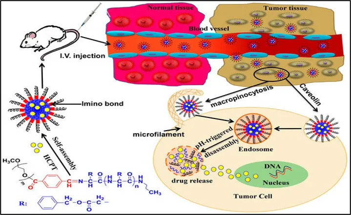
Fig 2: PH responsive model
Summery table:1
|
Stimuli Type 24 |
Nanocarrier |
Cargo |
Cell Line/ Cancer Model |
Main Results |
|
pH-responsive |
cRGD-Dex-DOX/HDZ |
RGD-modified dextran-hydrazone-doxorubicin |
4T1 |
Most prominent antitumor effect with the fewest side effects compared to controls |
|
pH-responsive |
Gd-DTPA/CaP |
Hybrid calcium phosphate (CaP); Gd-diethylenetri-aminepentaacetic acid |
C26 |
Effectively killed cancer cells without damaging healthy tissues |
Cancer Theranostic Applications of pH-Responsive Nanocarriers
pH-responsive nanocarriers selectively release anticancer drugs in the acidic tumor microenvironment, thereby increasing drug accumulation at the tumor site while minimizing toxicity to healthy tissues. This targeted drug delivery approach enhances therapeutic efficacy and reduces systemic side effects associated with conventional chemotherapy. For example, cRGD-Dex-DOX/HDZ nanoparticles demonstrated significant antitumor activity in the 4T1 breast cancer model with fewer side effects compared to conventional treatment.
Stimulus-responsive nanocarriers can carry imaging agents such as fluorescent dyes, MRI contrast agents, or nanoparticles, enabling visualization and monitoring of tumors. These systems improve diagnostic accuracy and allow real-time assessment of disease progression and treatment response.
For example, Gd-DTPA/CaP hybrid nanoparticles have been investigated for tumor imaging and image-guided cancer therapy.
Stimulus-responsive nanocarriers can simultaneously deliver therapeutic drugs and imaging agents, allowing real-time diagnosis, treatment, and monitoring of cancer progression. This integrated strategy, known as theranostics, improves treatment precision and supports personalized cancer therapy.
For example, Gd-DTPA/CaP nanoparticles have shown potential as theranostic platforms by combining imaging and therapeutic functions within a single nanosystem.
Advantages
Targeted and controlled drug release.
Reduced side effects and systemic toxicity.
Improved drug stability and bioavailability.
Enhanced therapeutic efficacy.
Suitable for teranostic applications.
Application 18
Redox-Responsive Polymeric Nanocarriers
Glutathione (GSH) levels are higher in tumor cells than in healthy cells, resulting in a reducing intracellular environment. To achieve targeted medication delivery, redox-responsive nanocarriers utilize this discrepancy.
Mechanism:
With disulfide bonds (–S–S–), which are stable in blood circulation but are broken down by the high GSH concentration in tumor cells, these nanocarriers are often created. Disulfide bond cleavage causes the nanocarrier to disintegrate or disassemble, allowing the loaded medication to be released in a controlled manner. In polymeric nanocarriers, disulfide bonds may be present in the side chain, main chain, or cross-linked structure. This redox-triggered medication release increases medication buildup in cancer cells, improves therapeutic effectiveness, and lowers toxicity to healthy tissues.17
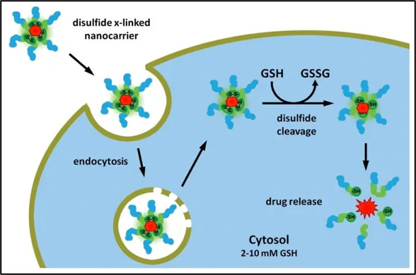
Fig 3: redox mechanism19
Summary: 2
|
Stimuli Type
|
Nanocarrier
|
Cargo |
Cell Line/ Cancer Model |
Main Results |
|
Redox-responsive
|
DHA2-SS |
Dihydroartemisinin (DHA) |
HepG2; H22 tumor
|
Demonstrated improved therapeutic efficacy and safety compared with free DHA |
|
Redox-responsive
|
Transferrin-Gated Mesoporous Silica Nanoparticles |
Anticancer drug-loaded mesoporous silica nanoparticles |
Cancer cells |
Achieved redox-responsive and targeted drug delivery with enhanced intracellular drug |
Redox-Responsive Nanocarriers Used in Cancer Theranostic Applications
Targeted Drug Delivery: Due to increased GSH levels, drugs are specifically released inside cancer cells.
Deliver imaging agents for cancer detection and monitoring: Theranostics is a combination of therapy and diagnosis. This is done by delivering medication and imaging in the same platform.
Gene delivery: The effective delivery of genes and nucleic acids to cancer cells.
Targeted Intracellular Drug Release: Boosts treatment effectiveness and minimizes off-target reactions.
Advantages
Highly specific drug release in cancer cells.
Improved therapeutic efficacy.
Reduced toxicity to healthy tissues.
Controlled and on-demand drug release.
Light responsive nanocarriers
Light-responsive nanocarriers are intelligent medication delivery systems that release drugs at the target site in response to light irradiation. They are often created using organic materials (carbon-based nanoparticles, fluorophores, and photosensitizers) and inorganic materials (quantum dots, gold nanoparticles, and upconversion nanoparticles).
Mechanism:
Upon exposure to a specific wavelength of light (UV, visible, or near-infrared), the nanocarrier undergoes structural changes or generates heat/reactive oxygen species (ROS), which triggers a regulated drug release. In addition to causing the destruction of cancer cells, light may also induce photothermal therapy (PTT) or photodynamic therapy (PDT).18
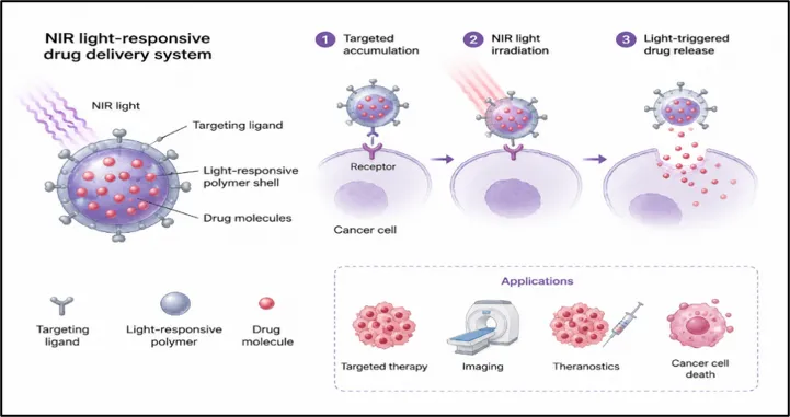
Fig 4 light responsive model
|
Stimuli Type
|
Nanocarrier
|
Cargo |
Cell Line/ Cancer Model |
Main Results |
|
Light-responsive |
Gold Nanoparticle-Based Nanocarriers |
Anticancer drugs and photothermal agents |
HeLa |
Generated heat under NIR irradiation, leading to efficient tumor cell destruction |
Cancer Theranostic Applications
Advantages
Accurate spatial and temporal management of medication administration.
Non-invasive stimulation.
Enhanced therapeutic efficacy.
Comparison Table: 3
|
Parameter |
Light-Responsive Nanocarriers |
pH-Responsive Nanocarriers |
Redox-Responsive Nanocarriers |
|
Stimulus |
External light (UV, visible, NIR) |
Acidic tumor microenvironment |
High intracellular glutathione (GSH) levels |
|
Mechanism of Drug Release |
Light-induced structural change, heat generation, or ROS production |
Protonation or cleavage of pH-sensitive bonds in acidic conditions |
Cleavage of disulfide bonds by GSH |
|
Type of Stimulus |
Exogenous |
Endogenous |
Endogenous |
|
Target Site |
Light-irradiated tumor region |
Acidic tumor tissues and endosomes |
Tumor cells with high GSH concentration |
|
Advantages |
Precise activation, non-invasive control, supports PDT/PTT |
Simple design, targeted release, reduced systemic toxicity |
Efficient intracellular drug release, enhanced tumor targeting |
Prospects for the future
Intelligent Nanocarriers That Respond to Light
Future research will concentrate on enhancing tumor specificity, photothermal and photodynamic efficiency, and systems that respond to near-infrared (NIR) light with deeper tissue penetration. It is anticipated that integration with imaging modalities would facilitate real-time cancer diagnosis and treatment monitoring.
Smart Nanocarriers that Respond to pH
Better selective medication delivery in acidic tumor microenvironments and lower systemic toxicity will be possible thanks to advancements in pH-sensitive materials. Therapeutic outcomes may be improved by targeting and imaging ligands integrated with pH-responsive and multifunctional platforms.
Nanocarriers that respond to redox signals
Heightened intracellular glutathione levels are anticipated to be used in future redox-responsive systems for precise drug release. Their efficacy in treating metastatic and resistant tumors may be enhanced by combining them with gene therapy, immunotherapy, and combination treatments.
Applications of Theranostic Cancer Treatment
Personalized cancer therapy has a bright future with the creation of multipurpose theranostic nanocarriers that can simultaneously diagnose, deliver medication in a targeted manner, and monitor treatment. Better biocompatibility, scalability, and long-term safety evaluation will be necessary for clinical translation.26
CONCLUSION
Targeted cancer therapy and diagnostics may find creative applications for stimulus-responsive smart nanocarriers. To get regulated drug release, they answer particular inputs like pH, redox state, and light. These nanocarriers lower overall toxicity and improve drug buildup at tumor sites. While redox-responsive carriers use high intracellular glutathione levels, pH-responsive systems leverage the acidic tumor microenvironment. Light-sensitive nanocarriers give exact control over the activation of therapy. Combining diagnostic and therapeutic tasks helps real-time tracking of treatment. These tools help to reduce negative effects and raise treatment success. Multi-stimuli-responsive devices improve treatment results and targeting accuracy even more. Still existing, though, are problems with safety, stability, and high-volume manufacturing. More research is needed to solve these restrictions and increase clinical utility. Nanotechnology advances are projected to hasten their integration into clinical application. Personalized cancer treatment and precision medicine will both benefit much from stimulus-responsive intelligent nanocarriers going forward.
REFERENCES


Kishor Deshmukh, Dr. Anil Jadhav, Dr. Atul Bendale, Prajwal Aher, Stimulus-Responsive Smart Nanocarriers (Light, PH, Redox): Mechanisms and Cancer Theranostic Applications, Int. J. of Pharm. Sci., 2026, Vol 4, Issue 6, 6196-6209. https://doi.org/10.5281/zenodo.20835795
 10.5281/zenodo.20835795