

We use cookies to ensure our website works properly and to personalise your experience. Cookies policy
Department of Pharmaceutics, G.P. Pharmacy College.
Narcolepsy, a chronic neurological disorder characterized by excessive daytime sleepiness and disrupted night-time sleep, significantly impacts the quality of life for affected individuals. While oral medications have been the mainstay of treatment, they often present challenges related to fluctuating drug levels, potential side effects, and adherence. In response, the development of transdermal drug delivery systems has emerged as a promising avenue for providing sustained and controlled therapeutic benefits. This review delves into the intricate realm of pharmaceutical formulation and manufacturing aspects that underpin the creation of effective transdermal patches for narcolepsy. The skin, acting as a sophisticated portal, presents both opportunities and obstacles for drug delivery. Overcoming the stratum corneum's barrier function requires meticulous formulation design, encompassing the selection of appropriate penetration enhancers, the development of advanced drug reservoirs, and the optimization of adhesive technologies. Furthermore, the precise control of drug release kinetics is paramount in narcolepsy, where consistent wakefulness is crucial. This necessitates the exploration of innovative manufacturing techniques, including microneedle fabrication, polymer matrix engineering, and advanced coating processes.
"Narcolepsy, a chronic neurological disorder characterized by excessive daytime sleepiness and disrupted night-time sleep, significantly impacts the quality of life for affected individuals. While oral medications have been the mainstay of treatment they often present challenges related to fluctuating drug levels, potential side effects, and adherence. In response, the development of transdermal drug delivery systems has emerged as a promising avenue for providing sustained and controlled therapeutic benefits. This review delves into the intricate realm of pharmaceutical formulation and manufacturing aspects that underpin the creation of effective transdermal patches for narcolepsy.[1]
The skin, acting as a sophisticated portal, presents both opportunities and obstacles for drug delivery. Overcoming the stratum corneum's barrier function requires meticulous formulation design, encompassing the selection of appropriate penetration enhancers, the development of advanced drug reservoirs, and the optimization of adhesive technologies. Furthermore, the precise control of drug release kinetics is paramount in narcolepsy, where consistent wakefulness is crucial. This necessitates the exploration of innovative manufacturing techniques, including microneedle fabrication, polymer matrix engineering, and advanced coating processes.[2]
Formulation Aspects for Transdermal Delivery
The development of effective transdermal drug delivery systems (TDDS) requires meticulous consideration of the physicochemical properties of the drug as well as the challenges posed by the skin’s natural barrier. An ideal drug candidate for transdermal delivery should exhibit high potency, ensuring therapeutic efficacy at low doses suitable for the limited drug-loading capacity of patches.
A molecular weight under 500 Daltons is preferred, as smaller molecules traverse the skin more efficiently. Lipophilicity, expressed by a log P value between 1 and 3, provides an optimal balance between aqueous and lipid solubility, enhancing skin permeation while avoiding excessive accumulation in the lipid layers.
Additionally, drugs with low melting points generally exhibit better solubility in formulation matrices, simplifying patch fabrication. Skin tolerance is another essential consideration; the drug and its excipients must be non-irritating and non-sensitizing to minimize local reactions. A wide therapeutic index offers dosing flexibility and reduces systemic toxicity risks, which is particularly beneficial in chronic conditions such as narcolepsy. Chemical and physical stability throughout manufacturing, storage, and application phases is also crucial, as degradation can reduce efficacy and safety. Furthermore, pharmacokinetic properties that support sustained drug release and the maintenance of therapeutic plasma levels are ideal for ensuring prolonged symptom control without frequent dosing.[3]
In the treatment of narcolepsy, several drugs have shown potential for transdermal administration. Modafinil, a widely used wakefulness-promoting agent, meets many transdermal criteria due to its low molecular weight and moderate lipophilicity, supporting effective skin penetration and sustained action. Armodafinil, the pharmacologically active R-enantiomer of modafinil, shares similar properties but may offer enhanced potency and prolonged effects, making it another attractive candidate. Sodium oxybate (GHB), despite its therapeutic utility in reducing cataplexy and daytime sleepiness, poses formulation challenges due to its high water solubility and low lipophilicity. Nonetheless, advanced delivery approaches such as prodrugs or chemical enhancers could improve its dermal permeability. Pitolisant, a selective histamine H3 receptor inverse agonist, offers a novel mechanism of action and moderate molecular weight, rendering it a promising molecule for transdermal innovation.[4]
Despite promising candidates, several formulation challenges must be addressed to ensure effective transdermal therapy. The primary obstacle is the stratum corneum, the outermost layer of the skin, which serves as a formidable barrier to drug permeation. Overcoming this requires strategic enhancement techniques. Chemical penetration enhancers can temporarily disrupt the lipid matrix of the stratum corneum to increase permeability, while iontophoresis employs mild electrical currents to facilitate the movement of charged or polar drugs across the skin. Microneedle arrays offer a minimally invasive alternative by creating microchannels that bypass the stratum corneum without reaching nerve endings, thus enhancing delivery without pain. Additionally, liposomal and nanoparticulate carriers can encapsulate drugs, improving their solubility, stability, and controlled release while enhancing permeation.
Stability and compatibility are also pivotal formulation concerns. Transdermal systems must be designed to protect the drug from environmental degradation caused by factors such as temperature, humidity, pH, and light exposure. Chemically, the drug must remain stable when combined with excipients, as undesirable interactions can lead to degradation or loss of efficacy. Physical incompatibilities, such as phase separation or crystallization within the patch matrix, can alter drug release kinetics. Furthermore, all components of the transdermal system—including adhesives, backing layers, and permeation enhancers—must be dermatologically safe to prevent irritation, sensitization, or allergic reactions upon application. Therefore, an integrated understanding of both drug characteristics and formulation science is essential for advancing transdermal delivery as a viable strategy in the management of narcolepsy.
Enhancers and Carriers in Transdermal Delivery
To overcome the inherent barrier properties of the stratum corneum and ensure effective drug penetration, transdermal delivery systems often incorporate permeation enhancers and specialized carriers. Permeation enhancers function by transiently modifying the skin’s barrier to facilitate the transport of therapeutic molecules. These enhancers are broadly categorized into chemical and physical types. Chemical enhancers primarily act by disrupting the lipid architecture of the stratum corneum, thus increasing its permeability. Common examples include alcohols such as ethanol and isopropanol, which enhance both drug solubility and skin permeability through lipid disruption. Fatty acids, notably oleic acid, can reversibly fluidize stratum corneum lipids to create temporary channels for drug diffusion. Surfactants like sodium lauryl sulfate (SLS) increase skin permeability by altering protein and lipid structures, particularly benefiting the delivery of hydrophilic drugs. Another potent enhancer, Azone, facilitates drug diffusion by inserting into the lipid bilayers and reducing their resistance to molecular movement. Complementing chemical methods, physical enhancement techniques utilize external stimuli to breach the skin barrier directly. Microneedles create microscopic channels that bypass the stratum corneum, enabling painless and efficient delivery of drugs. Iontophoresis employs a mild electrical current to propel charged molecules across the skin, while ultrasound-based sonophoresis uses high-frequency sound waves to temporarily disorganize the lipid matrix and enhance permeation.[5]
Alongside these enhancers, carriers and polymers are critical components in modern transdermal systems, offering drug stability, controlled release, and improved bioavailability. Carriers such as hydrogels are composed of hydrophilic polymer networks that swell upon hydration, forming a moist matrix ideal for drug encapsulation and sustained release. Their versatility makes them suitable for both hydrophilic and lipophilic drugs. Liposomes, composed of biocompatible phospholipid bilayers, encapsulate active compounds and facilitate deeper skin penetration while protecting the drug from enzymatic degradation. Nanoparticles, including solid lipid nanoparticles (SLNs) and nanostructured lipid carriers (NLCs), provide an advanced platform for controlled release, enhanced permeation, and improved drug targeting. These nanosystems can be engineered to optimize particle size, surface charge, and composition for maximal therapeutic effect. Polymers play a structural and functional role in transdermal formulations. For instance, polyvinyl alcohol (PVA) is valued for its excellent film-forming capacity and biocompatibility, making it ideal for patch matrices.
Polyethylene glycol (PEG) contributes to patch flexibility and skin hydration, enhancing user comfort and drug diffusion. Chitosan, a natural polysaccharide derived from chitin, offers the added benefit of mucoadhesion and intrinsic permeation-enhancing effects, particularly useful for bioadhesive transdermal systems.Collectively, these enhancers and carriers form the foundation of sophisticated transdermal delivery strategies, enabling the effective administration of drugs for chronic conditions like narcolepsy while improving patient compliance and therapeutic consistency.[6]
Mechanisms of Transdermal Drug Delivery
The success of transdermal drug delivery relies on a comprehensive understanding of the skin’s complex architecture and the pathways available for drug permeation. The skin is a multilayered organ that acts as the body's primary barrier against environmental insults while selectively regulating the penetration of external substances, including therapeutic agents. The outermost layer, the epidermis, serves as the skin’s primary protective interface. Within the epidermis lies the stratum corneum, a formidable barrier composed of terminally differentiated keratinocytes embedded in a lipid matrix. This structure resembles a “brick and mortar” system, where the keratin-filled cells (bricks) are surrounded by intercellular lipids (mortar), forming a highly hydrophobic and compact layer.
The stratum corneum is the principal obstacle to transdermal drug delivery due to its dense, non-living structure. Beneath the epidermis lies the dermis, a vascularized connective tissue matrix containing blood vessels, nerve endings, and lymphatics. Once a drug successfully penetrates the stratum corneum, it enters the dermis where it can be absorbed into the systemic circulation. Below the dermis is the hypodermis or subcutaneous tissue, primarily composed of adipose tissue and connective fibers. While this layer does not directly influence drug permeation, it provides mechanical support and aids in thermal regulation, indirectly impacting the pharmacokinetics of transdermally delivered agents.[7]
Drugs administered via the transdermal route may follow one or more permeation pathways to reach systemic circulation. The most prominent route is through the stratum corneum, which offers two possible paths: the intercellular route, wherein the drug diffuses around the corneocytes through the lipid-rich matrix, and the transcellular route, which involves sequential passage through the keratin-filled cells and their surrounding lipid domains. The intercellular pathway is generally favored by lipophilic drugs due to the lipid-dense environment, while the transcellular path requires drugs to alternate between hydrophilic and lipophilic domains, posing greater challenges to permeation. In addition to these main routes, hair follicles provide an accessory pathway for drug penetration. Though hair follicles occupy a relatively small fraction of the skin's surface area, they bypass the stratum corneum and are associated with sebaceous glands, which can facilitate the entry of larger molecules and hydrophilic drugs. This follicular route is particularly valuable for targeting localized or systemic effects in formulations involving nanoparticles or other carriers designed to localize within follicular openings. Sweat glands, while less significant in terms of surface area and permeability, represent a minor adjunct pathway for drug delivery, especially under conditions that increase sweat production or with formulations designed to exploit these eccrine ducts. Together, these transdermal pathways underscore the importance of drug physicochemical properties, formulation strategies, and enhancer technologies in optimizing delivery efficiency through the skin’s robust defensive layers.[8]
Structure of skin:
The epidermis comprises several distinct layers, each playing a vital role in skin function and integrity. The stratum basale (or basal layer) is the deepest layer, consisting of a single row of mitotically active basal cells that continuously divide to replenish the skin. These basal keratinocytes are anchored to the basement membrane and give rise to all the upper layers. This layer also houses melanocytes, which produce melanin for pigmentation, and Merkel cells, which contribute to touch sensation. Above it lies the stratum spinosum, often referred to as the prickle cell layer due to the desmosomal connections between keratinocytes that appear spiny under a microscope. [9]
This layer provides structural support and houses Langerhans cells, which are key players in the skin’s immune defense. Moving outward, the stratum granulosum or granular layer is where keratinocytes begin to flatten and accumulate keratohyalin granules, crucial for keratin formation. Here, lipid secretion also occurs, contributing to the epidermal water barrier.
Just above is the stratum lucidum, a thin, transparent layer found only in thick skin—such as the palms of the hands and soles of the feet. It consists of several layers of dead, flattened keratinocytes and provides an additional barrier for high-friction areas. Together, these layers work in harmony to protect the body from mechanical injury, microbial invasion, and water loss. [10]
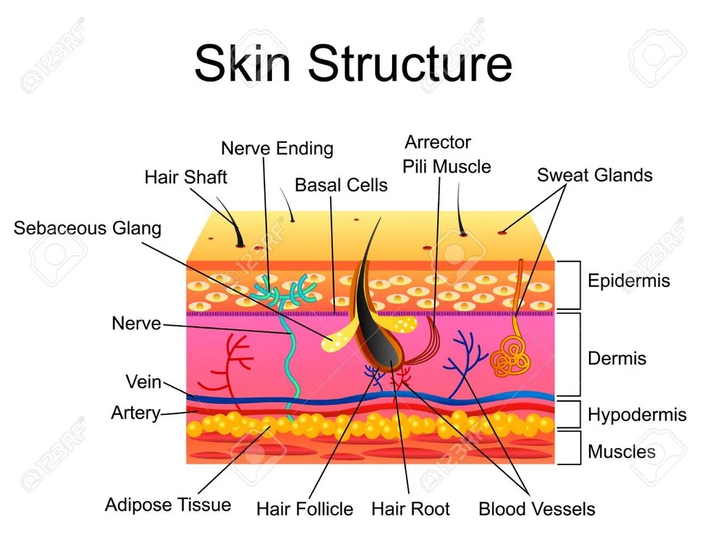
Figure 1: Structure of Skin
The stratum corneum is the outermost layer of the epidermis and serves as the body’s primary barrier against the external environment. It is composed of 15–20 layers of flattened, dead keratinocytes known as corneocytes. These cells are embedded in a lipid matrix made up of ceramides, cholesterol, and free fatty acids, which together form a “brick-and-mortar” structure—corneocytes as the bricks and lipids as the mortar. This structure provides a robust, impermeable shield that protects underlying tissues from mechanical stress, microbial invasion, chemical irritation, and excessive water loss. Since the cells in this layer are anucleated (without nuclei), they are biologically inactive, but structurally vital for skin integrity.[11]
The stratum corneum also plays a critical role in maintaining skin hydration and regulating transepidermal water loss (TEWL). The lipids between the cells act as a waterproof seal, while natural moisturizing factors (NMFs) within corneocytes help retain moisture. Desquamation—the process of shedding dead cells from the surface—is tightly regulated to balance cell renewal and barrier function. Disruption in this layer can lead to various skin disorders, such as eczema, psoriasis, and ichthyosis, where the barrier becomes compromised. Moreover, the stratum corneum is a major focus in transdermal drug delivery, as overcoming its barrier function is essential for effective drug penetration. Overall, despite its non-living nature, the stratum corneum is indispensable in preserving homeostasis and protecting the body.[12]
Transdermal Systems and Technologies
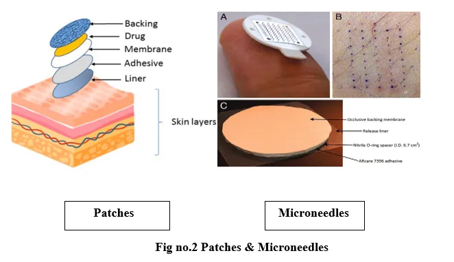
1.Patches
Transdermal patches are adhesive drug delivery systems engineered to transport therapeutic agents through the skin into systemic circulation. These systems consist of several key components that work in unison to ensure effective drug administration. The backing layer serves as a protective barrier, providing mechanical support and shielding the patch from external elements. The drug reservoir contains the active pharmaceutical ingredient, which can be formulated as a gel, liquid, or solid. In reservoir-type patches, a control membrane is included to regulate the drug release rate. The adhesive layer not only ensures the patch adheres securely to the skin but may also incorporate the drug in certain designs. Prior to application, a release liner covers the adhesive surface and is removed to activate the patch. There are primarily two types of transdermal patches: matrix patches, where the drug is uniformly dispersed within a polymeric matrix, and reservoir patches, where the drug is stored in a distinct liquid or gel reservoir beneath a rate-controlling membrane.[13]
The use of transdermal patches offers several therapeutic advantages. They enable controlled and sustained drug release over an extended duration, enhancing treatment efficacy and patient convenience. Additionally, they are non-invasive, reduce the frequency of dosing, and minimize adverse effects associated with oral or injectable routes. Transdermal systems are widely employed in clinical settings for nicotine replacement therapy, hormone replacement, and pain management.[14]
2.Gels and Creams
Gels and creams are topical semi-solid formulations widely used to facilitate the diffusion of drugs through the skin layers. Gels are typically clear, jelly-like preparations, often water-based, that offer uniform drug distribution and ease of application. They are known for their cooling effect and are generally less greasy compared to ointments, making them more cosmetically acceptable. In contrast, creams are emulsions composed of oil and water, either as oil-in-water or water-in-oil systems. These formulations enhance skin hydration and drug penetration, and their composition can be adapted for either lipophilic or hydrophilic drugs, depending on the emulsion type. Both gels and creams offer several advantages in transdermal drug delivery. They are versatile, allowing for localized or systemic delivery, and can be tailored to specific therapeutic needs. Moreover, these formulations are user-friendly, easy to apply, and generally well-tolerated, making them especially suitable for use on sensitive skin areas.[15]
3.Microneedles
Microneedles are microscopic, needle-like structures designed to enhance transdermal drug delivery by penetrating the outermost layer of the skin. These projections typically range from 50 to 900 micrometers in length and are engineered to create temporary microchannels that bypass the stratum corneum, the primary barrier to drug permeation. Microneedles are categorized into different types based on their structure and mechanism of drug delivery. Solid microneedles are used to pre-treat the skin by creating microchannels through which topical drugs can later be applied. Dissolving microneedles, made from biodegradable polymers, encapsulate drugs within the needle matrix and dissolve upon insertion, releasing the therapeutic agents directly into the skin. Hollow microneedles are equipped with internal channels that allow direct infusion of drugs into the skin layers. These systems offer significant advantages for enhancing drug delivery. By forming microscopic pores, microneedles enable efficient drug transport while being painless and minimally invasive. They are suitable for a wide range of applications, including the delivery of vaccines, therapeutic drugs, and even cosmetic agents, offering a promising alternative to conventional transdermal systems.[16]
Nanocarrier Techniques for Transdermal Drug Delivery
Nanocarriers have emerged as advanced drug delivery platforms that significantly enhance the permeability and therapeutic efficacy of drugs administered via the transdermal route. These nanoscale systems encapsulate or bind the drug, facilitating its transport across the stratum corneum — the major barrier of the skin. Their flexibility, controlled release properties, and potential for targeting specific tissues make them highly suitable for conditions requiring sustained drug delivery, such as narcolepsy.[17,18,19]
1. Liposomes
Liposomes are microscopic spherical vesicles composed of one or more phospholipid bilayers enclosing an aqueous core. Due to their structural similarity to biological membranes, liposomes can merge with the stratum corneum lipids, facilitating drug diffusion into deeper layers of the skin. They can encapsulate both hydrophilic and lipophilic drugs, offering protection from enzymatic degradation and enhancing stability. Furthermore, liposomes are biocompatible and biodegradable, making them a safe option for transdermal delivery. However, their penetration capability through intact skin is limited, and they are prone to instability, such as aggregation and oxidation during storage.[20,21,22,23]
2. Niosomes
Niosomes are vesicular carriers made from non-ionic surfactants and cholesterol, structurally similar to liposomes but more stable. They offer several advantages over liposomes, including improved shelf life, chemical stability, and cost-effectiveness. Niosomes enhance drug penetration and retention in the skin layers by modifying the stratum corneum’s structure.
They are capable of carrying a wide range of drugs, both hydrophilic and lipophilic, and are particularly useful in transdermal systems where controlled and targeted delivery is desired. Their use in delivering CNS-acting drugs via the skin has been gaining attention due to their low toxicity and high encapsulation efficiency.[24,25,26,27]
3. Ethosomes
Ethosomes are soft, malleable lipid vesicles that contain high concentrations of ethanol, which is the key enhancer in this system. Ethanol disrupts the lipid organization of the stratum corneum, increasing its permeability and fluidity. This allows the ethosomes to penetrate more efficiently and carry both hydrophilic and lipophilic drugs into deeper layers or even systemic circulation. Ethosomes are especially effective for transdermal delivery of large molecules and poorly permeable drugs. However, the high ethanol content, while beneficial for permeation, may occasionally lead to skin irritation or dryness upon prolonged use.[28,29,30,31]
4. Transfersomes
Transfersomes are ultra-deformable, elastic vesicles composed of phospholipids and edge activators like sodium cholate or Tween 80. These edge activators make the vesicle highly flexible, enabling it to squeeze through the narrow pores and intercellular gaps of the skin barrier without rupturing. Transfersomes can transport drugs more effectively than traditional vesicles, making them highly suitable for systemic delivery through the skin. Their ability to deliver large biomolecules and poorly soluble drugs has led to growing interest in their application for central nervous system disorders, including narcolepsy, where sustained drug release is critical.[32,33,34,35]
5. Solid Lipid Nanoparticles (SLNs)
Solid Lipid Nanoparticles (SLNs) are submicron colloidal carriers composed of solid lipids that remain solid at both room and body temperatures. These carriers provide a matrix for drug entrapment, allowing for controlled release and improved drug stability. SLNs are known for their low toxicity, ability to protect labile drugs from degradation, and potential to provide long-term storage stability. However, they often suffer from low drug loading capacity and the possibility of drug expulsion during lipid crystallization, which can limit their effectiveness in some formulations.[36,37,38,39]
6. Nanostructured Lipid Carriers (NLCs)
Nanostructured Lipid Carriers (NLCs) represent the next generation of lipid nanoparticles designed to overcome the limitations of SLNs. They are made by blending solid lipids with liquid lipids, creating an imperfect matrix that enhances drug loading and reduces the risk of drug expulsion. NLCs offer improved skin hydration, prolonged drug release, and enhanced permeation across the skin barrier. Their structural flexibility makes them ideal for transdermal delivery of chronic medications, including those used in narcolepsy, where maintaining consistent plasma drug levels is essential for effective symptom control.[40,41,42,43]
Preclinical and Clinical Studies in Transdermal Drug Delivery
The development of transdermal drug delivery systems (TDDS) necessitates rigorous preclinical and clinical evaluations to ensure their efficacy, safety, and pharmacokinetic suitability. Preclinical studies begin with in vitro models, which serve as a foundational step in screening and optimizing formulations. Among the most widely used techniques are skin diffusion studies, often employing Franz diffusion cells, where excised human or animal skin is mounted between donor and receptor compartments. These studies enable quantitative assessment of drug permeation across skin layers, evaluation of release kinetics, and estimation of the potential for systemic drug delivery.[44]
Additionally, artificial skin models such as Epidermis and Skin Ethic—composed of synthetic or bioengineered constructs—mimic the stratum corneum and viable epidermis, offering ethical and reproducible alternatives for assessing drug penetration, irritation potential, and formulation behaviour under controlled conditions.[45]
Following promising in vitro results, animal models are employed to understand the pharmacokinetics, systemic absorption, safety, and efficacy of transdermal formulations in a biological context. Rodents such as rats and mice are commonly used for preliminary studies, providing data on systemic bioavailability and therapeutic effects. However, due to anatomical and permeability differences, these models often require correlation with larger animals. Pigs, particularly miniature swine, are favoured in dermal studies because their skin closely resembles human skin in structure, lipid content, and thickness. Likewise, hairless guinea pigs are valuable in transdermal research due to their minimal fur barrier and well-characterized permeability profiles. Data generated from these models assist in refining formulations and dose predictions prior to human trials.[46,47]
Clinical studies play a pivotal role in validating the therapeutic promise of transdermal delivery in human populations. Clinical trials for narcolepsy-related TDDS have explored transdermal delivery of wake-promoting agents such as modafinil and armodafinil. These trials assess multiple endpoints including pharmacokinetics, therapeutic efficacy, and patient-reported outcomes. Notably, transdermal systems have demonstrated sustained drug release, resulting in stable plasma drug concentrations and reduced dosing frequency, which are particularly beneficial for managing chronic conditions like narcolepsy that require long-term therapy. Patients using transdermal formulations often report improved wakefulness, reduced daytime sleepiness, and greater adherence compared to oral dosing, likely due to the non-invasive, convenient nature of patches or gels.[48,49]
From a safety standpoint, the most frequently reported adverse effects include mild skin irritation or sensitization at the application site. Clinical findings emphasize the importance of selecting biocompatible polymers, using non-irritating excipients, and ensuring formulation stability under physiological conditions. Continued innovations in formulation science and clinical validation strategies will enhance the development of safer, more effective transdermal therapies for narcolepsy and other central nervous system disorders.[50]
Case Study 1: Coexistence of Narcolepsy Type I and Psychosis in an Adolescent
This case study examines a rare and complex presentation involving the coexistence of narcolepsy type I (NT1) and psychosis in a 14-year-old girl. The case highlights the diagnostic and therapeutic challenges that arise when managing comorbid neurological and psychiatric conditions in adolescents. The patient initially presented with symptoms of excessive daytime sleepiness, impaired nighttime sleep, binge eating, and notable weight gain. She also had a history of attention-deficit/hyperactivity disorder (ADHD) and a family history of schizophrenia, which further complicated the clinical picture.[51]
She was diagnosed with narcolepsy type I, characterized by excessive daytime sleepiness, cataplexy, and hallucinations, with an underlying etiology linked to hypocretin deficiency and HLA-DQB1*06:02 positivity. Treatment began with modafinil and sodium oxybate to manage narcolepsy symptoms. However, the patient subsequently developed psychotic symptoms, necessitating a change in medication. She was switched to haloperidol, which effectively managed the psychosis but led to a worsening of narcoleptic symptoms.
This case underscores the complex interplay between narcolepsy and psychosis, which poses significant challenges for both diagnosis and treatment. The need for careful clinical monitoring is emphasized, particularly when balancing therapies that may have opposing effects on the coexisting conditions. In conclusion, this rare case of concurrent NT1 and psychosis in an adolescent highlights the importance of understanding potential shared pathophysiological mechanisms and developing integrated treatment strategies for such comorbid presentations.
Case Study 2: Familial Narcolepsy in a Young Woman and Her Father
This case study presents the familial occurrence of narcolepsy, a chronic neurological disorder marked by excessive daytime sleepiness, cataplexy, and REM sleep abnormalities. Due to symptom overlap with other neurological and psychiatric conditions, narcolepsy is often misdiagnosed, delaying appropriate treatment. The subject of this case is a 22-year-old female who exhibited classical symptoms of narcolepsy, including persistent daytime drowsiness, cataplexy, and sleep paralysis. A detailed medical history revealed that her father exhibited similar symptoms, prompting further investigation into a potential genetic link.[52]
Both the patient and her father were diagnosed with narcolepsy and tested positive for the HLA-DQB1*0602 allele, a genetic marker commonly associated with narcolepsy type I. Treatment with modafinil, a wakefulness-promoting agent, and venlafaxine, an antidepressant effective in suppressing cataplexy, led to significant symptomatic improvement in both individuals.
This case underscores the critical importance of an accurate and timely diagnosis, especially in the presence of a familial pattern. Recognizing genetic predispositions such as the HLA-DQB1*0602 allele can aid in early identification and management, ultimately improving patient outcomes in narcolepsy.[53]
Case Study 3: Health and Social Outcomes in Elderly Patients with Narcolepsy with Cataplexy
This case study investigates the long-term health and social outcomes of older adults diagnosed with narcolepsy with cataplexy (NC), a chronic neurological disorder characterized by excessive daytime sleepiness and sudden muscle weakness due to hypocretin neuron deficiency. Affecting approximately 0.045% of the population, NC has been well-studied in younger populations, but limited research exists on its progression and impact in older adults. The objective of this study was to evaluate the health status, cognitive function, social engagement, and symptom progression in individuals over 60 years of age with NC, compared to age- and sex-matched controls.[54]
The study involved 42 NC patients aged 60 and above and 46 control participants, with a mean age of 71.9 years. Findings revealed that NC patients experienced higher rates of hypertension and diabetes, indicating a greater burden of physical comorbidities. Interestingly, cognitive function did not significantly differ between the NC group and the control group, and levels of social engagement were comparable. However, depression scores were elevated in the NC group, suggesting a greater psychological impact.[55]
In discussion, the results demonstrate that while cognition and social activity remain preserved, elderly NC patients show signs of poorer physical health and are more prone to depressive symptoms, highlighting the need for ongoing clinical monitoring and support. The study concludes that although older patients with NC maintain mental sharpness and social connectivity, they are at an increased risk for comorbid physical conditions, necessitating targeted healthcare strategies to improve quality of life in this population.[56]
Regulatory and Commercial Aspects of Transdermal Drug Delivery
The successful development and commercialization of transdermal drug delivery systems (TDDS) require compliance with comprehensive regulatory frameworks established by major health authorities such as the U.S. Food and Drug Administration (FDA) and the European Medicines Agency (EMA). The FDA offers detailed guidance on the quality and performance standards for transdermal and topical delivery systems. These include specifications for drug release profiles, product stability, skin compatibility, and residual drug content, which refers to the amount of drug remaining in the patch after use—a critical safety concern. Additionally, developers must demonstrate adhesive performance over the intended wear period and ensure that the formulation does not cause irritation or sensitization. Similarly, the EMA provides regulatory directives that emphasize the pharmaceutical development, manufacturing processes, and quality control strategies specific to transdermal patches. EMA guidelines also outline expectations for the dissolution characteristics, skin permeation efficiency, and composition of transdermal systems to ensure consistent therapeutic performance.[57,58]
The regulatory approval process for TDDS follows a structured pathway that includes preclinical studies, where drug safety and efficacy are validated through in vitro testing and animal models, followed by clinical trials to evaluate pharmacokinetics, therapeutic benefits, and tolerability in human subjects. Upon successful trial outcomes, a regulatory dossier is submitted, containing detailed data on formulation development, manufacturing protocols, stability profiles, and clinical evidence. Approval does not mark the end of regulatory oversight; post-marketing surveillance is mandatory to monitor real-world safety and effectiveness, particularly concerning adverse skin reactions and product reliability.[59,60]
From a commercial perspective, the transdermal drug delivery market is expanding rapidly due to the increasing demand for non-invasive, patient-friendly, and controlled-release formulations. Transdermal technologies are especially promising in managing chronic conditions such as narcolepsy, where sustained drug delivery and improved patient adherence are crucial. Innovations such as microneedle arrays, iontophoretic devices, and nanocarrier-based systems are propelling the field forward, offering new possibilities for enhanced skin permeation and targeted delivery. However, commercialization is not without challenges. Manufacturing complexities such as ensuring uniform drug distribution, maintaining adhesive integrity, and achieving product stability present significant hurdles. Scalability remains a concern, as translating lab-scale formulations to mass production while maintaining batch-to-batch consistency demands advanced manufacturing infrastructure and quality control measures.[61,62,63]
In addition to technical challenges, regulatory compliance continues to be demanding, particularly regarding residual drug levels, skin tolerability, and device reliability. The high cost of research and development, coupled with the need for specialized manufacturing equipment, represents a substantial investment. Therefore, companies must balance cost-efficiency with innovation to ensure the final product is not only effective and compliant but also affordable and competitive in the pharmaceutical market. Despite these challenges, the increasing adoption of TDDS reflects strong commercial potential, particularly for central nervous system disorders like narcolepsy, where improved delivery mechanisms can significantly enhance quality of life for patients.[64,65]
Methods of Preparation for Transdermal Patches
Hot-Melt Extrusion Method
The hot-melt extrusion method involves heating a mixture of the drug and polymer to a molten state and then extruding it through a mold to form thin films. This process eliminates the need for solvents, making it more environmentally friendly and cost-effective. Once the extruded film cools, it can be cut into patches. This method ensures a uniform distribution of the drug within the polymer matrix, making it ideal for controlled release formulations. It is particularly useful for drugs with low melting points, offering precise drug loading and consistency in the final product.[66,67]
Compression Molding Method
In the compression molding method, a pre-prepared drug-polymer mixture is placed into a mold and subjected to heat and pressure to form the transdermal patch. The method allows for excellent control over the thickness and uniformity of the patch, which is essential for achieving the desired drug release profile.This technique is especially suitable for matrix-type patches, where the drug is uniformly distributed throughout the polymer matrix. The compression molding method is also highly scalable, making it suitable for industrial production while ensuring high precision in the final product.[68,69]
Gel or Polymer Solution Casting Method
The gel or polymer solution casting method involves dissolving or dispersing the drug in a polymer solution or gel, which is then poured onto a flat surface or mold and allowed to dry. The dried mixture forms a flexible, thin film, which can be cut into patches. This method is particularly useful for drugs that require a hydrogel or polymer matrix to control the release rate. By adjusting the concentration of the polymer or the type of gel used, the release profile can be finely tuned. This technique is also cost-effective and easily scalable for larger batches, making it popular in both research and commercial applications.[70,71]
Microwave-Assisted Method
The microwave-assisted method utilizes microwave energy to heat the drug-polymer mixture quickly and uniformly, facilitating the dissolution and incorporation of the drug within the polymer matrix. This method provides faster processing times compared to traditional heating techniques and ensures a homogeneous distribution of the drug throughout the polymer. It is especially beneficial for drugs that require rapid dissolution or for those that are poorly soluble. The microwave-assisted method is energy-efficient, and its ability to produce uniform patches makes it ideal for formulations needing precise drug control.[72,73]
Spray Drying Method
In the spray drying method, a drug-polymer solution is sprayed into a heated chamber where the solvent rapidly evaporates, leaving behind small, uniform particles of drug dispersed within the polymer. This technique results in the formation of microstructured films that can be molded into patches. Spray drying is highly effective for improving the bioavailability of drugs with low solubility and for achieving controlled release.
This method allows for precise control over the drug distribution within the patch, making it a preferred choice for formulations that require a sustained and steady release of the active ingredient.[74,75]
Solvent casting method
The solvent casting method is one of the most widely adopted techniques for formulating transdermal patches due to its simplicity and effectiveness. In this method, a polymer is first dissolved in a suitable organic solvent—commonly acetone, ethanol, or dimethyl sulfoxide (DMSO)—to form a uniform solution. Once a clear, homogenous polymeric solution is achieved, the active pharmaceutical ingredient (API) is incorporated and thoroughly mixed to ensure uniform drug distribution. The resulting mixture is then cast onto a flat surface, such as a glass plate or Teflon mold, forming a thin film. The thickness of this film can be adjusted by varying the volume of the casting solution and the method used for spreading. The casted film is left to dry under controlled environmental conditions—either in ambient air, desiccators, or vacuum ovens—to allow complete evaporation of the solvent. This step is crucial to prevent surface defects like air bubbles or thickness inconsistencies, which could affect the final patch performance.[76,77]
After the solvent has fully evaporated, the resulting film is carefully peeled off and cut into uniform patches of the desired size and shape. At this stage, additional layers such as the backing membrane and release liner are laminated to provide structural support and protect the drug-loaded adhesive surface. The solvent casting method offers several advantages, including cost-effectiveness, ease of scale-up, and excellent control over drug distribution and release kinetics. Parameters such as polymer type, drug-to-polymer ratio, and solvent selection can be manipulated to optimize the release profile. However, the method also presents challenges—particularly the use of volatile organic solvents, which raise environmental and toxicity concerns, and the need for precise drying conditions to avoid residual solvent retention. Despite these limitations, solvent casting remains a highly versatile and effective method for developing transdermal delivery systems with consistent and controlled drug release.[78,79]
Evaluation and Characterization of Transdermal Patches
The evaluation and characterization of transdermal patches are essential for ensuring their safety, efficacy, and consistency in drug delivery. The following parameters are crucial for assessing the performance of these patches:
Physical and Mechanical Properties
The physical and mechanical properties of a transdermal patch are key to ensuring its functionality during wear. These properties include thickness, weight uniformity, tensile strength, and peel adhesion, which determine the patch's durability, comfort, and ability to stay attached to the skin. Thickness is a critical parameter, as uniformity ensures consistent drug release and performance across the entire patch. It is typically measured using digital calipers or micrometers. Weight uniformity is also evaluated to ensure that the drug content is evenly distributed throughout the patch. Tensile strength and elongation tests assess the patch's ability to withstand stress and deformation without tearing, which is important for maintaining integrity during normal use. Peel adhesion testing ensures the patch adheres well to the skin without causing irritation, while being easy to remove when necessary.[80]
Drug Content Uniformity
One of the most important aspects of transdermal patch evaluation is ensuring drug content uniformity. It is critical that the drug is uniformly distributed within the polymer matrix to ensure that each part of the patch delivers the intended dose. To evaluate this, samples from different areas of the patch are extracted using a suitable solvent, such as ethanol or methanol, and the drug content is quantified using techniques like high-performance liquid chromatography (HPLC) or UV spectrophotometry. This ensures that the drug concentration is consistent throughout the patch and that there is no significant variation between samples, which could lead to inconsistent drug delivery and therapeutic outcomes.[81]
In Vitro Drug Release Studies
In vitro drug release studies are conducted to determine the drug release profile from the transdermal patch. This is typically done using Franz diffusion cells, which simulate the skin as a barrier. In this setup, the patch is applied to a membrane representing the skin, with the receptor compartment filled with a release medium, such as phosphate-buffered saline (PBS). Over time, samples are taken from the receptor compartment, and the concentration of the drug is measured using techniques like HPLC or UV spectrophotometry. These studies help to assess the release kinetics of the drug, which is essential for ensuring that the patch delivers the drug at a controlled and sustained rate. The release pattern is often modeled to determine if the patch follows zero-order, first-order, or Higuchi models, which indicate the nature of the drug release mechanism.[82,83]
Skin Permeation Studies
The ability of the drug to permeate the skin is one of the most critical characteristics of transdermal patches. Skin permeation studies are conducted to measure how much of the drug from the patch passes through the skin barrier and reaches the systemic circulation. In these studies, Franz diffusion cells are used, where the patch is placed on a membrane that simulates human skin. Cumulative drug permeation is measured over time, and the permeation rate is calculated. This helps assess the efficiency of the patch in delivering the drug into the bloodstream. Parameters such as lag time, which refers to the time it takes for the drug to begin permeating the skin, are also measured. The permeation rate is influenced by factors such as molecular size, solubility, and the use of penetration enhancers.[84,85]
In Vivo Studies
In vivo studies are crucial for confirming the therapeutic effectiveness and safety of the transdermal patch in real biological conditions. These studies typically involve administering the patch to animal models (e.g., rats, rabbits) or human volunteers and measuring the drug's concentration in the bloodstream at various time intervals. Pharmacokinetic parameters, such as Cmax (maximum concentration), Tmax (time to reach maximum concentration), and half-life, are determined to evaluate the systemic bioavailability of the drug. These studies also assess the comfort and wearability of the patch, ensuring that it adheres well to the skin and does not cause irritation or discomfort. In vivo evaluations also include skin irritation tests, where the patch is applied to the skin for a specified duration, and the skin is monitored for redness, swelling, or other signs of irritation.[86,87]
Stability Testing
Stability testing is vital for determining the shelf-life of transdermal patches and ensuring that they maintain their physical integrity and drug release properties over time. Patches are stored under various environmental conditions (e.g., at different temperatures and humidity levels), and their appearance, drug content, and release profile are periodically assessed. Stability studies are designed to simulate real-life conditions, ensuring that the patch retains its efficacy and safety throughout its intended shelf life. Patches should not exhibit any significant changes in physical characteristics such as color, texture, or drug content, and the release rate should remain consistent throughout the study period.[88]
Future Prospects in Transdermal Drug Delivery
The landscape of transdermal drug delivery is rapidly evolving, driven by significant advancements in material science, nanotechnology, and personalized medicine. One of the most promising frontiers is the development of smart patches, which incorporate miniaturized biosensors and microprocessors to monitor physiological parameters such as temperature, hydration, or drug plasma levels. These patches can dynamically adjust drug release based on real-time data, offering a highly personalized therapeutic approach.Such systems have the potential to revolutionize the management of chronic conditions by improving both efficacy and adherence. Another exciting innovation lies in the realm of nano-formulations, including liposomes, nanoemulsions, nanoparticles, nanocrystals, and dendrimers, which significantly enhance drug solubility, stability, and permeability through the skin. These nano-based carriers allow for targeted drug delivery, reduce systemic side effects, and offer controlled or sustained release profiles, making them ideal for transdermal applications.
Microneedle arrays are also undergoing transformation, with next-generation designs focusing on dissolving microneedles, which release drugs as they degrade within the skin, and hydrogel-forming microneedles, which absorb interstitial fluid to form channels for continuous drug release. These minimally invasive systems enhance drug permeation while maintaining patient comfort and safety. Future innovations are likely to focus on more effective permeation enhancers that are potent yet cause minimal skin irritation, and biodegradable, biocompatible polymers that reduce the risk of allergic reactions or long-term dermal damage. Additionally, there is a growing emphasis on formulation stability, particularly in addressing environmental sensitivities such as temperature, pH, and humidity, to ensure consistent therapeutic outcomes. Cost efficiency is another area poised for improvement, with ongoing efforts to optimize manufacturing processes, simplify patch architecture, and scale production to make advanced transdermal systems economically viable and accessible.[89]
From a research standpoint, novel drug candidates are being actively explored to expand the therapeutic reach of transdermal systems, particularly in the treatment of complex central nervous system disorders like narcolepsy. Hypocretin agonists, which aim to address the core orexin deficiency underlying narcolepsy, are emerging as potential candidates for transdermal development due to their targeted action. Similarly, next-generation histamine H3 receptor antagonists, building on the foundation laid by pitolisant, are being examined for enhanced efficacy and suitability for skin-based delivery. Additionally, antidepressants used in the treatment of cataplexy, a common symptom of narcolepsy, are being reformulated for transdermal use to achieve sustained therapeutic effects with fewer systemic side effects.[90]
Long-term research is essential to support the clinical adoption of these innovations. Extended safety and efficacy studies are needed to assess the chronic use of transdermal systems and to understand potential adverse reactions such as skin sensitization or systemic toxicity over time. Another crucial area of study is patient compliance, with research focusing on user experiences, ease of application, and long-term adherence to transdermal regimens, especially in populations with chronic conditions. Additionally, advances in dermatokinetics, such as microdialysis, confocal microscopy, and laser scanning techniques, are offering new insights into how drugs migrate and interact within the skin layers. These tools can help refine drug formulation and predict in vivo behavior more accurately. Collectively, these future directions hold the potential to establish transdermal drug delivery not only as a convenient alternative but as a sophisticated and adaptable platform for next-generation therapeutics.[91]
CONCLUSION
This review points out the crucial parameters that govern transdermal drug delivery in the treatment of narcolepsy. The key points are the selection of potent drugs with good properties such as modafinil, armodafinil, and pitolisant, which have good pharmacokinetic profiles for prolonged release. It is challenging to break the barrier of the skin's stratum corneum, but approaches like penetration enhancers, microneedles, and nanocarriers provide solutions. Pharmacological technological development, including intelligent patches and nanotechnology formulation, improves patient compliance and drug penetration, ensuring regulatory approval with EMA and FDA. Advanced research and development with new drugs as candidates for narclepsy treatment, i.e., hypocretin agonists, as well as advance skin models, will further narrow the delivery platform strategies. Transdermal technologies are poised with very high patient compliance improvement along with personalized and non-invasive narcolepsy therapies.
REFERENCES


Kaviyarasan P., V. Parthasarathi, Dr. S. Thenmozhi, The Skin as a Gateway: Transdermal Drug Delivery Approaches for Narcolepsy Management, Int. J. of Pharm. Sci., 2026, Vol 4, Issue 11, 2537-2554. https://doi.org/10.5281/zenodo.24300653
 10.5281/zenodo.24300653