

We use cookies to ensure our website works properly and to personalise your experience. Cookies policy
1Professor and HOD, Department of Pharmaceutical Chemistry, Crescent College of Pharmaceutical Sciences, Payangadi P O, Kannur-670358, Kerala, India, 670358.
2Associate Professor, Department of Pharmaceutical Chemistry, Crescent College of Pharmaceutical Sciences, Payangadi P O, Kannur-670358, Kerala, India, 670358.
3,4Department of Pharmaceutical Chemistry, Crescent College of Pharmaceutical Sciences Payangadi P O, Kannur-670358, Kerala, India, 670358.
Diabetes mellitus is a common metabolic disorder characterized by elevated blood glucose levels. Inhibition of carbohydrate-digesting enzymes such as ?-amylase is an important approach for controlling postprandial glucose levels. The present study aimed to design, synthesize, and evaluate arylidene thiazolidine-2,4-dione (TZD) derivatives as potential ?-amylase inhibitors. Different derivatives were designed by introducing suitable substituents into the TZD nucleus. Molecular docking studies were performed using AutoDock Vina to investigate their binding interactions with ?-amylase. Among the designed compounds, TZ1 and TZ2 showed favourable docking scores and important interactions with the enzyme's active site. The selected compounds were synthesized using suitable methods and characterized using spectral techniques. Their in-vitro ?-amylase inhibitory activity was evaluated to assess their antidiabetic potential. The results indicated that TZ1 and TZ2 exhibited moderate inhibitory activity against ?-amylase. Overall, the study suggests that arylidene TZD derivatives may serve as promising lead compounds for developing novel ?-amylase inhibitors. Further biological evaluation and safety studies are required to establish their therapeutic potential.
Diabetes Mellitus, characterized by abnormal glucose metabolism, is a major global health concern with increasing prevalence1. Current therapeutic options for diabetes management are associated with limitations, such as adverse effects and suboptimal efficacy. Therefore, there is an urgent need to discover new anti-diabetic agents that can effectively modulate key molecular targets involved in glucose homeostasis2. As per International Diabetes Federation (IDF) report, it is estimated that 537 million adults are living with diabetes and 6.7 million deaths have reported in 2021. It is predicted that diabetes cases will rise to 643 million by 2030 and 783 million by 2045.
Diabetes Mellitus (DM), is a metabolic disease characterized by high blood sugar resulting from defects in insulin secretion or insulin action or both. High blood sugar levels in diabetic patients are associated with long term damage, dysfunction and failure in various organs, especially the kidneys, nerves, heart, blood vessels and eyes. The imbalance between insulin levels and sensitivity leads to a functional deficit in insulin and insulin resistance is multifactorial, but it usually develops from obesity and age, as people with Type 2 diabetes suffer from obesity and their fat level is high and is mostly distributed in the abdominal area3.
The word diabetes was coined by ‘Aretaeus of Cappadocia’, (81-138 A.D.). This consequent began from the Greek “diabainein”. It literally means “was passing through” or “syphon”. During that time, diabetes condition of patients was indicated by one of the most important symptoms i.e. too much production of urine. In addition, in 1674 Thomas Willis used supplementary word of “mellitus” to the diabetes disease. The supplementary word mellitus was obtained from Latin word meaning “honey”, an indication of the condition during that time was sweet taste of urine owing to presence of glucose4,5.
Type 1 diabetes results from autoimmune destruction of the pancreatic beta-cells. Markers of immune destruction of the beta-cells are present at the time of diagnosis in 90% of individuals and include antibodies to the islet cell (ICAs), to glutamic acid decarboxylase (GAD65), tyrosine phosphatases IA-2 and IA-2b, ZnT8, and insulin auto-antibodies (IAAs). Individuals may convert to negative if only one marker is positive, but individual risk of developing type 1 DM increases with the number of positive markers. This form of diabetes usually occurs in children and adolescents; it can occur at any age. Type 2 Diabetes is characterized by insulin resistance and, at least initially a relative deficiency of insulin secretion. The plasma insulin concentration (both fasting and meal-stimulated) usually is increased although “relative” to the severity of insulin resistance, the plasma insulin concentration is insufficient to maintain normal glucose homeostasis. Most individuals with type 2 diabetes exhibit intra-abdominal (visceral) obesity, which is part of the “ectopic fat” deposition pattern closely related to the presence of insulin resistance. Type 2 diabetes has a strong genetic predisposition. Gestational diabetes mellitus (GDM) is defined as glucose intolerance which is first recognized during pregnancy. In most women who develop GDM, the disorder has its onset in the third trimester of pregnancy. GDM can be classified into two types, (A1GDM), which is diet controlled gestational diabetes that can be controlled without medication and responds to nutritional therapy and A2GDM. Insulin resistance to the placental hormone secretion, as lactogen human is the main hormone associated with increased insulin resistance in gestational diabetes, while other hormones associated with the development of this disease are growth hormone, prolactin and progesterone, which contribute to stimulating insulin resistance and high blood sugar during pregnancy6.
Symptoms of type 1 diabetes include extreme hunger, increased thirst, unintentional weight loss, frequent urination, blurry vision, tiredness and type 2 diabetes include increased hunger, increased thirst, increased urination, blurry vision, tiredness7. Diabetes complications are common among patients with type 1 or type 2 diabetes but, at the same time, are responsible for significant morbidity and mortality. The chronic complications of diabetes are broadly divided into micro vascular and macro vascular, with the former having much higher prevalence than the latter8. Microvascular complications include diabetic nephropathy, diabetic neuropathy, diabetic retinopathy and macrovascular complications include cardiovascular, cerebrovascular and peripheral artery diseases9.
Diabetes is the most common metabolic disorder, with an extremely serious effect on health systems worldwide. The first and most important mechanism is a deficiency of insulin secretion. The second is an impairment of insulin action (insulin resistance), which should normally lead to a compensatory increase in insulin secretion (hyperinsulinemia)10. Hyperglycemia is the main hallmark of diabetes11. The pathophysiology of diabetes involves intricate interactions between insulin secretion and action, with distinct mechanisms leading to beta-cell destruction in type 1 diabetes and the dual effects of insulin resistance and progressive beta-cell dysfunction in type 2 diabetes12. Insulin resistance in muscle, liver and beta-cell failure is the core pathophysiologic defects in type 2 diabetes13. Insulin resistance is an integral pathophysiological feature of type 2 diabetes mellitus. Insulin resistance occurs with the development of upper-body (central or visceral) obesity and not lower-body or generalized obesity14,15.
Intensive lifestyle intervention, including physical activity and individualized medical nutrition treatment (MNT), have the proven to be highly effective at preventing diabetes. Exercise, diet or a combination of both can reduce T2D risk by 30-60 %16,17,18. Risk factors of diabetes can be categorized into two categories: irreversible risk factors, which include race, familial grouping (inbreeding), genetics, age and gender, and modifiable risk factors, which include obesity, physical inactivity, quantity and quality of fats and carbohydrates consumed, intake of certain nutrients, and dietary intake19.
Pharmacological treatment for diabetes includes sulphonylureas, biguanides (Metformin, a biguanide, primarily decrease hepatic glucose production by inhibiting gluconeogenesis)20, Thiazolidinediones (Thiazolidinediones are a group of insulin sensitizing drugs that have primary application in the treatment of T2DM)21, Alpha-glucosidase inhibitors (these drugs act by competitively inhibiting alpha-glucosidase enzyme and decreasing carbohydrate absorption from the small intestine, thus reducing postprandial glucose levels)22, Sodium-glucose co-transporter-2 inhibitors (SGLT-2 inhibitors promote renal excretion of glucose and thereby modestly lower elevated blood glucose levels)23.
Thiazolidine-2,4-dione (TZD) is a flexible pharmacophore that contains a five-membered ring with a 2-oxygen atom with double bond 2,4- position and one nitrogen atom as well as Sulphur containing in the heterocyclic compound. A famous electron-rich nitrogen transporter combines invigorating electronic properties with the prospective for elemental applications. Thiazolidine-2,4-dione analogues have been synthesized using a variety of methods, all of which have shown to have a strong biological effect24.Thiazolidinediones are five-membered, heterocyclic compounds that possess a number of pharmacological activities such as antihyperglycemic, antitumor, antiarthritic, ant-inflammatory and antimicrobial. It is used in the production of drugs for diabetes mellitus (type 2) treatment. It belongs to the so-called glitazone drugs such as rosiglitazone, pioglitazone, lobeglitazone, and troglitazone25.
The antidiabetic potential of the synthesized compounds was investigated through molecular docking studies and in vitro biological evaluation. Molecular docking was performed against α-amylase to investigate the binding interactions, binding affinity and potential inhibitory activity of the synthesized derivatives. The docking results provided insights into the interactions of the compounds with important amino acid residues in the active site of the target enzyme. The in vitro α-amylase inhibition assay was carried out to evaluate the ability of the synthesized compounds to inhibit enzymatic activity, which is an important approach for controlling carbohydrate digestion and postprandial blood glucose levels.
The results obtained from the molecular docking and biological evaluation provided comparative insights into the antidiabetic potential of the synthesized arylidene thiazolidine-2,4-dione derivatives. The findings highlight the potential of structural modifications in arylidene thiazolidine-2,4-dione derivatives to influence their enzyme-binding interactions and biological activity. Overall, the study integrates structure-guided design, synthesis, molecular docking and in vitro evaluation to explore the potential of these derivatives as α-amylase inhibitors and provides a basis for further investigation of their antidiabetic properties.
REVIEW OF LITERATURE
1. Venkatesh et al., (2026) studied the synthesis, computational analysis, and biological evaluation of novel thiazolidine-2, 4-dione derivatives. The study aimed to develop new bioactive compounds due to the increasing problem of antibiotic resistance. The biological potential of the synthesized derivatives was evaluated for their anti-inflammatory activity using an enzyme immunoassay targeting inflammatory biomarkers such as TNF-α, IL-1β, and malondialdehyde (MDA). The study showed that the thiazolidine-2, 4-dione scaffold can be modified for the development of biologically active derivatives. It also demonstrated that combining experimental synthesis, biological evaluation, and molecular docking helps in identifying promising lead compounds.
2. Shinde et al., (2026) reviewed the synthesis, structure–activity relationships, biological activities and computational investigations of thiazolidine-2,4-dione derivatives. The authors emphasized that structural modifications of the TZD nucleus can influence binding affinity, selectivity and biological efficacy. Introduction of different substituents, particularly aromatic and heteroaromatic groups, can modify the electronic, steric and lipophilic characteristics of the molecules. Electron-donating and electron-withdrawing substituents may consequently produce differences in biological activity and target binding. The introduction of an arylidene group at the 5-position extends the conjugated system of the TZD nucleus and allows different substituents to be incorporated into the aromatic ring. The combination of synthetic modification, SAR analysis, molecular docking and in-vitro biological evaluation provides a systematic approach for the identification of promising TZD derivatives.
3. Srinivasa et al., (2026) conducted a study entitled “synthesis and antidiabetic evaluation of novel 2,4-thiazolidinedione derivatives targeting key carbohydrate-digesting enzymes” to develop and evaluate novel 2,4-thiazolidinedione derivatives as potential antidiabetic agents. The study involved the design, synthesis, characterization, molecular docking and biological evaluation of five derivatives (D1-D5). The molecular docking results revealed that compounds D1 and D5 exhibited favourable binding affinities against human maltase-glucoamylase (PDB ID: 3TOP), with binding energies of -7.74 and -7.40 kcal/mol, respectively. The in-vitro enzyme inhibition studies demonstrated that compounds D1 and D5 exhibited significant inhibitory activity against α-amylase and α-glucosidase. The authors concluded that additional toxicological studies and clinical investigations are necessary to establish their therapeutic potential.
4. Earati et al., (2025) reported a study on the design and synthesis of thiazolidine-2, 4-dione-biphenyl derivatives. The main objective of the study was to evaluate anti-cancer activity, the work is relevant to anti-diabetic research because thiazolidine-2, 4-dione is an important pharmacophore showing anti-diabetic activity. The study includes the modification of the TZD nucleus with biphenyl moiety which can produce compounds with significant biological activity. The authors also perform molecular docking studies, which showed favourable interaction of the compounds with target protein. The presence of the TZD pharmacophore, with different aromatic substituents, provides an opportunity to modify the physicochemical and biological properties of the compound. Overall, the study supports the importance of further structural modification of thiazolidine-2, 4-dione derivatives. Introducing different aromatic substituent into TZD scaffold may help in identifying novel compounds with improved activity and potential application in the management of diabetes.
5. Gharge et al., (2024) reported the design and synthesis of novel 2, 4-thiazolidinedione derivatives and evaluated their potential as anti-diabetic agents. The synthesized compounds demonstrated promising anti-diabetic activity through mechanisms involving PPAR-γ modulation and glucose uptake. Molecular docking studies with PPAR-γ (PDB ID: 5U5L) supported favourable interactions of the active derivatives with the target receptor. The study also demonstrated that structural modification of the 2, 4-thiazolidinedione scaffold can produce compounds with multiple pharmacological activities. These findings support the use of the 2, 4-thiazolidinedione nucleus as a promising scaffold for the development of novel anti-diabetic agents and provide a rationale for further investigation of appropriately substituted TZD derivatives against carbohydrate-hydrolysing enzymes such as α-amylase and α-glucosidase.
6. Najmi et al., (2023) synthesized a series of novel benzylidene-thiazolidine-2, 4-dione derivatives as potential partial agonists of peroxisome proliferator-activated receptor gamma (PPAR-γ) for anti-diabetic activity. Five derivatives (5a–e) were synthesized and characterized using spectroscopic techniques. The synthesized compounds were evaluated for their in vivo anti-diabetic activity using streptozotocin-induced diabetic mice, while molecular docking and molecular dynamics studies were performed to investigate their interactions and stability with the PPAR-γ receptor. Compounds 5d and 5e showed promising anti-diabetic activity comparable to the standard drug rosiglitazone. Molecular docking revealed that compounds 5c and 5d exhibited favourable binding energies of −10.1 and −10.0 kcal/mol, respectively. The study suggested that benzylidene-thiazolidine-2, 4-dione derivatives are promising scaffolds for the development of novel anti-diabetic agents targeting PPAR-γ.
7. Sameeh et al., (2022) synthesized and evaluated a series of thiazolidinedione derivatives for their potential anti-diabetic and antioxidant activities. The synthesized compounds were characterized by spectral techniques and investigated computationally using density functional theory (DFT) to study their molecular properties and reactivity. Molecular docking studies were performed against peroxisome proliferator-activated receptor gamma (PPAR-γ) and α-amylase to determine the possible binding interactions of the synthesized derivatives with the respective targets. The compounds were also evaluated in vitro for α-amylase inhibitory and radical-scavenging activities. The study suggested that thiazolidine-2,4-dione derivatives possess significant potential as anti-diabetic agents, particularly through α-amylase inhibition and antioxidant activity.
8. Thari et al., (2022) synthesized and investigated two novel thiazolidine-2, 4-dione derivatives for their potential anti-diabetic activity. The synthesized thiazolidine-2, 4-dione derivatives were evaluated in vitro for α-amylase and α-glucosidase inhibitory activities. Both compounds demonstrated inhibitory potential against the two carbohydrate-digesting enzymes. Molecular docking studies further indicated that the compounds could interact favourably with important residues present within the active sites of α-amylase and α-glucosidase, with low predicted binding energies. The study demonstrated that incorporation of suitable substituents into the thiazolidine-2, 4-dione scaffold can produce compounds with promising carbohydrate-hydrolyzing enzyme inhibitory activity. The authors suggested that these derivatives could serve as promising leads for the development of new α-amylase and α-glucosidase inhibitors for potential ant diabetic applications.
MATERIALS AND METHODS
2.1. MATERIALS
The chemicals and reagents used in the study were purchased from Sigma Aldrich, Alchemy lab solutions, Edappally, Kochi. The melting points of the synthesized compounds were determined using the open capillary tube method. IR spectra were recorded on Perkins Elmer FTIR spectrophotometer and values are expressed in cm-1. 1H NMR (at 400 MHz) and 13C-NMR (101 MHz) spectra were recorded on JEOL FTNMR spectrophotometer (JSS College of Pharmacy, SS Nagar, Mysore, Karnataka) and the chemical shifts are reported in parts per million (δ value), taking TMS (δ 0 ppm for 1H NMR) as the internal standard.
2.2 IN-SILICO STUDY
All computational analysis was carried out on a Windows 10 Pro OS platform on a Desktop with an Intel (R) Pentium (R) CPU J3710 @ 1.60GHz and 4 GB RAM.
2.3 PHARMACOKINETIC STUDY BY SWISSADME
SwissADME is free web-based tool used to calculate physicochemical properties Molecular weight, Molar refractivity, Polar surface area), pharmacokinetic properties (P-gp, CYP inhibition), drug-likeness of potent molecule. It also provides predictive models such as boiled-egg model, which helps to predict human intestinal absorption (HIA) and blood brain barrier (BBB) of a drug molecule. In this model, the white region indicates the probability of intestinal absorption and yolk region indicates high probability of brain penetration26.
Lipinski’s rule of five
Lipinski’s rule of five is a tool widely used to evaluate the ADME properties and to predict the drug likeness of a compound. The rule is based on the following criteria:
Hydrogen bond acceptors (HBA) ≤ 5 – Includes oxygen and nitrogen that can accept hydrogen bonds27.
2.4 IN-SILICO TOXICITY PREDICTION – PROTOX.3.0
Toxicity evaluation is one of the main steps in drug and chemical design. Protox 3.0 is free software available for access in organic chemistry portal. Protox 3.0 models provide several advantages over existing computational models. It includes chemical, molecular target knowledge, metabolism and the adverse outcome pathways28. Using this tool, mutagenicity, tumorigenicity, skin irritation and reproductive effects can be calculated.
Toxicity classes are defined according to the globally harmonized system of classification of labelling of chemicals (GHS). LD50 values are given in [mg/kg]:
Class I: Fatal if swallowed (LD50≤)
Class II: Fatal if swallowed (5<LD50≤2000)
Class III: Toxic if swallowed (50 <LD50≤300)
Class IV: Harmful if swallowed (300<LD50≤2000)
Class V: May be harmful if swallowed (2000<LD50≤2000)
Class VI: Non-toxic (LD50>5000) 29.
PASS Online
Prediction of Activity Spectra for Substance (PASS) is a computer programme that allows to estimate the probable profile of biological activity of a drug-like organic compound based on its structural formula30. It provides an output information as a list of predicted types of activity with estimated probability for each type of activity ‘to be active’ Pa and ‘to be inactive’ Pi, which may vary from 0-1.
If Pa > 0.7, the changes of finding experimental activity are rather high.
If 0.5 < Pa < 0.7, the compounds will be less similar to known pharmaceutical agents.
If Pa < 0.5, the compounds found to be a new chemical class for biological activity31.
2.5 MOLECULAR DOCKING
Molecular docking is a computational tool used to predict the preferred binding orientation of one molecule (ligand) to another (receptor), when both interact with each other to form a stable complex32. Molecular docking is performed by AutoDock Vina. The 3D crystallographic structure of protein is downloaded from protein data bank (PDB ID: 1B2Y).
AutoDock Vina is an open-source molecular docking software used for docking analysis. PyMOL was used to visualize the 3D structure of the protein and generate high-quality images.
PyRx was used for performing and analyzing the molecular docking studies33,34.
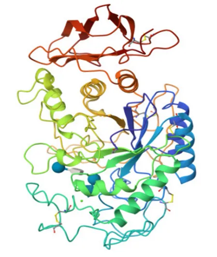
Figure 1: Protein (PDB ID: 1B2Y)
Steps in molecular docking:
Protein preparation
Structure of the protein downloaded from PDB database is unsuitable for docking studies. Protein preparation is done in Biovia Discovery Studio which involves cleaning, repairing, and preparing the protein structure for analysis. Manually manipulating the structure by removing unwanted molecules like water or co-factors, all of which are crucial for downstream tasks like molecular docking and simulations. Unwanted components are removed by selecting them in the hierarchy in the view and deleting them35.
Ligand preparation
The 2D structure of ligands is drawn using ACD lab ChemDraw Ultra 12.0 and generated the smiles notation of the compounds. This smiles notation is being converted into 3D PDB format with the help of freely accessible online SMILES Translator36.
Docking using Autodock Vina
Docking studies were carried out using PyRx, where both the ligand and the receptor were loaded into navigation work place. Then the protein was prepared and converted into macromolecule and the derivative was converted into ligand molecule. After the preparation, click on AutoDock Vina Wizard start button and adjust the grid size. After completion of the process, docking value is expressed in terms of binding affinity (Kcal/mol) along with RMSD upper bound and lower bound value. AutoDock Vina converts the PDB file into PDBQT format. Further analysis was performed by adding polar contacts to find out the types of amino acid interaction involved in ligand-receptor binding37.
2.6 CHEMISTRY
1. General procedure for the Synthesis of 5-(4-methoxybenzylidene)-thiazolidine-2,4- dione.
Thiazolidine-2,4-dione (1.17 g, 0.01 mol) was added in a solution of 4-methoxy benzaldehyde (1.36 g, 0.01 mol) in ethanol (20 mL). Catalytic amount of piperidine (0.2 mL) was added to reaction mixture and the resulting mixture was refluxed for about 6-8 hours at 70-80℃ using heating mantle. Upon completion of the reaction, monitored by TLC using ethyl acetate and hexane (4:6), the reaction mixture was allowed to cool to room temperature. The resulting solid was filtered and washed with cold ethanol, then dried and further purified by recrystallization from ethanol38.
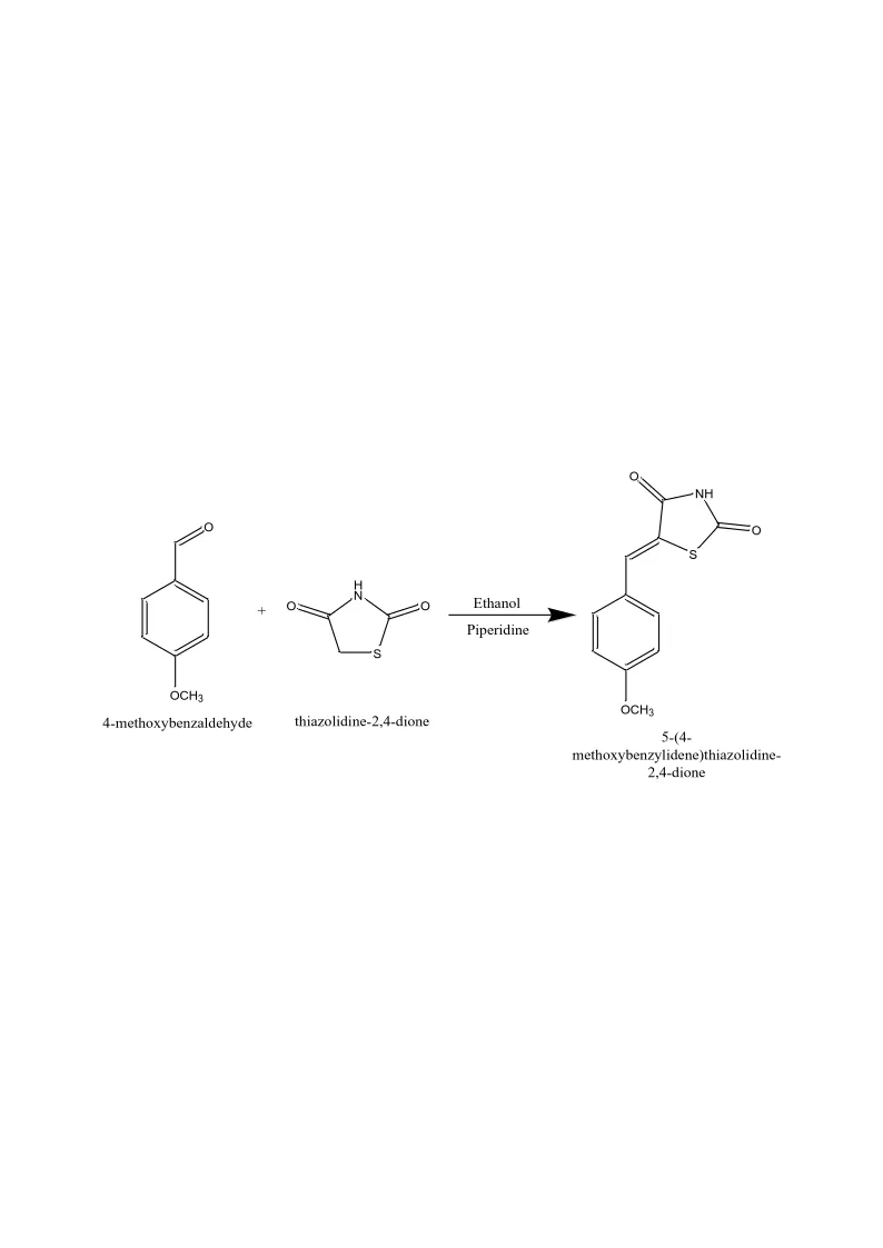
Figure 2: Synthesis of 5-(4-Methoxybenzylidene)-Thiazolidine-2,4- Dione.
2. General procedure for the synthesis of 3- [(substituted phenylamino) methyl]-5-(4-methoxybenzylidene)-thiazolidine-2,4-dione.
Formaldehyde (0.01 mol) was added in a solution of 5-(4-methoxybenzylidene)-thiazolidine 2,4-dione (0.005 mol) in DMF. The reaction mixture was stirred at room temperature for about 30 min. The solution of aryl amine (0.005 mol) in DMF was added to the above reaction mixture and then catalytic amount of conc. HCl (3-5 drops) was added. The reaction mixture was re- fluxed for 10-14 h at 120℃ and then, cooled at 2-8℃ for about 24 hrs. The reaction mixture was poured into crushed ice and the resulting solid was filtered, washed with cold water. The product was dried and recrystallized from ethanol. Completion of the reaction was monitored by TLC using an eluent, a mixture of solvents, toluene: ethylacetate (4:2)39.

Figure 3: Synthesis of 3- [(Substituted Phenylamino) Methyl]-5-(4-Methoxybenzylidene)-Thiazolidine-2,4-Dione.
2.7 REACTANTS
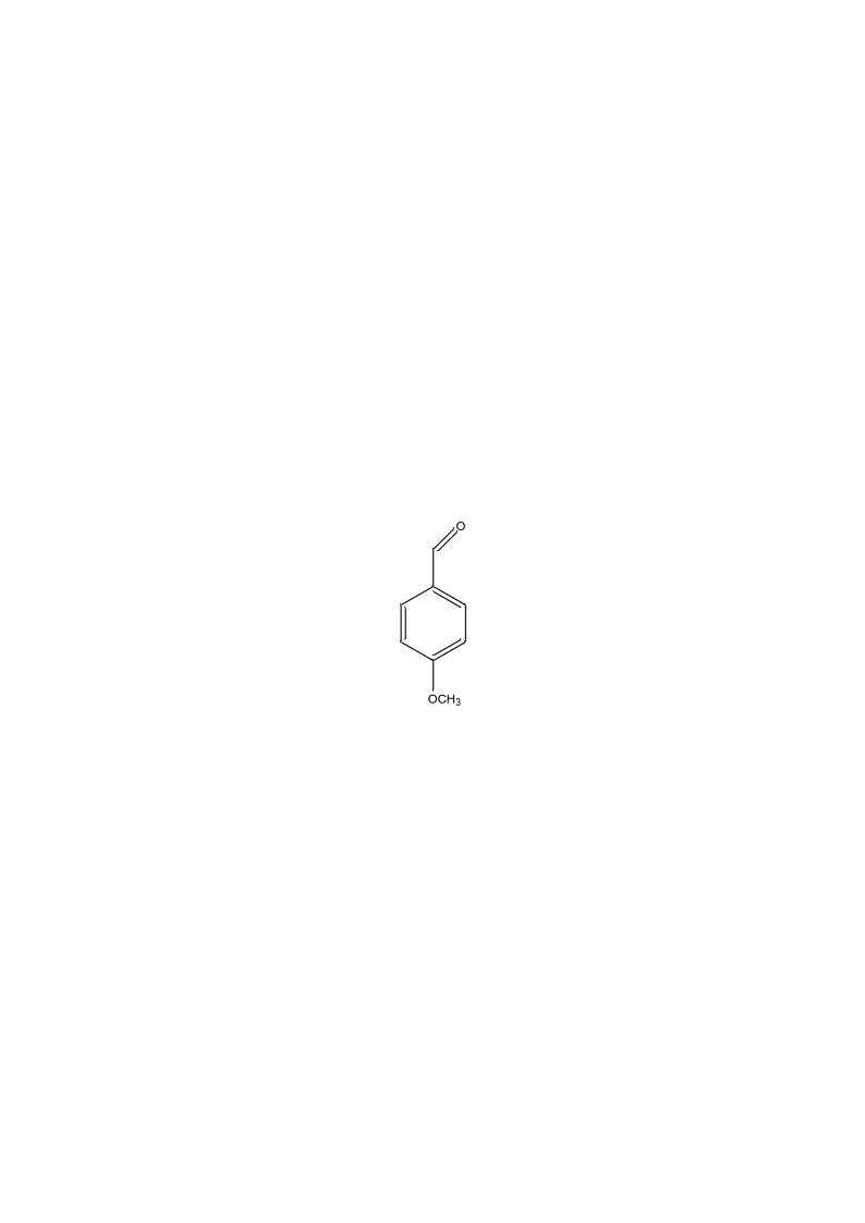
4-methoxybenzaldehyde
4-methoxybenzaldehyde
Chemical formula: C8H8O2
Molecular weight: 136.15
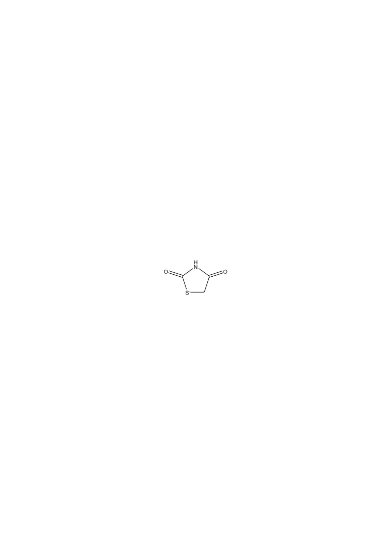
Figure 4: 4-Methoxybenzaldehyde
1,3-thiazolidine-2,4-dione
Chemical formula: C3H3NO2S
Molecular weight: 117.1220
5-(4-methoxybenzylidene)-thiazolidine-2,4-dione
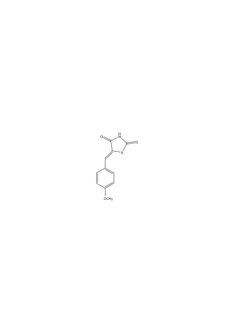
Figure 5: 1,3-Thiazolidine-2,4-Dione
5-(4-methoxybenzylidene)-thiazolidine-2,4-dione
Chemical formula: C11H9NO3S
Molecular weight: 235.26

Figure 6: 5-(4-Methoxybenzylidene)-Thiazolidine-2,4-Dione
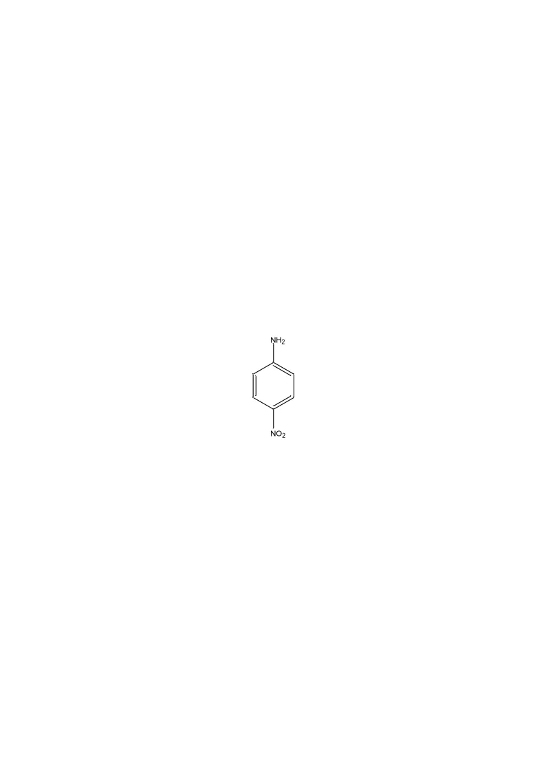
Aniline derivatives:
Aniline 4-nitroaniline
Chemical formula: C6H7N Chemical formula: C6H6N2O2
Molecular weight: 93.13 Molecular weight: 138.13
(a) (b)
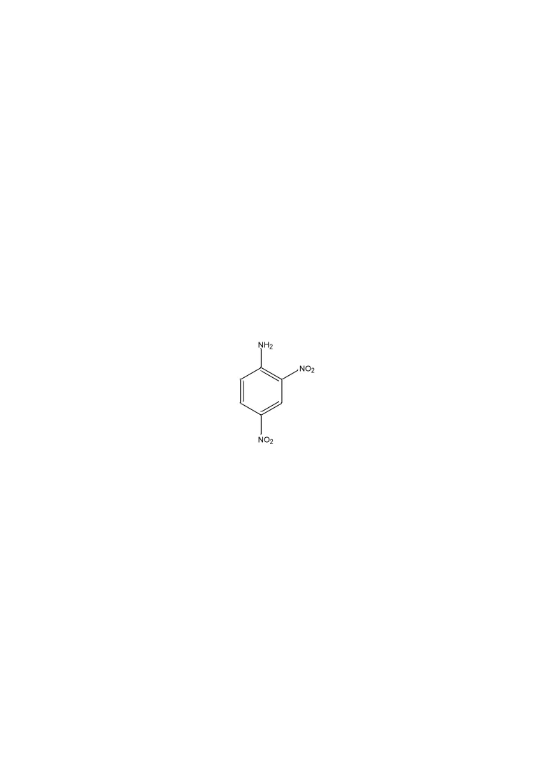

2,4-dinitroaniline 4-methoxyaniline
Chemical formula: C6H5N3O4 Chemical formula: C7H9NO
Molecular weight: 183.12 Molecular weight: 123.16
(c) (d)
Figure 7: (A) Aniline, (B) 4-Nitroaniline, (C) 2,4-Dinitroaniline, (D) 4-Methoxyaniline
2.8 STRUCTURE OF SYNTHESIZED COMPOUNDS
TZ1
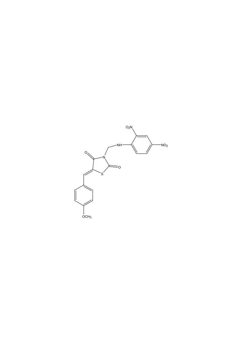
3-(((2,4-dinitrophenyl) amino) methyl)-5-(4-methoxybenzylidene) thiazolidine-2,4-dione
Chemical formula: C18H14N4O7S
Molecular weight: 430.39
TZ2
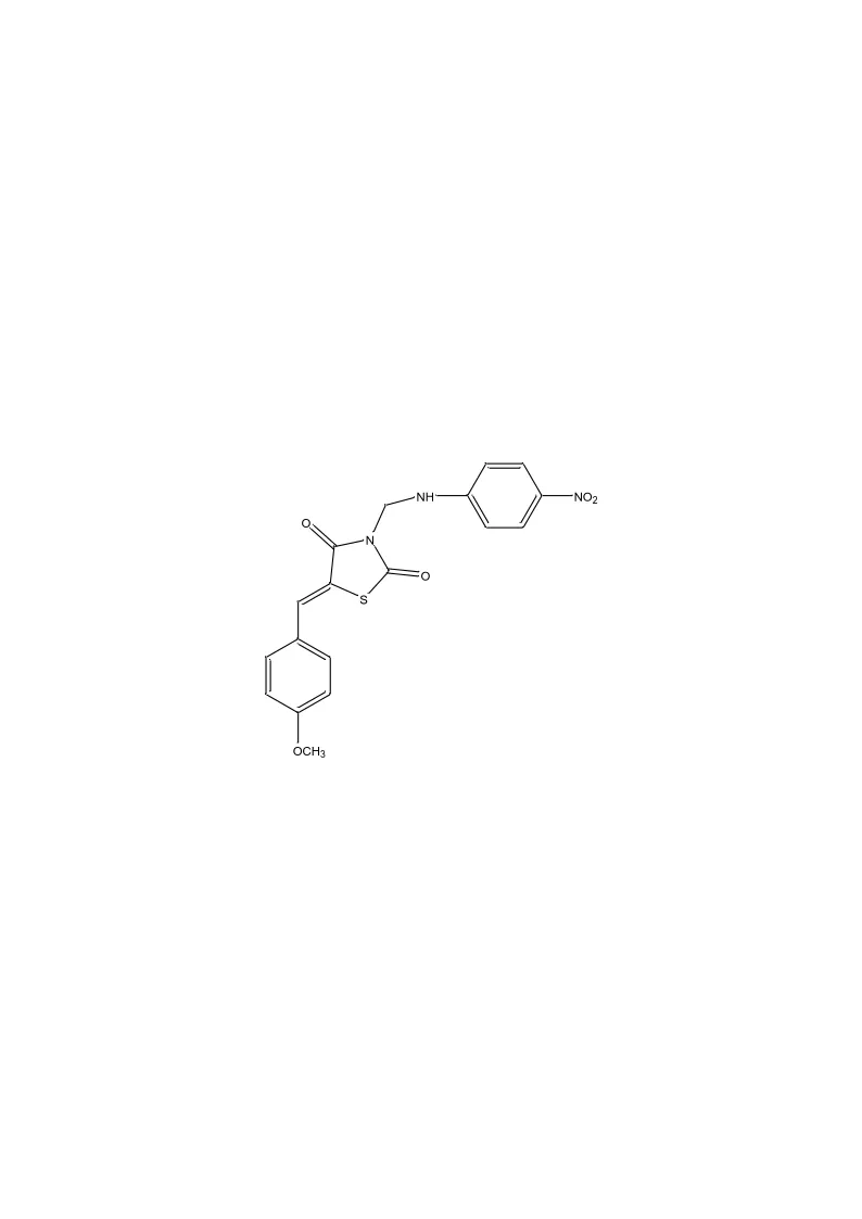
Figure 8: 3-(((2,4-Dinitrophenyl) Amino) Methyl)-5-(4-Methoxybenzylidene) Thiazolidine-2,4-Dione
5-(4-methoxybenzylidene)-3-(((4-nitrophenyl) amino) methyl) thiazolidine-2,4-dione
Chemical formula: C18H15N3O5S
Molecular weight: 385.39
Figure 9: 5-(4-Methoxybenzylidene)-3-(((4-Nitrophenyl) Amino) Methyl) Thiazolidine-2,4-Dione
2.9. CHARACTERIZATION
Melting point
The melting point is an important physicochemical property that describes the temperature at which a compound changes from solid to liquid state. The melting point of the synthesized compounds was determined by open capillary tube method. In this method, the sample is introduced into the capillary tube and the temperature is gradually increased until the sample is visibly melted completely40.
Thin layer chromatography
TLC is used to monitor the completion of chemical reaction. In this, the reactants and products are dissolved in a solvent in which it is soluble. A pre-coated silica gel GF plate was used as the stationary phase with a suitable mobile phase. A single principal spot for the product and the absence of secondary spots for parent compounds and intermediates confirmed the purity of the product. The spots are viewed under UV chamber41.
IR spectroscopy
Infrared Spectroscopy deals with the frequencies of bond vibration in a molecule. The main use is to identify the functional groups. IR spectroscopy is an important tool in the structural elucidation of organic compounds. In IR spectroscopy, there are two regions namely functional group and fingerprint region in which finger print region is used to compare the two compounds. Infrared spectrum shows percentage transmittance versus frequency expressed as wave numbers.
NMR spectroscopy
Nuclear Magnetic Resonance (NMR) spectroscopy is an important analytical technique used in the structural elucidation of organic molecules. It involves the interaction of the electromagnetic radiation and the proton of a nucleus of an atom when placed under an externally applied static magnetic field. NMR spectra provide the detailed information about a molecule’s structure. The chemical shift is used to predict the number of protons with refers to TMS as standard. The NMR spectra are recorded on JEOL FTNMR spectrometer at 400 MHz (13C NMR) and 101 MHz (1H NMR). DMSO is used as a solvent42.
2.10 IN-VITRO ANTI-DIABETIC ASSAY
α-Amylase inhibition assay:
Principle:
The α-amylase inhibition assay is based on the ability of α-amylase to hydrolyze starch into reducing sugars, mainly maltose. The assay is performed using Miller’s DNSA (3,5-dinitrosalicylic acid) colorimetric method.
When starch is incubated with α-amylase, the enzyme breaks down the starch and release maltose. The reducing sugars formed, react with DNS reagent under heating conditions, producing an orange-red colored compound. The intensity of color is proportional to the amount of reducing sugar produced and is measured at 540 nm using a UV-Visible spectrophotometer.
The compound which acts as α-amylase decreases the hydrolysis of starch and consequently reduces the formation of maltose. Lower absorbance indicates greater inhibition of α-amylase activity.
Procedure:
The anti-diabetic activity of the test samples was determined using the method described by the Worthington Enzyme Manual with slight modifications43.
The anti-diabetic activity of the test samples was determined according to the method described in the Worthington Enzyme Manual with slight modifications (Worthington, 1993; Kwon et al., 2006). In brief, 1mL of 0.02 M sodium phosphate buffer (pH 6.9 with 0.006 M NaCl) containing 0.5 mg/mL of a- amylase enzyme and different concentration of the test sample as enzyme inhibitor were pre-incubated at 37oC for 10 min. After the pre-incubation, 500 µL of a 1% starch solution in 0.02 M sodium phosphate buffer (pH 6.9) was added to each tube and incubated at room temperature for 5 mins. The reaction was stopped using 1.0 mL of dinitrosalicylic acid (DNSA) reagent. The test tubes were incubated in a boiling water bath for 5 min and then cooled to room temperature. The volume of the reaction mixture was made up to 5 mL by adding distilled water, and the absorbance was measured at 540 nm using UV-Visible spectrophotometer. The absorbance was compared with the controls and blank that contained buffer instead of test sample.
Percentage inhibition=((B-A)×100)/(B-C)
Calculation:
C – Absorbance of the Control with starch and without alpha amylase.
B – Absorbance of the Control with starch and alpha amylase.
A – Absorbance of the Test44.
RESULTS AND DISCUSSION
3.1 CHEMISTRY
The synthesized compound (TZ1 & TZ2) was obtained good yields (70-80%). Spectroscopic
data confirmed the proposed structures.
NMR of TZ1:
1H-NMR (400 MHz, DMSO-D6) δ 12.48 (s, 1H), 8.78 (d, J = 2.6 Hz, 1H), 8.37 (s, 2H), 8.15 (dd, J = 9.4, 2.7 Hz, 1H), 7.73 (s, 1H), 7.54 (d, J = 8.7 Hz, 2H), 7.12-7.07 (m, 3H), 3.82 (s, 3H).
13C-NMR (101 MHz, DMSO-D6) δ 168.0, 167.5, 161.0, 149.8, 135.1, 132.1, 131.8, 129.3, 128.7, 125.5, 123.4, 120.3, 119.8, 114.9, 55.5, 40.1, 39.9, 39.7, 39.5, 39.3, 39.1, 38.9.
The 1H NMR spectrum of TZ1 showed a characteristic signal at 12.48 ppm, which may be assigned to the NH proton of the thiazolidine-2,4-dione ring. Signals observed in the region of 7.07–8.78 ppm were attributed to aromatic and other deshielded protons, supporting the presence of an aromatic system. A signal at approximately 3.82 ppm was consistent with the presence of a methoxy group.
The 13C NMR spectrum of TZ1 exhibited signals at 168.03 and 167.54 ppm, corresponding to the two carbonyl carbons of the thiazolidine-2,4-dione ring. The signal at 160.98 ppm was consistent with an aromatic carbon attached to an electron-donating oxygen-containing substituent. The signal at 55.49 ppm supported the presence of a methoxy carbon. Signals in the region of 114.90-149.84 ppm was assigned to aromatic and other unsaturated carbons. Overall, the observed NMR signals support the proposed structure of TZ1.
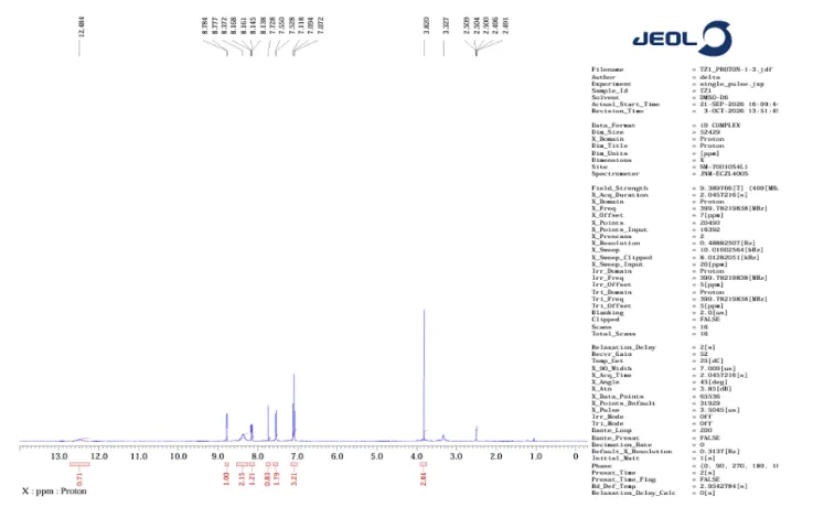
Figure 10: 1H NMR of TZ1
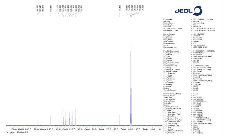
Figure 11: 13C NMR of TZ1
NMR of TZ2:
1H-NMR (400 MHz, DMSO-D6) δ 12.50 (s, 1H), 7.94 (dd, J = 7.1, 2.1 Hz, 1H), 7.75 (s, 1H), 7.56 (dd, J = 6.5, 2.4 Hz, 2H), 7.10 (dd, J = 6.8, 2.1 Hz, 2H), 6.71 (s, 1H), 6.59 (dd, J = 7.1, 2.1 Hz, 1H), 3.82 (s, 3H).
13C-NMR (101 MHz, DMSO-D6) δ 168.1, 167.6, 161.0, 155.8, 135.6, 132.1, 131.8, 126.5, 125.5, 120.3, 114.9, 112.4, 55.5, 40.1, 39.9, 39.7, 39.5, 39.3, 39.1, 38.9.
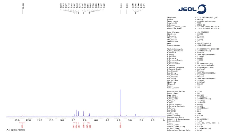
Figure 12: 1H NMR of TZ2
The 1H NMR spectrum of TZ2 showed a characteristic signal at 12.50 ppm, which may be assigned to the NH proton of the thiazolidine-2,4-dione ring. Signals observed in the region of 6.58–7.95 ppm were attributed to aromatic and other unsaturated protons. A signal at approximately 3.82 ppm was consistent with the presence of a methoxy group.
The 13C NMR spectrum of TZ2 exhibited signals at 168.07 and 167.56 ppm, corresponding to the two carbonyl carbons of the thiazolidine-2,4-dione ring. Signals at 161.01 and 155.76 ppm were consistent with deshielded aromatic or heteroaromatic carbons. The signal at 55.51 ppm supported the presence of a methoxy carbon. Additional signals in the region of 112.41–135.63 ppm were assigned to aromatic and other unsaturated carbons. Overall, the observed NMR signals provide preliminary support for the proposed structure of TZ2.
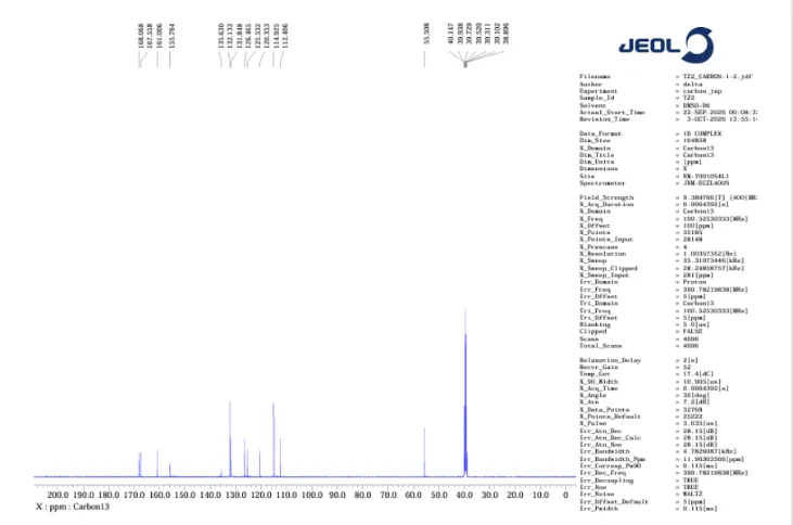
Figure 13: 13C NMR of TZ2
IR SPECTRA:
3331.71 (N–H), 3007.67 (C–H), 1681.13 (C=O), 1626.07 (C=C), 1583.71 (C=C), 1509.22 (NO₂), 1303 (NO₂), 1255.14 (C–O–C), 1177.14 (C–O), 1144.49 (C–N), 629.65 (C–S).
The FTIR spectrum of TZ1 exhibited a characteristic absorption band at 3331.71 cm⁻¹ corresponding to N–H stretching. The absorption bands in the region of 1734–1681 cm⁻¹ indicate carbonyl (C=O) stretching vibrations. Bands observed at 1626.07 and 1583.71 cm⁻¹ are attributed to aromatic ring vibrations. The bands around 1509 and 1303 cm⁻¹ suggest the asymmetric and symmetric stretching vibrations of nitro (NO₂) groups, respectively. Absorption bands in the region of 1255–1144 cm⁻¹ indicate C–O–C stretching vibrations.
3230.955 (N–H), 3007.679 (C–H), 1681.784 (C=O), 1587.193 (C=C), 1507.945 (NO₂), 1253.615 (C–O–C), 1176.881 (C–O), 1144.329 (C–N), 629.773 (C–S).
The FTIR spectrum of TZ2 exhibited characteristic absorption bands at 3230.95 cm⁻¹ (N–H stretching), 3007.67 cm⁻¹ (aromatic C–H stretching), and 1681.78 cm⁻¹ (carbonyl stretching). Bands observed in the region of 1587.193 cm⁻¹ corresponds to aromatic ring vibrations, while absorptions between 1258.61 and 1144.329 cm⁻¹ indicate C–O–C and C-O stretching. The observed spectral features are generally consistent with the functional groups present in the proposed molecular structure.
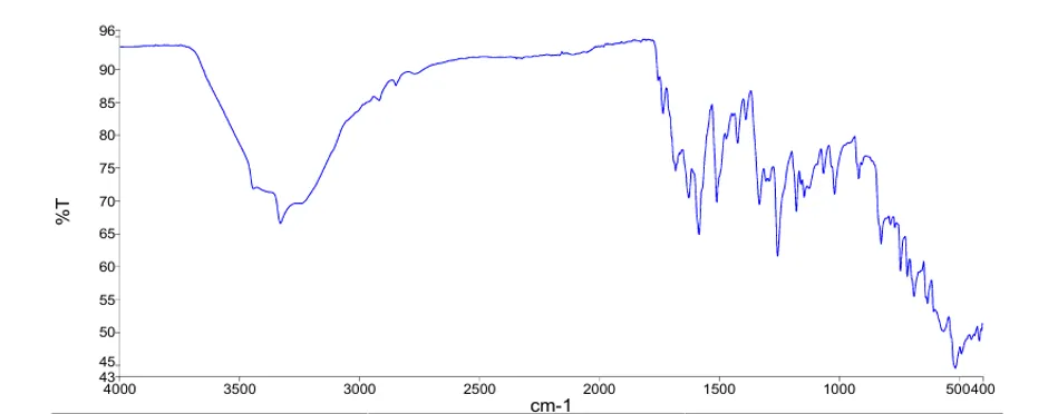
Figure 14: IR Spectrum of TZ1
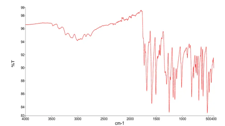
Figure 15: IR Spectrum of TZ2
3.2 PHARMACOKINETIC STUDY BY SWISSADME
Table 1: Pharmacokinetic Study by SwissADME
| Compound | Pharmacokinetic properties | |
|---|---|---|
| TZ1 | ||
| TZ2 | ||
| TZ3 | ||
| TZ4 |
3.3 IN-SILICO TOXICITY PREDICTION – Protox.3.0:
The Protox 3.0 web-based tool was used to predict the potential toxicity of the designed compounds. It estimates the toxicity of chemical molecules and classifies them according to the Globally Harmonized System (GHS) for the classification. The predicted toxicity is expressed in terms of median lethal dose (LD50), measured in mg/kg, which indicates the dose required to cause death in 50% of the test population.
LD50 values are given in [mg/kg]:
Table 2: Toxicity Prediction by PROTOX.3.0
| Compound Id | Protox toxicity | |
|---|---|---|
| TZ1 | ||
| TZ2 | ||
| TZ3 | ||
| TZ4 |
3.4 DOCKING RESULTS
The designed compounds were docked into the active site of the protein (PDB ID:1B2Y) using AutoDock Vina. The compounds were evaluated based on their docking scores and the interactions formed with important amino acid residues present in the binding pocket. The most favorable and stable pose was selected based on the docking score and multiple interactions observed. The thiazolidine-2,4dione ring and substituted aromatic portions contributed to the interaction with the binding site. Among the docked molecules, TZ1 exhibited the highest binding affinity towards the target protein, followed by TZ2 which showed comparatively lowest binding affinity.
Table 3: Docking Results
| Sl. No | Compound | Structure | Docking score | Interacting residue |
|---|---|---|---|---|
| 1 | TZ1 | -9.1 | HIS A: 305 ILE A: 235 LYS A: 200 ALA A: 198 LEU A: 162 TYR A: 151 |
|
| 2 | TZ2 | -8.5 | ILE A: 235 HIS A: 201 LYS A: 200 ALA A: 198 LEU A: 162 GLN A: 63 |
|
| 3 | TZ3 | -8.2 | HIS A: 305 ILE A: 235 GLU A: 233 LYS A: 200 ALA A: 198 LEU A: 162 |
|
| 4 | TZ4 | -8.3 | ILE A: 235 HIS A: 201 LYS A: 200 LEU A: 165 TYR A: 151 GLN A: 63 |
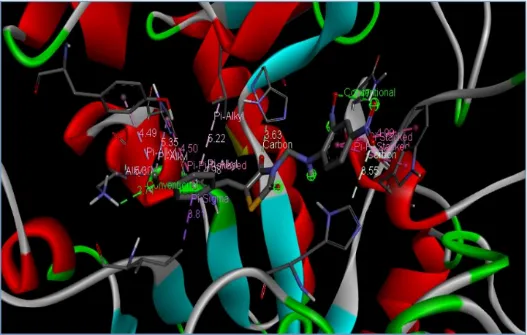
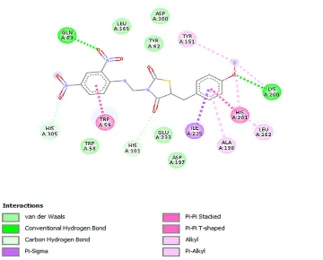
Figure 16: (A) Binding Interaction of TZ1, (B) Binding Pattern of TZ1 in Binding Pocket
(a) (b)
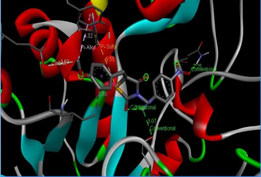
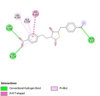
Figure 17: (A) Binding Interaction of TZ2, (B) Binding Pattern of TZ2 in Binding Pocket
(a) (b)
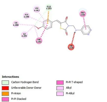
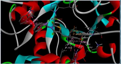
Figure 18: (A) Binding Interaction of TZ3, (B) Binding Pattern of TZ3 in Binding Pocket
(a) (b)
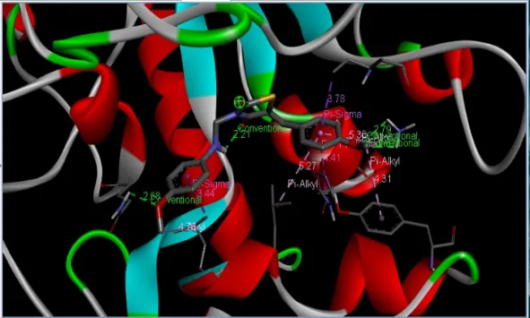
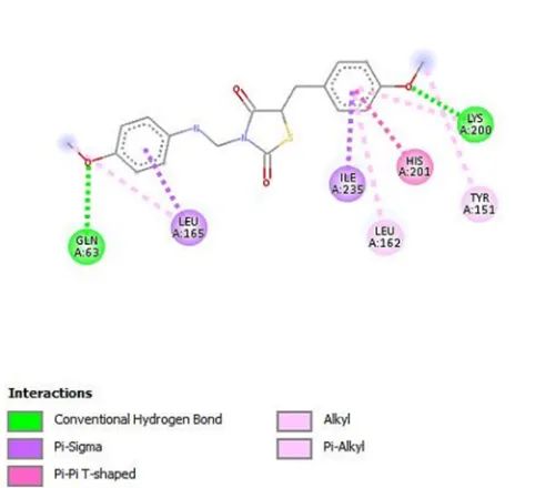
Figure 19: (A) Binding Interaction of TZ4, (B) Binding Pattern of TZ4 in Binding Pocket
(a) (b)
3.5 BIOLOGICAL EVALUATION
α-Amylase inhibition assay:
The compound with the highest docking scores (TZ1 & TZ2) was selected for in-vitro studies to assess its anti-diabetic activity. This colorimetric assay done using Miller's method i.e. the DNS Method (3,5-dinitrosalyclic acid reagent) involves conversion of alpha amylase in to maltose. Maltose released from starch is measured by the reduction of 3,5-dinitrosalicylic acid and the solution is quantified by measuring absorbance at 570 nm using an ELISA plate reader.
RESULTS:
Table 4: Assay Result and Percentage Inhibition for Various Concentration of Standard
| Standard | Concentration (µg/ml) |
Absorbance at 540 nm | Percentage Inhibition (%) |
|---|---|---|---|
| Blank | - | 0.782 | - |
| Control | - | 0.024 | - |
| Acarbose | 6.25 | 0.721 | 9.01 |
| 12.5 | 0.679 | 15.21 | |
| 25 | 0.530 | 37.22 | |
| 50 | 0.317 | 68.69 | |
| 100 | 0.178 | 89.22 | |
| IC 50 | 36.03 μg/ml | ||
Table 5: Assay Result and Percentage Inhibition for Various Concentrations of TZ1 & TZ2
| Sample code | Concentration (μg/ml) | Absorbance at 540nm | Percentage Inhibition (%) |
|---|---|---|---|
| Blank | - | 0.798 | - |
| Control | - | 0.020 | - |
| TZ1 | 6.25 | 0.770 | 3.59 |
| 12.5 | 0.706 | 11.82 | |
| 25 | 0.537 | 33.54 | |
| 50 | 0.296 | 64.52 | |
| 100 | 0.133 | 85.47 | |
| IC 50* | 50.45μg/ml | ||
| TZ2 | 6.25 | 0.711 | 4.75 |
| 12.5 | 0.659 | 11.43 | |
| 25 | 0.495 | 32.51 | |
| 50 | 0.257 | 63.11 | |
| 100 | 0.115 | 81.36 | |
| IC 50* | 52.53μg/ml | ||
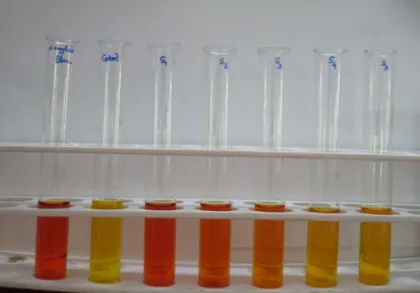
The in-vitro evaluation of TZ1 and TZ2 demonstrated moderate α-amylase inhibitory activity, with IC₅₀ values of 50.45 µg/mL and 52.53 µg/mL, respectively. TZ1 exhibited slightly better inhibitory activity than TZ2, as indicated by its lower IC₅₀ value. These findings suggest that both compounds may have potential as antidiabetic agents.
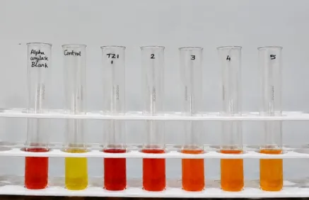
(a) (b)
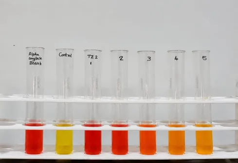
Figure 20: Visual Observation of Α-Amylase Inhibition Assay
(c)

Figure 21: Percentage Inhibition vs Concentration Graph of TZ1
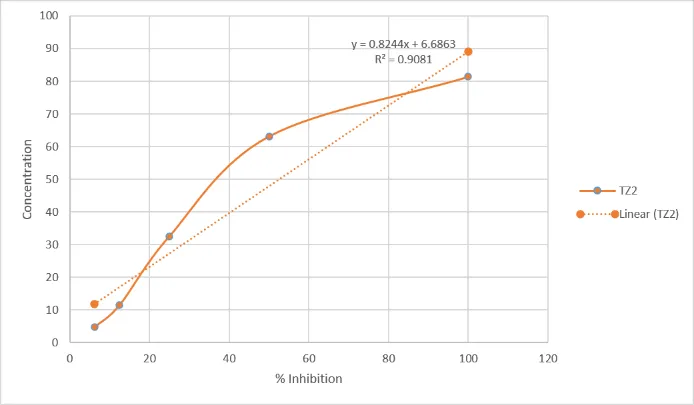
Figure 21: Percentage Inhibition vs Concentration Graph of TZ2
5. ACKNOWLEDGEMENT
It gives me immense pleasure to express my sincere gratitude to the college management and guide Dr. Sreena K, Professor and HOD Department of Pharmaceutical Chemistry, Crescent College of Pharmaceutical Sciences, Payangadi for providing the facilities, valuable guidance and encouragement throughout the research work and for the successful completion of my project work.
6. CONFLICT OF INTEREST
We declare that we have no conflict of interest.
REFERENCES

Sreena K., Ashique P., Fathimathul Rifa Rasheed, Fathimath Sana A., Structure-Guided Design, Synthesis, Molecular Docking and In-Vitro Evaluation of Arylidene Thiazolidine-2,4-Dione Derivatives, Int. J. of Pharm. Sci., 2026, Vol 4, Issue 11, 2452-2473. https://doi.org/10.5281/zenodo.24279024
 10.5281/zenodo.24279024