

We use cookies to ensure our website works properly and to personalise your experience. Cookies policy
Department of Chemistry, Marwari College, Darbhanga, Bihar 846004, India.
Guava (Psidium guajava L.) leaves are a potential source of bioactive secondary metabolites. This study compared the qualitative phytochemical profiles of guava leaf extracts prepared by ethanolic maceration and aqueous decoction and evaluated the antibacterial activity of the aqueous extract against selected bacterial strains. Fresh leaf material was extracted using 96% ethanol or water. Both extracts were screened qualitatively for tannins, saponins, flavonoids, alkaloids, terpenoids, steroids and cardiac glycosides. Antibacterial activity of the aqueous extract was assessed by the disc diffusion method, while the minimum inhibitory concentration (MIC) and minimum bactericidal concentration (MBC) were determined by broth microdilution and subculture, respectively. Both extracts gave positive reactions for all seven phytochemical groups. In the disc diffusion assay, the aqueous extract inhibited the tested Gram-positive bacteria, including resistant and wild-type isolates of Staphylococcus aureus and Staphylococcus pseudintermedius, as well as ?-haemolytic Streptococcus spp. No inhibition zones were detected against the tested Escherichia coli strains at the concentrations examined. At the highest concentration tested (11.4 mg/mL), the largest inhibition zone was observed against ?-haemolytic Streptococcus spp. (21.0 ± 2.0 mm). The MIC was reported as 6.8 mg/mL for all tested strains. The MBC was 6.8 mg/mL for the resistant S. pseudintermedius isolate, whereas it was greater than 11.4 mg/mL for the other strains evaluated. These findings provide preliminary evidence that guava leaf extracts contain several detectable phytochemical groups and that the aqueous extract exhibits in vitro activity against the tested Gram-positive bacteria. Further quantitative phytochemical analysis, confirmation of active constituents, and broader antibacterial and safety studies are required.
Antimicrobial resistance (AMR) is a major global public health concern that reduces the effectiveness of antimicrobial agents and complicates the treatment of infectious diseases. Resistance may develop through genetic mutations or the acquisition of resistance determinants and can subsequently spread among bacterial populations. The inappropriate and excessive use of antimicrobial agents contributes to the selection and dissemination of resistant microorganisms. A global systematic analysis estimated that bacterial AMR was directly attributable to approximately 1.27 million deaths in 2019, highlighting the urgent need for effective strategies to prevent and manage resistant infections1. These challenges have stimulated research into bioactive compounds from natural sources as potential candidates for developing new antimicrobial preparations. Nevertheless, plant-derived extracts require systematic scientific evaluation before their effectiveness and suitability for practical applications can be established.
Medicinal plants are important sources of secondary metabolites, including tannins, flavonoids, saponins, alkaloids and terpenoids, many of which have been investigated for their diverse biological activities. Among these plants, guava (Psidium guajava L.), belonging to the family Myrtaceae, is widely cultivated in tropical and subtropical regions, including India. Its leaves have attracted considerable research interest because of their diverse phytochemical composition and reported pharmacological properties. Previous studies have reviewed the nutritional composition, phytochemical profile and health-promoting properties of guava leaves, highlighting the presence of several phenolic compounds and other bioactive constituents2. Qualitative phytochemical investigations have also demonstrated the presence of saponins, tannins and flavonoids in guava leaf extracts3. Furthermore, chemical investigations have identified structurally diverse compounds in guava leaves, including flavanones and other aromatic metabolites, some of which have demonstrated antibacterial activity against selected microorganisms4.
Tannins, flavonoids and saponins are of particular interest in phytochemical investigations because of their diverse chemical properties and reported biological activities. Tannins can interact with proteins and other cellular components, potentially contributing to their antibacterial effects. Flavonoids have been investigated for their antioxidant and anti-inflammatory properties5, as well as their antibacterial activities through mechanisms involving bacterial cell membranes, nucleic acid synthesis and energy metabolism 6. Saponins are another important class of plant secondary metabolites that possess surface-active properties and may contribute to the biological activities of plant extracts. The reported presence of these phytochemical classes in guava leaves provides a rationale for examining their occurrence in extracts prepared under different extraction conditions. However, qualitative phytochemical screening indicates only the possible presence of particular compound classes and does not establish the identity, concentration or individual contribution of specific compounds to antibacterial activity.
The extraction method and solvent used are important factors influencing the recovery of phytochemicals from plant materials. Maceration is a conventional solid–liquid extraction technique in which plant material is soaked in a suitable solvent for a specified period, facilitating the dissolution and diffusion of extractable constituents into the solvent. This method is relatively simple and can generally be conducted at room temperature, although it may require longer extraction periods and considerable solvent volumes. In contrast, decoction involves boiling plant material in water to extract water-soluble constituents. Although heating may facilitate the extraction of certain compounds, prolonged exposure to elevated temperatures may alter or degrade heat-sensitive constituents. The selection of an appropriate extraction technique depends on several factors, including solvent properties, temperature, extraction duration and the chemical characteristics of the target compounds7,8. Since ethanol and water differ in polarity and solvent properties, ethanol maceration and aqueous decoction may produce extracts with different phytochemical profiles.
Previous experimental investigations have examined the antibacterial activity of guava leaf extracts against different bacterial species. Guava leaf extract has been investigated for its antibacterial activity against Escherichia coli9. Similarly, the antibacterial activity of a combination of guava leaf and Arumanis mango leaf extracts against Staphylococcus aureus has also been evaluated10. However, the latter investigation involved a combination of two plant extracts; therefore, its findings cannot be attributed exclusively to guava leaves. These studies provide a basis for further investigation, although the phytochemical composition and antibacterial activity of plant extracts may vary depending on the plant material, extraction conditions, extract concentration and test microorganism.
Therefore, the present study aimed to investigate the qualitative phytochemical composition of guava (Psidium guajava L.) leaf extracts prepared by ethanol maceration and aqueous decoction, with particular emphasis on tannins, flavonoids and saponins. The study also aimed to evaluate the antibacterial activity of the prepared extracts against selected bacterial strains. Comparing the two extraction methods may help determine whether differences in extraction conditions are associated with variations in phytochemical screening results and antibacterial activity. The findings may contribute to a better understanding of the potential of guava leaves as a source of plant-derived antibacterial constituents.
MATERIALS AND METHODS:
2.1. Research Design
The study employed a laboratory-based experimental approach with descriptive qualitative analysis to investigate the phytochemical constituents of guava leaves (Psidium guajava L.). Phytochemical screening was performed to detect the presence of flavonoids, tannins, saponins, alkaloids, terpenoids, steroids and cardiac glycosides in extracts prepared using two extraction techniques: ethanol maceration and aqueous decoction. The extracts were evaluated using appropriate qualitative reagent tests based on the characteristic colour changes or other observable reactions associated with the target phytochemical groups.
2.2. Plant Materials and Reagents
Approximately 1 kg of fresh guava leaves was collected, of which separate 300 g portions were used for ethanol maceration and aqueous decoction. The reagents and materials included 96% ethanol (Merck), distilled water, concentrated hydrochloric acid (HCl; Merck), 1% ferric chloride (FeCl₃) solution, magnesium powder, Whatman filter paper, aluminium foil, a glass funnel and suitable laboratory glassware. Water was used as the extraction solvent for the decoction procedure.
2.3. Sample Preparation
Fresh 1kg guava leaves were washed thoroughly under running water to remove adhering dirt and other surface impurities. The cleaned leaves were cut into small pieces and crushed or blended without adding solvent to facilitate subsequent extraction. The prepared plant material was then used for ethanol maceration and aqueous decoction according to the respective procedures.
2.4. Extraction of Guava Leaves
2.4.1. Ethanol Maceration
Ethanolic extraction was carried out using the maceration technique. Briefly, 300 g of prepared guava leaf material was immersed in 96% ethanol and allowed to stand for 6 hours with intermittent stirring to facilitate solvent penetration and the extraction of soluble phytoconstituents. The mixture was filtered through Whatman filter paper using a glass funnel to separate the filtrate from the solid residue. The residue was re-extracted with fresh 96% ethanol, and the resulting filtrates were combined. The combined extract was kept for 24 hours and subsequently filtered again. Finally, the solvent was evaporated to obtain a concentrated guava leaf extract, following the reported extraction approach11.
2.4.2. Aqueous Decoction
For aqueous extraction, the prepared 300 gm guava leaf material was placed in a pot containing 500 mL of water. The mixture was heated over a medium flame for 15 minutes. After heating, the decoction was allowed to cool, and the liquid extract was separated from the residual plant material by filtration. The resulting aqueous extract was collected for subsequent phytochemical screening.
2.5. Qualitative Phytochemical Screening
The ethanol and aqueous extracts were subjected to qualitative phytochemical screening for flavonoids, tannins, saponins, alkaloids, terpenoids, steroids and cardiac glycosides.. The tests were based on characteristic reactions following the addition of appropriate reagents. The presence or absence of each phytochemical group was recorded according to the observed reaction.
Preliminary phytochemical screening of guava (Psidium guajava L.) leaf extracts was performed using qualitative chemical tests to identify tannins, saponins and flavonoids. The tests were conducted on both ethanolic extract and aqueous decoction, using characteristic colour changes and persistent foam formation as preliminary indicators of the respective phytochemical groups, following previously reported methods11.
2.5.1. Ferric chloride test for tannins:
Approximately 1 mL of the guava leaf extract, prepared by ethanol extraction or aqueous decoction, was transferred into a clean test tube. Two to three drops of 1% ferric chloride (FeCl₃) solution were added and mixed gently. The development of a bluish-black or green coloration was considered a preliminary indication of tannins and other phenolic compounds in the extract.
2.5.2. Foam test for saponins:
Approximately 2 mL of the guava leaf extract was transferred into a clean test tube, and distilled water was added in sufficient quantity to cover the sample. The mixture was boiled for 2–3 minutes and allowed to cool to room temperature. The tube was then shaken vigorously and left undisturbed. The formation of persistent foam that remained for approximately 30 minutes and did not disappear after the addition of one drop of 2 N hydrochloric acid (HCl) was considered indicative of the possible presence of saponins.
2.5.3. Shinoda test for flavonoids:
Approximately 2 mL of the guava leaf extract was transferred into a clean test tube and heated for 5 minutes. After cooling, a few drops of concentrated hydrochloric acid were added, followed by approximately 0.2 g of magnesium powder. Amyl alcohol was then added, and the mixture was shaken gently and allowed to separate into layers. The development of a red, yellow, or orange coloration in the amyl alcohol layer was considered a preliminary indication of flavonoids. The observations were recorded separately for the ethanol extract and aqueous decoction.
2.5.4. Dragendorff's test for alkaloids:
Approximately 2 mL of the guava leaf extract was transferred into a clean test tube. A few drops of dilute hydrochloric acid were added, followed by 2–3 drops of Dragendorff's reagent. The mixture was shaken gently and allowed to stand for a few minutes. The formation of an orange or reddish-brown precipitate was considered a preliminary indication of the presence of alkaloids. The test was performed separately using the ethanolic extract and aqueous decoction12.
2.5.5. Salkowski test for terpenoids:
Approximately 2 mL of the guava leaf extract was mixed with 2 mL of chloroform in a clean test tube. Concentrated sulfuric acid (approximately 1 mL) was carefully added along the inner wall of the test tube to form a separate lower layer. The development of a reddish-brown coloration at the interface between the two layers was considered a preliminary indication of terpenoids12.
2.5.6. Liebermann–Burchard test for steroids:
Approximately 1 mL of the guava leaf extract was taken in a clean test tube and mixed with 1 mL of chloroform. Subsequently, 2 mL of acetic anhydride was added, followed by 2–3 drops of concentrated sulfuric acid. The mixture was allowed to stand for a few minutes. The development of a blue-green or emerald-green coloration was considered a preliminary indication of steroidal compounds12.
2.5.7. Keller–Killiani test for cardiac glycosides:
Approximately 2 mL of the guava leaf extract was mixed with 2 mL of glacial acetic acid containing one drop of 1% ferric chloride solution. The mixture was carefully transferred into a clean test tube containing approximately 1 mL of concentrated sulfuric acid along its inner wall. The formation of a reddish-brown ring at the interface of the two layers, sometimes accompanied by a bluish-green coloration in the upper layer, was considered a preliminary indication of cardiac glycosides13.
2.6. Antimicrobial Activity
The antimicrobial efficacy of the extract was evaluated using the disc diffusion and broth microdilution methods. The disc diffusion assay was used to assess the inhibitory effect of different extract concentrations by measuring the diameter of the inhibition zones, whereas the broth microdilution method was employed to determine the minimum inhibitory concentration (MIC) and minimum bactericidal concentration (MBC). Aqueous dilutions of the extract were tested against the following bacterial strains: extended-spectrum β-lactamase-related resistant Escherichia coli ESBL (CMY-2), E. coli (ATCC 25922), methicillin-resistant Staphylococcus aureus MRSA (ATCC 43300), S. aureus (ATCC 23923), Staphylococcus pseudintermedius (B19 and B20), and β-haemolytic Streptococcus spp.
Table 1: Description of the Strains Tested Against the Guava Leaf Extract.
Strains Code Resistance Pattern
E. Coli (ESBL) CMY-2 Betalactamic resistance (Penicillins
and cephalosporporins up to third generation)
E. coli ATCC 25922 Wild type E. coli
S. aureus (MRSA) ATCC43300) Methicillin resistance
S. aureus ATCC23923 Wild type S.aureus
S. pseudintermedius B19 Resistance to Sulphametoxazole + trimetoprim
S. pseudintermedius B20 No expressed resistance to any tested antimicrobials Streptococcus spp. Beta – hemolysis - No expressed resistance to any tested antimicrobials
Legend: ESBL – Extended Spectrum Betalactamase; MRSA- Methicillin resistanct Staphylococcus aureus
2.6.1 Disk Diffusion Test
The bacterial strains were cultured on Brain Heart Infusion (BHI) agar and incubated at 35°C for 24 hours. Sterile filter paper discs (0.38 cm²) impregnated with varying concentrations of the extract (2.3, 4.6, 6.8, 9.1, and 11.4 mg/mL) were placed on Mueller–Hinton (MH) agar plates previously inoculated with bacterial suspensions standardized to 0.5 McFarland turbidity. The inoculated plates were then incubated at 35°C for 24 hours14 All experiments were performed in triplicate, with an impregnation volume of 6 µL per disc. Water and 10% methanol served as negative controls (blanks).
2.6.2. Broth Microdilution Assay for Minimum Inhibitory Concentration (MIC)
The broth microdilution assay was performed using a 96-well microtiter plate to determine the minimum inhibitory concentration (MIC) of the aqueous Psidium guajava leaf extract. The bacterial strains were cultured in Mueller–Hinton (MH) broth at 35 °C and adjusted to a turbidity equivalent to a 0.5 McFarland standard. Extract concentrations were selected based on the results of the disc diffusion assay.
The assay included wells containing the bacterial inoculum without extract as growth controls and wells containing MH broth, extract, and no bacterial inoculum as extract blanks. Imipenem was included as an antibiotic control. Bacterial growth was assessed by measuring turbidity using spectrophotometry at 655 nm15. The MIC was defined as the lowest concentration of the extract at which bacterial growth was not detected relative to the appropriate controls and extract blank.
2.6.3. Determination of Minimum Bactericidal Concentration (MBC)
The minimum bactericidal concentration (MBC) was determined by subculturing aliquots from the MIC assay wells that showed no visible evidence of bacterial growth. The aliquots were inoculated onto Brain Heart Infusion (BHI) medium and incubated at 36 °C for 24 hours. Following incubation, the plates were examined for bacterial growth. The MBC was recorded as the lowest extract concentration that prevented bacterial recovery under the specified subculture conditions14.
2.7. Statistical Analysis
The experimental data, including inhibition-zone diameters and turbidity measurements, were subjected to statistical analysis to evaluate differences among the tested conditions. Data normality was assessed using the Shapiro–Wilk test. Data that met the assumptions for parametric analysis were evaluated using analysis of variance (ANOVA), followed by Tukey’s post hoc test for multiple comparisons. Data that did not satisfy the assumptions for parametric analysis were analysed using the Kruskal–Wallis test. Statistical significance was set at a 95% confidence level, with (GraphPad Prism 7 statistical program) considered statistically significant.
RESULTS AND DISCUSSION:
3.1. Preparation of Leaf Extracts by Maceration and Decoction
Guava (Psidium guajava L.) leaf extracts were prepared using two extraction techniques: ethanolic maceration and aqueous decoction. For ethanolic extraction, 300 g of prepared guava leaf material was immersed in 96% ethanol for 6 hours with intermittent stirring. The mixture was subsequently filtered, and the residual plant material was re-extracted using fresh ethanol. The resulting filtrates were combined and kept for 24 hours, followed by a second filtration. Finally, the solvent was evaporated to obtain a concentrated ethanolic extract. The resulting extract was brown in colour, with a final volume of approximately 20 mL.
For aqueous extraction, 300 g of prepared guava leaf material was boiled in 500 mL of water for approximately 15 minutes. After cooling, the mixture was filtered to remove the residual plant material, yielding approximately 250 mL of dark-brown aqueous extract.
The differences in the appearance and final volume of the resulting extracts may be associated with the extraction solvent, extraction procedure, processing conditions, and solvent evaporation. The ethanolic extract was concentrated through solvent evaporation, whereas the aqueous decoction was collected following filtration without a reported concentration step. Ethanol and water differ in their polarity and ability to dissolve plant constituents, potentially resulting in differences in the composition of the extracted phytochemicals. The brown and dark-brown colouration may reflect the presence of extracted plant pigments and other soluble compounds. However, colour intensity alone cannot establish the concentration or diversity of phytochemicals present in an extract.
The higher volume obtained through decoction does not necessarily indicate greater extraction efficiency, as the initial solvent volumes, plant-to-solvent ratios, and extraction yields must also be considered. Further quantitative analysis would be required to compare the extraction efficiency of the two methods.
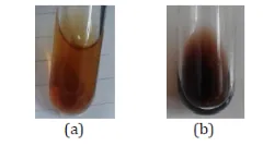
Figure 1: Guava (Psidium guajava) Leaf Extracts Obtained by (a) Ethanolic Maceration and (B) Aqueous Decoction.
3.2. Qualitative Phytochemical Screening
Qualitative phytochemical screening of the ethanolic maceration and aqueous decoction extracts of guava (Psidium guajava L.) leaves was performed to identify the major classes of secondary metabolites. Both extracts exhibited positive reactions for tannins, saponins, flavonoids, alkaloids, terpenoids, steroids, and cardiac glycosides, as indicated by the characteristic colour changes, precipitate formation, and other visual responses associated with the respective phytochemical tests.
The ferric chloride test produced a bluish-black or blackish-green coloration, suggesting the presence of tannin-related polyphenolic compounds. The formation of persistent foam in the foam test indicated the probable presence of saponins, while the development of a dark-red coloration in the Shinoda test suggested the presence of flavonoids. Alkaloids were preliminarily indicated by the formation of an orange or reddish-brown precipitate in Dragendorff's test. The Salkowski test produced a reddish-brown coloration at the interface of the two layers, suggesting the presence of terpenoid constituents. The Liebermann–Burchard test indicated steroidal compounds through the development of blue-green or emerald-green coloration. Similarly, the formation of a reddish-brown ring at the interface during the Keller–Killiani test suggested the possible presence of cardiac glycosides.
The characteristic positive reactions associated with the individual phytochemical screening procedures are presented in Table 2, whereas the qualitative experimental observations obtained for the ethanolic and aqueous extracts are summarized in Table 3.
Table 2: Qualitative Phytochemical Tests and Characteristic Positive Reactions in Guava (Psidium guajava L.) Leaf Extracts
| S.No. | Phytochemical group | Test performed | Positive observation |
|---|---|---|---|
| 1. | Tannins | Ferric chloride test | Bluish-black or green coloration |
| 2. | Saponins | Foam test | Persistent foam, approximately 1 cm in height, remaining stable for about 30 minutes and resistant to one drop of 2 N HCl |
| 3. | Flavonoids | Shinoda test | Red, pink, or orange coloration, depending on the flavonoids present and the test conditions |
| 4. | Alkaloids | Dragendorff's test | Formation of an orange or reddish-brown precipitate |
| 5. | Terpenoids | Salkowski test | Reddish-brown coloration at the interface of the two layers |
| 6. | Steroids | Liebermann–Burchard test | Development of blue-green or emerald-green coloration |
| 7. | Cardiac glycosides | Keller–Killiani test | Formation of a reddish-brown ring at the interface, sometimes accompanied by bluish-green coloration in the upper layer. |
Table 3: Qualitative Phytochemical Screening Results of Ethanolic Maceration and Aqueous Decoction Extracts of Guava (Psidium guajava L.) Leaves
| Phytochemical Group | Observation Reaction | Extraction Method | |
|---|---|---|---|
| Maceration | Decoction | ||
| Tannins | Blue or Blackish Green Colour | + | + |
| Saponins | Formation of Stable Foam | + | + |
| Flavonoid | Dark Red Colour | + | + |
| Alkaloids | Orange or reddish-brown precipitate | + | + |
| Terpenoids | Reddish-brown colouration at the interface | + | + |
| Steroids | Blue-green or emerald-green coloration | + | + |
| Cardiac glycosides | Reddish-brown ring at the interface | + | + |
Note: (+) indicates a positive qualitative test. The results shown must correspond to the observations recorded during the experiments.
The detection of these phytochemical groups suggests that guava leaves contain a diverse range of secondary metabolites with potential biological relevance. Tannins are polyphenolic compounds known for their ability to interact with proteins and other biological macromolecules. Flavonoids are widely investigated for their antioxidant properties and potential contribution to antimicrobial activity. Saponins possess surface-active characteristics and may interact with biological membranes. Alkaloids, terpenoids, steroids, and cardiac glycosides also represent structurally diverse groups of plant constituents that have attracted considerable attention in phytochemical and pharmacological investigations. Nevertheless, qualitative screening provides only preliminary evidence of their possible presence and does not establish the identity, concentration, or specific biological contribution of individual compounds.
The positive reactions observed in both extracts suggest that the ethanolic maceration and aqueous decoction procedures were capable of recovering detectable constituents belonging to the tested phytochemical groups under the experimental conditions employed. The extraction efficiency and phytochemical composition of plant extracts are influenced by several factors, including solvent polarity, extraction temperature, duration, and the chemical stability of individual constituents. Ethanol and water differ in their solvent properties and may therefore extract different proportions of phytochemicals. Furthermore, heating during aqueous decoction may facilitate the release of certain constituents from plant tissues, although prolonged exposure to elevated temperatures may affect the stability of heat-sensitive compounds.
Despite the similar qualitative screening profiles, the findings do not necessarily indicate identical phytochemical compositions or concentrations in the two extracts. Differences in the relative abundance of individual constituents may remain undetected through preliminary colour-based tests. Quantitative phytochemical estimation and advanced analytical techniques, such as high-performance liquid chromatography (HPLC) or liquid chromatography–mass spectrometry (LC–MS), would be required for more detailed characterization and confirmation of individual compounds.
Overall, the qualitative phytochemical screening provides preliminary evidence of the presence of several classes of secondary metabolites in the ethanolic maceration and aqueous decoction extracts of guava leaves. These findings provide a useful basis for further investigation of their physicochemical properties and potential biological activities, including antimicrobial applications.
3.3 Antimicrobial Evaluation
The disk diffusion assay revealed antibacterial activity against Gram-positive bacterial strains; however, no inhibition zones were detected against either sensitive or resistant strains of Escherichia coli at the concentrations tested (Table 4 and Figure 2). Similar observations have been reported by Araújo et al.16 and Kidaha et al.17, who also found no detectable inhibitory effect against E. coli. The comparatively lower susceptibility of Gram-negative bacteria to certain antimicrobial agents may be explained by the presence of an additional outer membrane, which serves as a protective permeability barrier and limits the penetration of antimicrobial compounds into bacterial cells.18
Table 4: Concentration-dependent Antibacterial Activity of Aqueous Psidium Guajava Leaf Extract Against Selected Bacterial Strains, Expressed as Inhibition Zone Diameter (mm; Mean ± SD).
| Concentration (mg/mL) | 2.3 | 4.6 | 6.8 | 9.1 | 11.4 |
|---|---|---|---|---|---|
| Staphylococcus aureus (resistant) | 14.0 ± 2.00ᵃ | 15.3 ± 2.31ᵃ | 16.7 ± 1.15ᵃ | 17.3 ± 2.31ᵃ | 18.0 ± 3.46ᵃ |
| Staphylococcus aureus (wild-type) | 9.3 ± 1.15ᵃ | 11.3 ± 1.15ᵃ | 14.0 ± 0.00ᵇ | 14.7 ± 1.15ᵇ | 16.0 ± 0.00ᵇ |
| Staphylococcus pseudintermedius (resistant) | 13.3 ± 1.15ᵃ | 15.3 ± 1.15ᵇ | 15.3 ± 1.15ᵇ | 16.0 ± 0.00ᵇ | 18.0 ± 0.00ᵇ |
| Staphylococcus pseudintermedius (wild-type) | 13.3 ± 1.15ᵃ | 15.3 ± 1.15ᵇ | 15.3 ± 1.15ᵇ | 16.0 ± 0.00ᵇ | 18.0 ± 0.00ᶜ |
| Streptococcus beta-hemolytic (wild-type) | 14.7 ± 1.15ᵃ | 18.7 ± 1.53ᵃ | 18.0 ± 4.0ᵃ | 19.3 ± 3.06ᵃ | 21.0 ± 2.0ᵃ |
Note: Equal letters do not differ significantly between the concentrations (p > 0.05). Different letters differ significantly between the concentrations (p < 0.05).
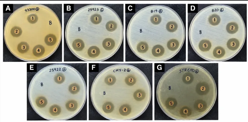
Figure 2: Inhibition Zones Produced by Psidium Guajava Aqueous Extract at Concentrations Ranging from 2.3 to 11.4 mg/ml (1–5), With the Blank Control (B), Against Different Bacterial Strains: (A) Resistant Staphylococcus Aureus; (B) Wild-Type S. Aureus; (C) Resistant Staphylococcus Pseudintermedius; (D) Wild-Type S. Pseudintermedius; (E) Wild-Type Escherichia Coli; (F) Resistant E. Coli; and (G) Wild-Type Β-Haemolytic Streptococcus Spp.
At the highest tested concentration (11.4 mg/mL), the aqueous extract of Psidium guajava exhibited varying degrees of antibacterial activity against all tested Gram-positive bacterial strains. The maximum inhibition zone (21.0 ± 2.00 mm) was observed against sensitive β-hemolytic Streptococcus, followed by resistant Staphylococcus aureus (18.0 ± 3.46 mm), sensitive S. pseudintermedius (18.0 ± 0.00 mm), resistant S. pseudintermedius (17.3 ± 1.15 mm), and sensitive S. aureus (16.0 ± 0.00 mm). Overall, the inhibition zones showed an increasing trend with increasing extract concentrations across the tested Gram-positive bacterial strains. A similar concentration-dependent antibacterial effect was reported by Bolzan et al.19, who investigated the activity of ethanolic P. guajava leaf extract against microorganisms associated with the canine oral microbiota.
The antibacterial potential of aqueous P. guajava leaf extracts against S. aureus has also been documented in previous investigations. However, these studies reported inhibitory effects at concentrations of 50.0 mg/mL20 and 62.5 mg/mL16, both substantially higher than the maximum concentration employed in the present study (11.4 mg/mL). Such differences in the observed antibacterial activity may be related to variations in the phytochemical profiles and concentrations of bioactive constituents, particularly phenolic compounds, which are known to contribute to antibacterial effects. Additionally, differences in extraction procedures, plant material composition, extract preparation, and experimental conditions may influence antibacterial outcomes and should be considered when comparing results across different studies.
Based on the disk diffusion results, concentrations of 6.8 and 11.4 mg/mL were selected for further evaluation using minimum inhibitory concentration (MIC) and minimum bactericidal concentration (MBC) assays. The broth microdilution assay demonstrated inhibition of bacterial growth in all tested strains at both concentrations. Accordingly, the MIC was determined to be 6.8 mg/mL for all tested strains under the experimental conditions employed. This value was comparable to that reported by Metwally et al. 21 (5.25 mg/mL) for sensitive S. aureus, higher than the value reported by Sanches et al.22 (500 μg/mL) for sensitive S. aureus, and lower than the concentrations reported by Araújo et al. 16 (62.5 mg/mL) for both sensitive and resistant S. aureus and by Ratnakara et al.23 (30–40 mg/mL) for sensitive S. aureus. These differences highlight the potential influence of extract composition and experimental methodology on the antibacterial activity of P. guajava.
The minimum bactericidal concentration (MBC) assay demonstrated variation in the bactericidal activity of the aqueous Psidium guajava extract among the tested bacterial strains. The resistant strain of Staphylococcus pseudintermedius exhibited an MBC of 6.8 mg/mL, whereas the MBC values for all other tested strains exceeded the maximum concentration evaluated (11.4 mg/mL) (Figure 3). These findings indicate differential susceptibility among the bacterial strains, with resistant S. pseudintermedius showing comparatively greater susceptibility to the aqueous extract under the experimental conditions.
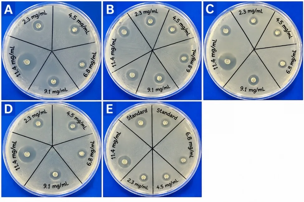
Figure 3: Determination of Minimum Bactericidal Concentration (MBC) by Subculturing Aliquots from the Broth Microdilution Assay onto Brain Heart Infusion (BHI) Agar Plates. the Representative Plates Show Bacterial Growth Patterns for: (A) Resistant Staphylococcus Aureus; (B) Wild-Type Staphylococcus Aureus; (C) Resistant Staphylococcus Pseudintermedius; (D) Wild-Type Staphylococcus Pseudintermedius; and (E) Wild-Type Β-Haemolytic Streptococcus Spp.
Previous investigations have reported antibacterial activity of aqueous P. guajava extracts, particularly against Gram-positive bacteria, although the extent of inhibition varies considerably among bacterial strains. Differences in the inhibitory concentrations reported across studies may be associated with variations in the phytochemical composition of the extracts, particularly their total phenolic and tannin contents. Such variations may result from differences in extraction procedures, geographical origin, environmental conditions, and other factors influencing the accumulation of secondary metabolites in plant materials. Similar considerations regarding the phytochemical composition and biological activities of different P. guajava extracts have been discussed by Raj et al. 20 and Biswas et al.24. Similar findings regarding the bactericidal activity of aqueous Psidium guajava leaf extract have also been reported by Pereira et al.25, who investigated its activity against susceptible and antimicrobial-resistant bacterial strains. Their study highlighted variations in bactericidal susceptibility among the tested bacterial isolates, supporting the importance of evaluating MBC alongside inhibitory activity when assessing the antibacterial potential of plant extracts.
Furthermore, Moorthy et al.26, highlighted the potential contribution of tannins to the antimicrobial properties of P. guajava extracts.
The influence of plant parts and extraction solvents on antibacterial activity was demonstrated by Sanches et al. 22. They investigated aqueous extracts prepared from the leaves, roots, and stem bark of P. guajava and reported varying levels of activity against Staphylococcus aureus, with minimum inhibitory concentration (MIC) values of 500, 125, and 250 μg/mL, respectively. In contrast, these extracts exhibited comparatively limited activity against Gram-negative bacteria, including Escherichia coli and Pseudomonas aeruginosa, for which the MIC values exceeded 1000 μg/mL. Their findings also indicated that hydroethanolic extracts exhibited greater antibacterial activity than aqueous extracts, emphasizing the importance of solvent selection in extracting bioactive phytochemicals from medicinal plants.
Beyond the investigation of crude plant extracts, the identification and evaluation of individual bioactive constituents are important for understanding their antimicrobial potential and possible interactions. Certain phytochemicals may exhibit additive or synergistic effects when used in combination 27. Several flavonoids, including luteolin, morin, naringin, rutin, and quercetin, have demonstrated antibacterial activity against a range of Gram-positive and Gram-negative bacteria, including S. aureus and E. coli 28. Similarly, Amin et al.29 reported combined inhibitory effects of morin, rutin, and quercetin against methicillin-resistant S. aureus (MRSA). Their investigation also demonstrated additive antibacterial effects when these flavonoids were combined with conventional antibiotics against the tested isolates. These observations suggest that plant-derived flavonoids may warrant further investigation as potential components of complementary antimicrobial strategies.
The present study provides experimental evidence of the antibacterial activity of aqueous P. guajava leaf extract against several bacterial strains, including resistant isolates of S. aureus and Staphylococcus pseudintermedius. The observed activity is particularly noteworthy in view of the medical and veterinary significance of these organisms and the increasing concern regarding antimicrobial resistance. However, the bactericidal response differed among the tested strains, as reflected by the variation in their MBC values. Although the findings indicate the potential of aqueous guava leaf extract as a source of antibacterial phytochemicals, further investigations are required to establish the consistency and reproducibility of its activity against a broader range of antimicrobial-resistant isolates.
The increasing prevalence of antimicrobial resistance has compromised the effectiveness of several conventional antimicrobial agents, highlighting the importance of exploring alternative sources of antibacterial compounds30,31. MRSA represents a major public health concern because of its association with difficult-to-treat infections and its capacity to spread in both healthcare and community settings32,33. Turner et al. 34 further emphasized the wider public health implications of MRSA, particularly in relation to its persistence, transmission, and clinical management. The emergence and spread of multidrug-resistant bacterial pathogens also contribute to substantial healthcare and economic burdens, reinforcing the need for continued antimicrobial research.
Against this background, systematic investigations of medicinal plants and their bioactive constituents may contribute to the identification of promising candidates for future antimicrobial development. Plant-derived compounds offer a diverse range of chemical structures and biological activities that may provide leads for developing alternative or complementary approaches to infection management. Nevertheless, their potential therapeutic applications require comprehensive evaluation, including investigations of antimicrobial efficacy, mechanisms of action, toxicity, pharmacological properties, and interactions with existing antibiotics.
CONCLUSIONS:
The phytochemical screening of Psidium guajava L. leaf extracts obtained through maceration and aqueous decoction revealed the presence of tannins, saponins, flavonoids, alkaloids, terpenoids, steroids and cardiac glycosides.. Tannins were indicated by the development of a bluish-black or green coloration, saponins by the formation of stable foam, and flavonoids by the appearance of a dark red coloration. These findings indicate that guava leaves contain phytochemical constituents of potential biological significance.
The present study also demonstrated the antibacterial activity of the aqueous P. guajava leaf extract against the tested sensitive and resistant Gram-positive bacterial strains. These findings highlight the potential of guava leaves as a source of naturally occurring antibacterial compounds and support further investigation into their activity against antimicrobial-resistant bacteria. However, additional studies are required to isolate and characterize the active constituents, elucidate their mechanisms of action, and evaluate their safety and efficacy. Further research may contribute to the development of plant-derived antibacterial agents as potential candidates for addressing the growing challenge of antimicrobial resistance.
5. ACKNOWLEDGEMENT:
The author sincerely acknowledges the Department of Chemistry, Marwari College, Darbhanga, Bihar, India, for providing the necessary laboratory facilities and academic support to carry out the present work. The author is also grateful to the laboratory staff for their assistance in conducting the experimental studies.
6. CONFLICT OF INTEREST:
The author declares no conflict of interest
7. FUNDING:
The author received no financial support for the research, authorship and publication of this article.
8. DECLARATION OF GENERATIVE AI AND AI-ASSISTED TECHNOLOGIES IN THE MANUSCRIPT PREPARATION PROCESS:
During the preparation of this manuscript, the author used ChatGPT (OpenAI) to assist with language improvement, grammar, readability, and clarity of the manuscript. The author reviewed and edited the AI-assisted content and takes full responsibility for the accuracy, integrity, interpretation, and final content of the manuscript. ChatGPT was not used to generate, analyze, or interpret the experimental data or to alter the primary research images presented in this manuscript.
REFERENCES

Rinky Kumari, Comparative Phytochemical Screening of Ethanolic and Aqueous Guava (Psidium Guajava L.) Leaf Extracts and Antibacterial Evaluation of the Aqueous Extract Against Selected Bacterial Strains, Int. J. of Pharm. Sci., 2026, Vol 4, Issue 11, 2485-2498. https://doi.org/10.5281/zenodo.24279408
 10.5281/zenodo.24279408