

We use cookies to ensure our website works properly and to personalise your experience. Cookies policy
M. Pharmacy. Department of Pharmaceutics, Sarojini Naidu Vanita Pharmacy MahaVidyalaya, Tarnaka, Hyderabad, Telangana, India.
The present study was undertaken to formulate and evaluate a floating mucoadhesive gastroretentive in-situ gel containing Litchi chinensis fruit extract for sustained antiulcer drug delivery. Gastroretentive drug delivery systems offer an approach to prolong the residence of a formulation in the stomach, thereby improving its contact with the gastric mucosa and providing sustained release of the incorporated therapeutic agent. In this study, sodium alginate was selected as the primary gelling polymer, while Carbopol 934 was incorporated to impart mucoadhesive properties. A total of nine formulations (F1–F9) were prepared by varying the concentrations of sodium alginate and Carbopol 934. Calcium carbonate was incorporated as a gas-generating agent and a source of calcium ions to facilitate floating and gel formation in the gastric environment. The prepared formulations were evaluated for physical appearance, pH, viscosity, gel strength, gelation time, floating behavior, ex-vivo mucoadhesive strength, drug content uniformity and in-vitro drug release. UV-visible spectrophotometry was employed for the estimation of the extract, with the analytical wavelength selected at 217 nm. In-vitro dissolution studies were performed using USP Type II dissolution apparatus in simulated gastric fluid (pH 1.2) for 10 hours. The prepared formulations exhibited pH values ranging from 4.12 to 4.71 and viscosity values from 328 to 672 cP. All nine formulations showed a floating duration exceeding 12 hours. Gelation time ranged from 32.8 to 43.2 seconds, while mucoadhesive strength varied from 8.4 to 15.6 g. The drug content was found to be between 84.56% and 93.23%. Among the formulations, F6 demonstrated the highest drug content (93.23%), mucoadhesive strength (15.6 g) and cumulative drug release (97.90%) at the end of 10 hours. In comparison, F9 exhibited the lowest drug release of 82.40%. Based on the evaluated physicochemical and in-vitro release characteristics, F6 was identified as the comparatively promising formulation
Gastroretentive drug delivery systems (GRDDS) are advanced oral drug delivery approaches designed to prolong the residence time of dosage forms in the stomach, thereby providing an opportunity for controlled drug release within the gastric environment. These systems may improve local drug availability, prolong contact with the gastric mucosa and facilitate sustained drug delivery for suitable therapeutic agents[1]. Prolonged gastric retention enhances bio-availability, minimizes systemic plasma concentration fluctuations, reduces adverse drug reactions, and enables localized treatment for gastric pathologies [2]. Candidate drugs for GRDDS formulations typically display poor distal intestinal permeability, possess a distinct narrow absorption window within the upper small intestine, or exhibit structural instability and poor solubility in the alkaline pH of the lower gastrointestinal tract [3].
1.1 Basic Physiology of the Gastrointestinal Tract
Anatomically, the human stomach is divided into three functional regions: the fundus, the body, and the antrum (pylorus). The proximal portion (fundus and body) functions primarily as a storage reservoir for ingested contents, whereas the distal antrum serves as the primary site for mechanical mixing and grinding, propelling gastric contents toward the pylorus.
Gastric motility patterns differ fundamentally between the fasted and fed states. In the fasted condition, an interdigestive cyclic electrical pattern called the Migrating Myoelectric Complex (MMC) or Interdigestive Myoelectric Complex (IMMC) repeats every 2 to 3 hours across the stomach and small intestine [4], [5]. The MMC is subdivided into four characteristic phases:
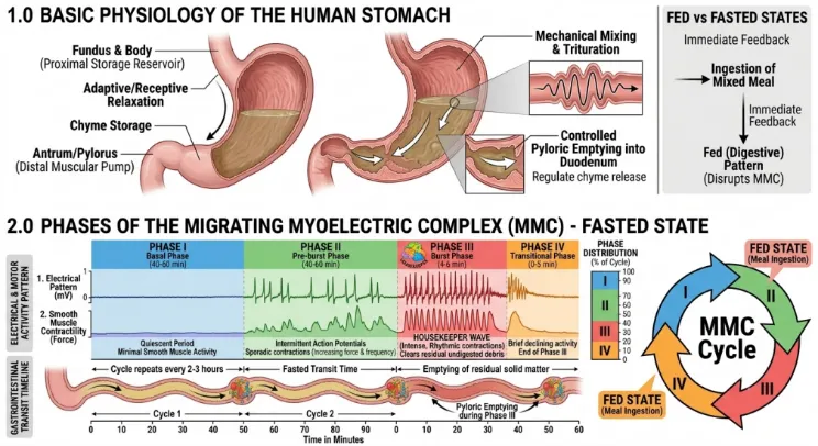
Figure 1: Comparative Gastrointestinal Motility
Dynamics and Motor Patterns.
Ingestion of a solid or mixed meal disrupts the cyclic fasting MMC, triggering the fed (digestive) motility pattern. This state exhibits persistent, irregular contractions similar to Phase II fasting activity, which reduce solid food particles to a suspension of less than 1 mm before emptying through the pylorus. The onset of the IMMC is delayed in the fed state, significantly slowing the gastric emptying rate. Gastric emptying can influence the residence time of conventional oral controlled-release dosage forms, potentially resulting in variable gastric transit and drug-release behavior.
1.2 Factors Affecting Gastric Retention Time (GRT)
Gastric retention time directly governs drug absorption efficacy, particularly for APIs targeting narrow absorption windows in the stomach or upper small intestine. Extending the residence time of the formulation near its primary absorptive membrane maximizes overall bioavailability. Under unformulated conditions, transit through the upper gastrointestinal tract is rapid, with typical gastric emptying occurring within 2 to 3 hours [6].
Gastric retention time is evaluated using direct imaging techniques (e.g., X-ray radiography, radiotelemetry, magnetic marker monitoring, and gamma-scintigraphy) and indirect methods (such as the hydrogen breath test and site-specific absorbed biomarkers) [7].
Gastric emptying rates and overall GRT are governed by two main categories: (i) pharmaceutical technology factors and (ii) intrinsic biological/physiological factors.
Pharmaceutical Technology Factors
Physiological and Biological Factors
1.3 Peptic Ulcer Disease
Historically, peptic ulcer disease (PUD) was managed through surgical interventions associated with high morbidity and mortality. The emergence of histamine H2-receptor antagonists (H2RAs) such as Cimetidine and Ranitidine in the 1970s transformed treatment paradigms, causing elective ulcer surgeries to drop by 85% [19]. Subsequent introduction of proton-pump inhibitors (PPIs) provided superior acid suppression without tachyphylaxis, achieving high healing rates for gastric and duodenal ulcers [20]. Despite these advancements and the discovery of Helicobacter pylori, PUD remains a substantial global health burden, costing billions annually and disproportionately affecting elderly populations [–[23]. Current clinical challenges include overcoming H. pylori eradication failure due to antibiotic resistance, preventing NSAID-induced mucosal damage, and managing non-NSAID, non-H. pylori ulcerations.
1.4 Pathophysiology of Peptic Ulcer Disease
Peptic ulceration develops from an imbalance between aggressive luminal factors (gastric acid, pepsin, NSAID exposure, H. pylori toxins) and mucosal protective barriers (mucus gel layer, bicarbonate secretion, endogenous prostaglandins, mucosal blood flow).
Table 1: Balance of Gastric Aggressive and Protective Factors in Peptic Ulcer Pathophysiology
| S. No | Aggressive Factors | Protective Factors |
|---|---|---|
| 1 | Gastric Acid & Pepsin Hypersecretion | Mucus & Bicarbonate Secretion Layer |
| 2 | Helicobacter pylori Cytotoxins | Mucosal Blood Flow & Perfusion |
| 3 | NSAIDs & Aspirin-induced COX-1 Inhibition | Endogenous Prostaglandins (PGE2, PGI2) |
1.5 In-Situ Gelling Systems
In-situ gelling systems undergo sol-to-gel phase transitions at the site of administration upon exposure to specific environmental triggers:
a) By Physical Change (Swelling and Diffusion)
b) By Chemical Change
Altering the ionic environment induces polymer cross-linking. Ion-sensitive polymers (e.g., sodium alginate, calcium alginate, gellan gum, low-methoxy pectin) undergo a phase transition in the presence of monovalent or divalent cations (Ca2+, Mg2+, Na+, K+). For instance, low-methoxy pectin cross-links with Ca2+ ions, while sodium alginate gels via interaction of polyvalent cations with guluronic acid (G-blocks) along the alginate chains.
c) By Physiological Changes
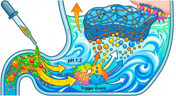
Figure 2: Mechanism of pH-Triggered In-Situ Gelation and Gas Generation in Gastric Environment
d) Dilution-Sensitive Systems
Polymers undergo sol-to-gel phase transitions when exposed to higher volumes of biological fluids
e) Electrical Signal-Sensitive Hydrogels
Polyelectrolyte hydrogels undergo swelling or deswelling in response to an applied electrical field due to directional migration of counter-ions.
f) Light-Sensitive Hydrogels
Photo None: Responsive Hydrogels Utilize Ultraviolet or Visible Light to Initiate Cross-Linking for Tissue Engineering and Targeted Drug Delivery.
g) Glucose-Sensitive Hydrogels
Self-regulating insulin delivery platforms incorporate glucose oxidase within pH-sensitive hydrogels. Glucose oxidation lowers microenvironmental pH, triggering hydrogel swelling and pulsatile insulin release. Alternative systems utilize competitive displacement of insulin from Concanavalin A binding sites by ambient glucose.
1.6 Litchi chinensis Sonn.
Litchi chinensis Sonn. (family Sapindaceae) is a commercially important fruit tree native to Southern China, currently cultivated across India, Vietnam, Thailand, Madagascar, and Mauritius [31], [32]. The tree reaches heights of 15 to 20 meters, yielding strong, corrosion-resistant timber suitable for structural uses. The fruit is oval to spherical with a red or purple-red scale-like pericarp enclosing a juicy edible aril and a brown seed [33], [34].
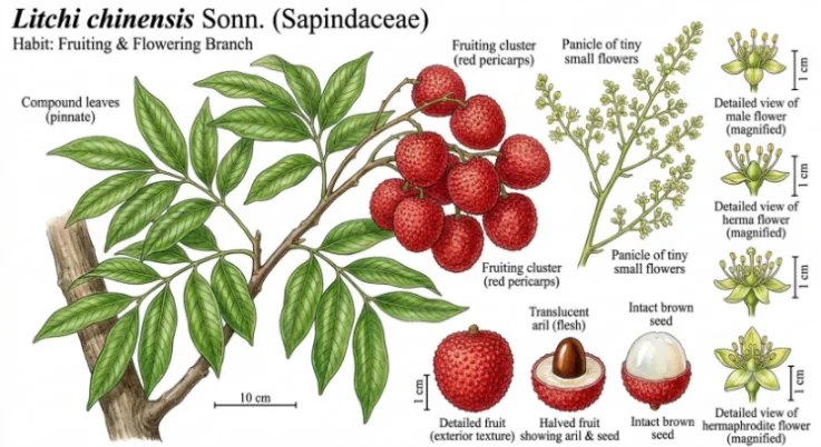
Figure 3: Botanical Profile and Fruit Pericarp of Litchi chinensis Sonn.
Due to rapid post-harvest degradation, fresh litchi fruit has a short shelf-life and is frequently processed into preserved fruit, jellies, jams, and juices [35], [36]. Litchi pulp is rich in glucose, fructose, dietary fibers, ascorbic acid, amino acids, and essential minerals [38], [39]. Ethnopharmacologically, the Chinese Pharmacopoeia (2020 edition) recognizes litchi seeds (Litchi Semen) for managing testicular swelling and hernia pain [37]. In traditional Indian medicine, ground seeds are used for intestinal helminthiasis and neuralgia, while the fruit pulp is used to treat anemia, asthma, and insomnia [38], [39].
Polysaccharides, flavonoids, and polyphenols isolated from Litchi chinensis exhibit significant biological properties, including antioxidant, immunomodulatory, hypoglycemic, and prebiotic activities [40]–[45]. Biocompatible and non-toxic litchi polysaccharides represent valuable natural polymers for functional foods and drug delivery systems.
1.7 Pharmacological Activities of Litchi chinensis
The reported pharmacological properties of Litchi chinensis provide a scientific basis for investigating its potential incorporation into gastroretentive drug delivery systems. However, the therapeutic efficacy of the prepared formulation depends on its actual pharmacological evaluation and cannot be established solely from the reported biological activities of the plant extract.
2.AIM AND OBJECTIVES
2.1 AIM
To formulate, develop, and evaluate a floating mucoadhesive gastroretentive in situ gel loaded with Litchi chinensis fruit extract using sodium alginate and Carbopol 934 for sustained antiulcer activity.
2.2 OBJECTIVES OF THE STUDY
1. To determine the maximum wavelength of absorption (λmax) of Litchi chinensis extract using UV-visible spectrophotometry and to develop a standard calibration curve in simulated gastric fluid.
2. To formulate and develop nine gastroretentive in-situ gel formulations (F1–F9) using different concentrations of sodium alginate and Carbopol 934.
4. To incorporate calcium carbonate (CaCO3) as a gas-generating agent and cross-linking cation source to ensure buoyancy and gel matrix rigidity.
5. To evaluate the physicochemical characteristics of the prepared formulations, including physical appearance, pH and viscosity.
6. To investigate the in-situ gelation characteristics of the formulations by determining gelation time and gel strength in simulated gastric fluid.
7. To evaluate the floating behavior of the formulations by determining floating lag time and floating duration.
8. To determine the ex-vivo mucoadhesive strength of the prepared formulations using goat gastric mucosa.
9. To determine the drug content of the prepared formulations using UV-visible spectrophotometry.
10. To investigate the in-vitro release profile of Litchi chinensis extract from the prepared formulations using USP Type II dissolution apparatus over a period of 10 hour.
MATERIALS AND METHODS
3.1 MATERIALS
| Chemical / Material | Functional Category |
|---|---|
| Litchi chinensis Fruit Extract | Active phytotherapeutic agent. |
| Sodium Alginate (NaAlg) | Primary Gelling Polymer |
| Carbopol 934 (CP-934) | Mucoadhesive Polymer / Carbomer |
| Calcium Carbonate (CaCO3) | Gas-generating agent and source of calcium ions for alginate gelation. |
| Trisodium Citrate | Sequestering / Complexing Agent |
| Tween 80 | Non-ionic Surfactant / Emulsifier |
| Liquid Paraffin | Oily Dispersed Phase Vehicle |
| Propylparaben | Antimicrobial Preservative |
| Hydrochloric Acid (0.1 N HCl) | Simulated Gastric Fluid (SGF, pH 1.2) |
| Deionized Water | Aqueous Solvent / Vehicle |
Table 2: List of Chemicals, Polymers, and Excipients Used in Formulation Development with Their Functional Roles.
3.2 INSTRUMENTATION:
| Equipment / Instrument |
|---|
| Double-Beam UV-Vis Spectrophotometer |
| Digital pH Meter |
| Brookfield Digital Viscometer |
| USP Dissolution Test Apparatus |
| Modified Physical Balance Set |
| Fourier transform infrared spectrophotometer |
Table 3: List of Equipment and Instruments Used in Formulation and Evaluation.
3.3 STANDARD OPERATING PROCEDURE & ANALYTICAL METHODOLOGY: UV-VIS SPECTROPHOTOMETRIC ESTIMATION OF LITCHI CHINENSIS EXTRACT IN 0.1 N HCl
1. Preparation of Stock Solutions
Step 1 — Primary Stock Solution (6000 µg/mL)
Step 2 — Secondary Stock Solution (600 µg/mL)
Step 3 — Intermediate Stock Solution (100 µg/ml)
Step 4 — Preparation of Scanning Solution (60 µg/ml) for λmax Determination
2. Determination of Wavelength of Maximum Absorption (λmax)
3. Preparation of Calibration Working Standards
A series of calibration standards ranging from 10 to 50 µg/ml was prepared using the intermediate stock solution (100 µg/ml).
The required volumes of the intermediate stock solution were transferred into separate 10 ml volumetric flasks and diluted to the mark with 0.1 N HCl.
Table 4: Dilution Scheme for the Preparation of Calibration Working Standards
| Target Concentration (µg/ml) |
Volume of 100 µg/ml Stock (ml) | Final Volume with 0.1 N HCl (ml) |
|---|---|---|
| 10 | 1.0 | 10.0 |
| 20 | 2.0 | 10.0 |
| 30 | 3.0 | 10.0 |
| 40 | 4.0 | 10.0 |
| 50 | 5.0 | 10.0 |
4. Absorbance Measurement and Calibration Curve Generation
Apply linear regression analysis to evaluate adherence to Beer-Lambert's law and determine the regression parameters:
3.4 FORMULATION DEVELOPMENT OF FLOATING MUCOADHESIVE IN-SITU GELS (F1 TO F9)
Nine formulations (F1–F9) of the gastroretentive floating mucoadhesive in-situ gel were developed by varying the concentrations of sodium alginate and Carbopol 934. Sodium alginate was used as the primary ion-sensitive gelling polymer, while Carbopol 934 was incorporated as a mucoadhesive polymer. Three concentrations of sodium alginate (750, 1000 and 1250 mg) and three concentrations of Carbopol 934 (250, 500 and 750 mg) were selected to prepare nine different polymer combinations. The quantities of the other formulation ingredients were maintained constant across all batches to investigate the influence of polymer concentrations on the physicochemical characteristics, gelation, floating behavior, mucoadhesion and drug release of the formulations.[58].
Table 5: Formulation Composition – F1 to F9
| Batch Code | Sodium Alginate (mg) | Carbopol 934 (mg) | Extract (mg) | Trisodium Citrate (mg) | CaCO3 (mg) | Tween 80 (mL) | Liq. Paraffin (mL) | Propyl-paraben (mg) |
|---|---|---|---|---|---|---|---|---|
| F1 | 750 | 250 | 100 | 200 | 1000 | 2.5 | 2.5 | 10 |
| F2 | 1000 | 250 | 100 | 200 | 1000 | 2.5 | 2.5 | 10 |
| F3 | 1250 | 250 | 100 | 200 | 1000 | 2.5 | 2.5 | 10 |
| F4 | 750 | 500 | 100 | 200 | 1000 | 2.5 | 2.5 | 10 |
| F5 | 1000 | 500 | 100 | 200 | 1000 | 2.5 | 2.5 | 10 |
| F6 | 1250 | 500 | 100 | 200 | 1000 | 2.5 | 2.5 | 10 |
| F7 | 750 | 750 | 100 | 200 | 1000 | 2.5 | 2.5 | 10 |
| F8 | 1000 | 750 | 100 | 200 | 1000 | 2.5 | 2.5 | 10 |
| F9 | 1250 | 750 | 100 | 200 | 1000 | 2.5 | 2.5 | 10 |
3.4.1 Step-by-Step Emulsion-Polymer Preparation Procedure
1. The required quantities of sodium alginate, Carbopol 934, calcium carbonate, trisodium citrate, and propylparaben were accurately weighed using an analytical balance. [59].
2. Sodium alginate and Carbopol 934 were dispersed in approximately 40 ml of deionized water containing trisodium citrate under continuous magnetic stirring at 70 °C until complete polymer hydration was achieved.
3. The polymeric dispersion was cooled to 40 °C, and calcium carbonate was added under slow agitation to ensure uniform dispersion [60].
4. Separately, an oil-in-water emulsion was prepared by mixing Tween 80 and liquid paraffin with water under high-speed stirring, followed by the homogeneous incorporation of Litchi chinensis extract [61].
5. The primary polymeric dispersion was mixed with the extract emulsion under continuous stirring to prevent air entrapment [62].
6. Propylparaben was added, the volume was adjusted to 60 ml with deionized water, and the final formulation was packed into amber glass bottles.
3.5 EVALUATION PARAMETERS
3.5.1 Visual Appearance and pH Measurement
All nine formulations (F1–F9) were visually examined for their color, clarity, homogeneity, presence of visible aggregates and any signs of phase separation. The pH of each formulation was measured at 25°C using a calibrated digital pH meter. [63].
3.5.2 Viscosity Determination
The rheological behavior and dynamic viscosity of the liquid formulations (sol state) were evaluated at 25 ± 0.5 °C using a Brookfield DV-II+ Pro digital rotational viscometer. Viscosity measurements were performed using spindle No. 4 at 50 rpm, and the readings were expressed in centipoise (cP).[64].
3.5.3 Gel Strength Measurement
Gel strength was evaluated to determine the mechanical resistance of the formed gel matrix. A measured quantity of formulation was exposed to simulated gastric fluid (SGF, pH 1.2) to initiate gel formation. A standard weight of 35 g was placed on the surface of the formed gel, and the time required for the weight to penetrate the specified depth of the gel matrix was recorded in seconds. A longer penetration time indicates greater resistance of the gel matrix to penetration under the specified experimental conditions.[65].
3.5.4 Gelation Time
Gelation time was evaluated to determine the time required for the liquid formulation to undergo a sol-to-gel transition upon exposure to simulated gastric fluid. A 5 mL portion of each formulation was introduced dropwise into 50 mL of simulated gastric fluid (0.1 N HCl, pH 1.2) maintained at 37 ± 0.5°C. The time required for the formation of a coherent gel structure was recorded in seconds.[66]
3.5.5 Floating Behavior (Floating Lag Time & Total Floating Duration)
To evaluate gastroretentive buoyancy, 10 mL of each formulation was placed in a USP dissolution vessel containing 900 mL of simulated gastric fluid (pH 1.2) maintained at 37 ± 0.5 °C with mild agitation (50 rpm). The floating lag time was recorded as the time required for the formulation to reach the surface of the dissolution medium. The total floating duration was determined by observing the period during which the formed gel remained continuously buoyant. Observations were continued for more than 12 hours.[67].
3.5.6 Ex-Vivo Mucoadhesive Strength
The mucoadhesive strength of the prepared formulations was evaluated using a modified physical balance method with freshly excised goat gastric mucosa. The gastric mucosal tissue was carefully collected and prepared for the adhesion study. A measured quantity of the gelled formulation was brought into contact with the mucosal surface under the specified experimental conditions. The force required to detach the formulation from the mucosal tissue was determined using the modified physical balance apparatus. The results were expressed in grams as the detachment load required to separate the formulation from the mucosal surface.[68].
3.5.7 Drug Content Uniformity
An accurately measured quantity of 5 mL of the prepared formulation was taken and diluted with simulated gastric fluid (SGF, pH 1.2). The resulting solution was mixed thoroughly to facilitate the extraction of the incorporated Litchi chinensis extract. The solution was filtered to remove insoluble formulation components, and the absorbance of the filtrate was measured using a UV-visible spectrophotometer at 217 nm against SGF as the blank. The concentration of the extract was determined using the previously established calibration curve, and the percentage drug content was calculated by comparing the experimentally determined extract content with the theoretical extract content present in the corresponding formulation sample.
3.5.8 In-Vitro Dissolution Studies
In-vitro dissolution studies were performed to evaluate the release of Litchi chinensis extract from the prepared gastroretentive in-situ gel formulations using a USP Type II (paddle) dissolution apparatus.
The dissolution study was carried out using 900 mL of simulated gastric fluid (SGF, pH 1.2), maintained at 37 ± 0.5°C, with a paddle rotation speed of 50 rpm. The dissolution experiment was continued for 10 hours.
Samples of 5 mL were withdrawn at predetermined time intervals of 1, 2, 4, 6, 8 and 10 hours. After each withdrawal, an equal volume of fresh SGF maintained at the same temperature was added to the dissolution vessel to maintain a constant dissolution volume. The withdrawn samples were filtered and analyzed using a UV-visible spectrophotometer at 217 nm.
The concentration of the released extract was determined using the previously established standard calibration curve. The cumulative amount of extract released was calculated by considering the quantity removed during previous sampling intervals, and the cumulative percentage drug release was determined. The release profiles of all nine formulations were compared to investigate the influence of polymer concentrations on the release behavior of the prepared formulations.
RESULTS AND DISCUSSION
4.1 Drug-excipient compatibility studies by FTIR spectroscopy
Fourier Transform Infrared (FTIR) spectroscopy is an analytical technique used to identify characteristic functional groups present in a substance by measuring the absorption of infrared radiation at different wavenumbers.
The technique is useful in formulation development because interactions between an active ingredient and pharmaceutical excipients may produce changes in characteristic absorption bands, including shifts in peak positions, changes in intensity or the appearance or disappearance of certain peaks.
In the present investigation, FTIR analysis was presented for both Litchi chinensis extract and the prepared formulation to examine their characteristic absorption bands and assess possible interactions between the extract and formulation ingredients.
The FTIR spectrum of Litchi chinensis extract shows the following characteristic absorption bands:
The formulation spectrum also exhibits absorption bands in several of the same regions as the extract spectrum. The presence of comparable bands suggests that characteristic functional groups of the extract may remain detectable after formulation.
However, a definitive conclusion regarding drug-excipient compatibility cannot be made merely because the spectra contain similar peaks. The individual excipient spectra, peak positions, possible shifts, intensity changes and a properly prepared comparison are needed.
FTIR analysis provides preliminary information regarding the chemical characteristics of the extract and the formulated system. The observed absorption bands indicate the presence of several functional groups associated with the phytochemical constituents of the extract.
The comparison of the spectra can help identify possible changes following incorporation into the polymeric formulation. Nevertheless, FTIR alone cannot establish the absence of all chemical interactions or prove long-term compatibility.
4.1 FTIR compatibility studies
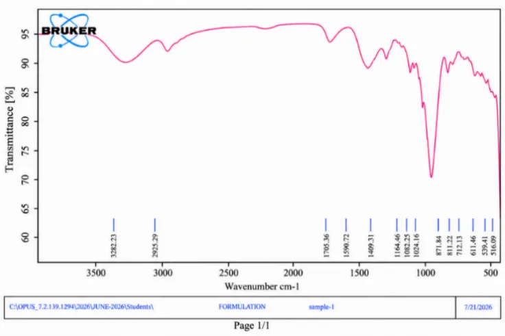
Figure 4: Fourier Transform Infrared Spectrum of Litchi chinensis extract.
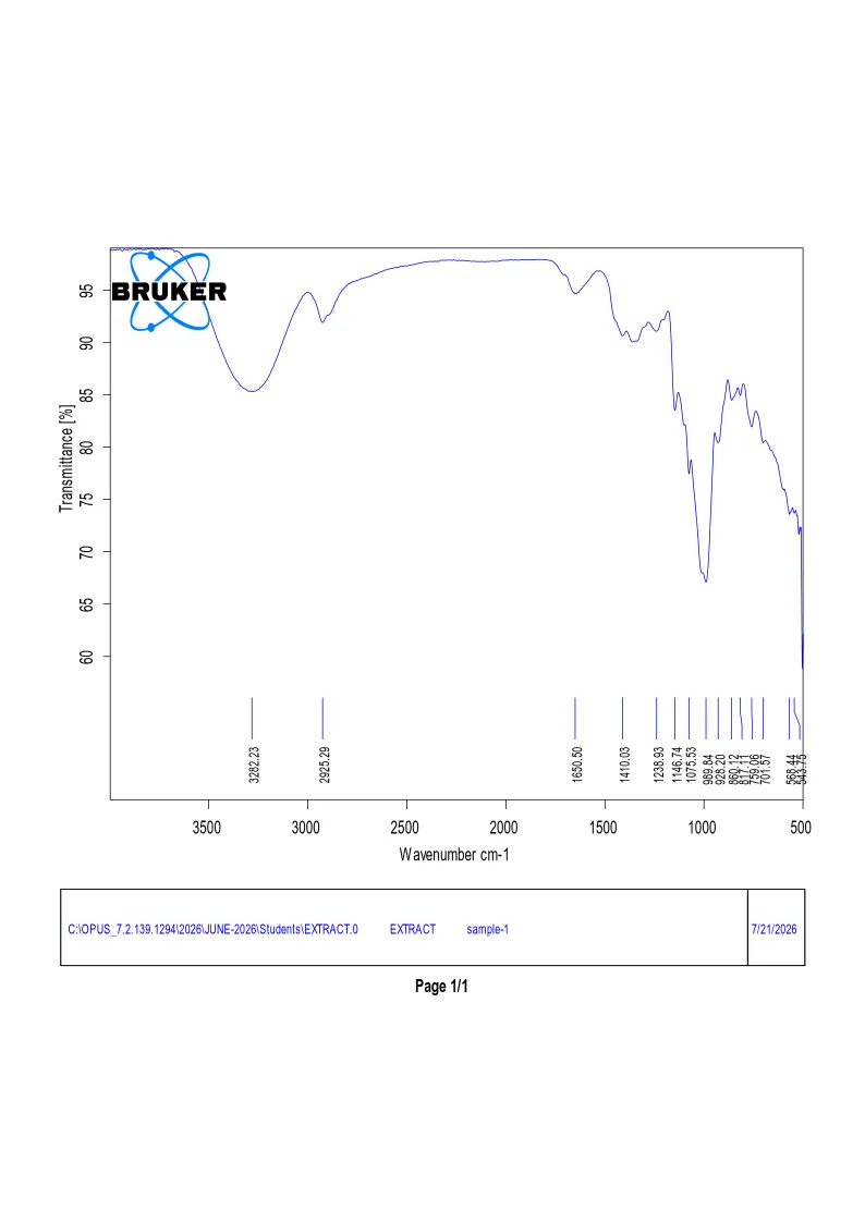
Figure 5: Fourier Transform Infrared Spectrum of Formulation
4.2 Development of the standard calibration curve
UV-visible spectrophotometry was employed to determine the absorbance characteristics of Litchi chinensis extract in 0.1 N HCl.
The method is based on the Beer–Lambert law, which describes the relationship between absorbance and concentration:
A= εbc
Where:
Initially, the absorption spectrum was recorded to identify the wavelength of maximum absorption (λmax). The maximum absorption was observed at 217 nm, which was subsequently selected for quantitative analysis.
A series of standard solutions with concentrations ranging from 10 to 50 µg/mL was prepared using the intermediate stock solution. Their absorbance values were measured at 217 nm.
The absorbance increased from 0.013 at 10 µg/mL to 0.051 at 50 µg/mL.
This progressive increase in absorbance with increasing concentration indicates a concentration-dependent response of the extract under the experimental conditions.
The calibration curve demonstrated an approximately linear relationship between concentration and absorbance.
The calculated coefficient of determination from the displayed data is approximately 0.9983, indicating a strong linear association within the tested concentration range.
The establishment of a calibration curve is important for the quantitative estimation of the extract during drug content determination and in-vitro dissolution studies.
The observed increase in absorbance with concentration supports the use of UV-visible spectrophotometry as a preliminary analytical approach for monitoring the extract.
However, the method should not be described as fully validated because parameters such as accuracy, precision, specificity, robustness and limits of detection and quantification have not been reported.
Additionally, the extract exhibits a secondary absorption feature around 268 nm in the spectrum on page 32. Since the primary absorption maximum is reported at 217 nm, the selected analytical wavelength should be justified and possible interference from formulation excipients should be considered.
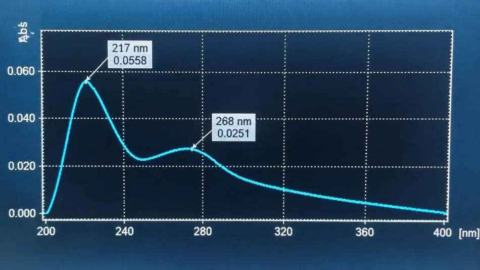
Figure 6: UV-Visible Absorption Spectrum of Litchi chinensis Extract Exhibiting a Characteristic Maximum Absorption Wavelength Λmax at 217 nm
4.2: Development of standard calibration curve
Table 6: Calibration Data of Litchi chinensis Extract at 217 nm.
| Concentration (µg/mL) | Absorbance (at 217 nm) |
|---|---|
| 0 | 0.003 |
| 10 | 0.013 |
| 20 | 0.024 |
| 30 | 0.032 |
| 40 | 0.041 |
| 50 | 0.051 |
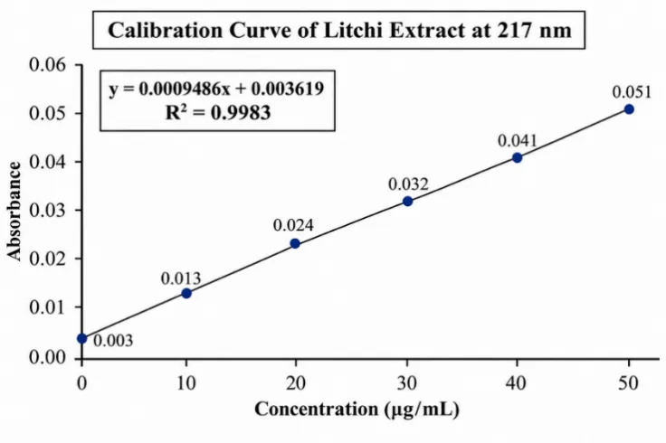
Figure 7: Standard Calibration Curve of Litchi Chinensis Extract at 217 nm.
4.3 Physicochemical evaluation of formulations F1–F9
The nine formulations were prepared by varying the concentrations of sodium alginate and Carbopol 934 while maintaining the other listed ingredients constant.
The formulations were evaluated for physical appearance, pH, viscosity, gel strength and gelation time.
4.3.1 Physical appearance
All nine formulations were reported to exhibit a clear, pale-yellow appearance.
The similar physical appearance of all batches indicates that the formulations had a comparable visual appearance following preparation.
The absence of visibly reported aggregates or phase separation suggests acceptable macroscopic uniformity at the time of observation.
However, visual examination alone cannot establish physical stability or prove that the extract is uniformly distributed throughout the formulation. Additional uniformity and stability assessments would be needed for that conclusion.
4.3.2 pH determination
The pH values ranged from 4.12 to 4.71.
F1 exhibited the lowest pH of 4.12, whereas F9 showed the highest pH of 4.71.
The pH values showed a gradual increase across the formulation batches.
This variation may be associated with changes in the concentrations of sodium alginate and Carbopol 934 and their interactions within the formulation system.
Both polymers contain ionizable functional groups that can influence the pH of the aqueous formulation.
The observed pH values indicate that the prepared formulations were mildly acidic before exposure to simulated gastric fluid.
Since the formulations are intended for oral administration and subsequent gelation in the stomach, their behavior following contact with acidic gastric conditions is particularly important.
The initial pH values alone do not establish the extent or efficiency of in-situ gel formation.
4.3.3 Viscosity determination
Viscosity was measured using a Brookfield digital viscometer under the specified experimental conditions.
The viscosity values ranged from 328 ± 4.5 cP to 672 ± 7.8 cP.
Viscosity is an important parameter in gastroretentive in-situ gel formulations because it influences the flow properties of the formulation before administration and may affect its ability to remain localized after gel formation.
An increase in polymer concentration generally increases the viscosity of the formulation because of greater polymer-chain interactions and the formation of a more structured polymeric dispersion.
The results indicate a general increase in viscosity with increasing polymer concentration.
I The first series, where Carbopol 934 was maintained at 250 mg, increasing sodium alginate from 750 mg to 1250 mg increased viscosity from 328 cP to 510 cP.
Similarly, at 500 mg Carbopol 934, viscosity increased from 456 cP to 618 cP as sodium alginate concentration increased.
At 750 mg Carbopol 934, viscosity increased from 502 cP to 672 cP.
These findings suggest that both sodium alginate and Carbopol 934 contributed to the viscosity of the formulations.
The higher viscosity of F9 indicates a more viscous liquid system. However, the highest viscosity does not automatically mean that F9 is the best formulation because viscosity must be considered alongside gelation, floating behavior, mucoadhesion and drug release.
4.3.4 Gel strength measurement
Gel strength was evaluated by allowing the formulation to form a gel in simulated gastric fluid and recording the time required for a standard weight to penetrate the gel matrix.
The reported values ranged from 28.4 ± 1.2 seconds to 48.8 ± 2.3 seconds.
Gel strength is an important characteristic because it indicates the mechanical resistance of the gel matrix after exposure to the gastric environment.
In the penetration-based method described in Chapter IV, a longer penetration time indicates greater resistance to penetration, provided the weight, penetration depth and test conditions remain identical.
The results show that F6 exhibited the highest penetration time, suggesting greater resistance to penetration among the tested formulations.
This may be associated with the combined contribution of sodium alginate and Carbopol 934 to the formation of a cohesive polymeric network.
The relatively high gel strength observed in F9 may also be attributed to its higher polymer content.
A sufficiently strong gel matrix is desirable because it may help maintain the structural integrity of the formulation during gastric residence.
However, excessive gel strength may also influence drug diffusion and release. Therefore, gel strength should be interpreted together with the dissolution results.
4.3.5 Gelation time
Gelation time was evaluated by introducing the liquid formulation into simulated gastric fluid maintained at 37 ± 0.5°C and recording the time required for the sol-to-gel transition.
The reported gelation times ranged from 32.8 ± 2.0 to 43.2 ± 2.1 seconds.
F1 exhibited the longest gelation time, whereas F6 showed the shortest gelation time.
Gelation time is an important parameter in gastroretentive in-situ gel systems because rapid gel formation following exposure to gastric fluid helps establish the gel matrix after administration.
The shorter gelation time observed in formulations with higher polymer concentrations may be associated with the availability of a greater quantity of polymer chains for matrix formation.
F6 demonstrated the shortest gelation time of 32.8 ± 2.0 seconds, indicating the fastest gel formation among the formulations according to the reported results.
The results suggest that the polymer combination in F6 provided a favorable balance for rapid gel formation under the tested conditions.
The gelation behavior may be influenced by the interaction between sodium alginate and calcium ions released from calcium carbonate in the acidic medium.
Carbopol 934 may additionally contribute to the cohesion and viscosity of the developing gel matrix.
Table 7: Physicochemical Evaluation of Gastroretentive In-Situ Gel Formulations F1–F9.
| Formulation | Appearance | pH | Viscosity (cP) | Gel Strength (sec) | Gelation Time (sec) |
|---|---|---|---|---|---|
| F1 | Clear, pale yellow | 4.12 | 328 ± 4.5 | 28.4 ± 1.2 | 43.2 ± 2.1 |
| F2 | Clear, pale yellow | 4.18 | 412 ± 5.2 | 33.1 ± 1.4 | 40.5 ± 1.8 |
| F3 | Clear, pale yellow | 4.25 | 510 ± 6.0 | 38.7 ± 1.8 | 37.2 ± 2.3 |
| F4 | Clear, pale yellow | 4.31 | 456 ± 4.8 | 34.6 ± 1.6 | 38.9 ± 1.5 |
| F5 | Clear, pale yellow | 4.44 | 524 ± 5.5 | 40.2 ± 2.0 | 35.4 ± 1.7 |
| F6 | Clear, pale yellow | 4.52 | 618 ± 7.1 | 48.8 ± 2.3 | 32.8 ± 2.0 |
| F7 | Clear, pale yellow | 4.58 | 502 ± 5.8 | 38.1 ± 1.9 | 36.8 ± 1.6 |
| F8 | Clear, pale yellow | 4.63 | 586 ± 6.3 | 43.5 ± 2.1 | 34.1 ± 1.9 |
| F9 | Clear, pale yellow | 4.71 | 672 ± 7.8 | 47.6 ± 2.5 | 34.2 ± 2.2 |
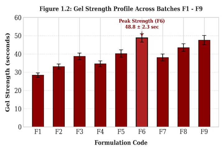
Figure 8: Gel Strength Profile Across Batches F1-F9
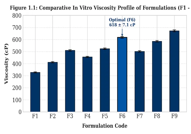
Figure 9: Comparative in Vitro Viscosity Profile of Formulation F1-F9
4.4 Evaluation of floating behavior, mucoadhesive strength and drug content
4.4.1 Floating lag time and floating duration
Floating behavior was evaluated by introducing the formulations into simulated gastric fluid maintained at 37 ± 0.5°C and observing the time required for the gel to reach the surface of the medium.
The floating lag time ranged from 38 to 48 seconds.
All nine formulations were reported to exhibit a floating duration exceeding 12 hours.
F6, F8 and F9 demonstrated the shortest floating lag time of 38 seconds.
Floating behavior is an important characteristic of gastroretentive drug delivery systems because it is intended to delay the movement of the dosage form from the stomach into the intestine.
The floating behavior of the present formulations may be attributed to the incorporation of calcium carbonate as a gas-generating agent.
Upon contact with hydrochloric acid in simulated gastric fluid, calcium carbonate reacts with the acidic medium and produces carbon dioxide.
CaCO3 + 2HCl → CaCl2 + H2O+CO2
The generated carbon dioxide may become entrapped within the developing polymeric gel matrix, reducing its apparent density and promoting flotation.
Sodium alginate contributes to the formation of the gel network, which may help retain the generated gas within the matrix. Carbopol 934 may further contribute to the structural cohesion of the gel.
The floating lag time results indicate that the formulations achieved flotation within approximately one minute under the experimental conditions.
All formulations were reported to remain floating for more than 12 hours, suggesting prolonged in-vitro buoyancy.
However, because all results are reported only as greater than 12 hours, it is not possible to determine which formulation had the longest actual floating duration.
It is also important to distinguish in-vitro floating duration from actual gastric residence time, which requires appropriate in-vivo assessment.
4.4.2 Ex-vivo mucoadhesive strength
Mucoadhesive strength was determined using a modified physical balance method with freshly excised goat gastric mucosa.
The results ranged from 8.4 g to 15.6 g.
Mucoadhesion refers to the ability of a polymeric formulation to adhere to a biological mucosal surface.
It is particularly important in gastroretentive formulations because adhesion to the gastric mucosa may improve local contact and potentially reduce premature removal of the formulation.
Carbopol 934 is a polyacrylic acid-based polymer containing carboxylic acid groups. These groups can participate in hydrogen bonding and other interactions with mucin present in the gastric mucus layer.
Sodium alginate may also contribute to the formation of a hydrated polymeric network that supports contact with the mucosal surface.
The results demonstrate an overall increase in mucoadhesive strength with increasing polymer concentration.
F6 exhibited the highest measured mucoadhesive strength of 15.6 g, followed by F9 at 15.2g.
The higher mucoadhesive strength of these formulations may be associated with increased polymer availability and the formation of a more cohesive gel matrix.
The findings suggest that the combination of sodium alginate and Carbopol 934 contributed to the mucoadhesive characteristics of the formulations.
However, the results represent ex-vivo adhesion measurements and do not independently establish prolonged gastric retention in living subjects.
4.4.3 Drug content uniformity
Drug content was evaluated by dissolving a measured quantity of the formulation in simulated gastric fluid, filtering the resulting solution and determining its absorbance spectrophotometrically.
The percentage drug content ranged from 84.56% to 93.23%.
Drug content uniformity is an important evaluation parameter because it indicates the amount of extract present in the formulation relative to the theoretical amount.
The results indicate variation in the percentage drug content among the nine formulations.
F6 exhibited the highest drug content of 93.23%, followed by F9 with 92.12%.
The comparatively higher values observed in these formulations may be associated with differences in polymer concentration and the ability of the polymeric system to maintain uniform dispersion of the extract.
However, the drug content values alone cannot establish the exact mechanism responsible for these differences.
The results indicate that the formulations contained measurable amounts of the incorporated extract, with F6 showing the highest reported percentage.
Table 8: Evaluation of Floating Behavior, Ex-Vivo Mucoadhesive Strength and Drug Content of Formulations F1–F9.
| Formulation | Onset of Flotation (sec) | Floating Duration | Mucoadhesive Strength (g) | Drug Content (%) |
|---|---|---|---|---|
| F1 | 48 | >12 h | 8.4 | 84.56 |
| F2 | 44 | >12 h | 9.1 | 86.34 |
| F3 | 40 | >12 h | 10.2 | 88.12 |
| F4 | 46 | >12 h | 10.8 | 85.78 |
| F5 | 42 | >12 h | 12.4 | 89.45 |
| F6 | 38 | >12 h | 15.6 | 93.23 |
| F7 | 40 | >12 h | 14.1 | 87.67 |
| F8 | 38 | >12 h | 14.8 | 90.34 |
| F9 | 38 | >12 h | 15.2 | 92.12 |
4.5 In-vitro dissolution studies
In-vitro dissolution studies were performed to investigate the release behavior of Litchi chinensis extract from the prepared gastroretentive in-situ gel formulations.
The study was carried out using USP Type II (paddle) dissolution apparatus containing 900 mL of simulated gastric fluid (pH 1.2).
The dissolution medium was maintained at 37 ± 0.5°C, and the paddle speed was maintained at 50 rpm.
Samples of 5 mL were withdrawn at 1, 2, 4, 6, 8 and 10 hours. An equal volume of fresh simulated gastric fluid was added after each withdrawal to maintain the dissolution volume.
The samples were filtered and analyzed spectrophotometrically at 217 nm.
The absorbance values were converted into extract concentrations using the calibration curve, and cumulative percentage release was calculated.
The dissolution results demonstrated a progressive increase in extract release with increasing dissolution time in all nine formulations.
At the end of the 10-hour study, the reported cumulative release values were:
F6 exhibited the highest cumulative drug release of 97.90%, whereas F9 showed the lowest release of 82.40% at the end of the study.
The dissolution profiles indicate that the release of Litchi chinensis extract from the prepared formulations was influenced by the polymer concentrations.
In the initial stages, the formulations exhibited varying release rates, which may be associated with differences in polymer hydration, gel formation and diffusion of the extract into the surrounding dissolution medium.
Following exposure to simulated gastric fluid, sodium alginate may undergo ion-induced gelation due to the availability of calcium ions released from calcium carbonate.
The resulting gel matrix can act as a diffusion barrier, influencing the movement of extract constituents from the formulation into the dissolution medium.
Carbopol 934 may further contribute to the viscosity and structural integrity of the gel matrix.
As the concentration of the polymers increases, the formation of a more structured polymeric network may increase resistance to diffusion and influence the rate of extract release.
This behavior can be observed particularly in F9, which contains the highest concentrations of both sodium alginate and Carbopol 934.
F9 exhibited the highest viscosity of 672 cP and the lowest cumulative drug release of 82.40% at 10 hours.
The relatively lower release may be associated with the formation of a more viscous and cohesive polymeric matrix, which could restrict the diffusion of extract constituents.
In comparison, F6 exhibited a cumulative release of 97.90% at 10 hours.
F6 contains 1250 mg of sodium alginate and 500 mg of Carbopol 934. Its comparatively lower Carbopol concentration than F9 may have resulted in differences in the structure and diffusional resistance of the gel matrix.
The results indicate that increasing polymer concentration does not necessarily produce a proportional increase in sustained-release performance.
Instead, the release behavior depends on the combined influence of polymer concentration, gel structure, hydration and diffusion.
F6 demonstrated the highest reported cumulative release while also exhibiting favorable viscosity, gel strength, gelation time, floating behavior and mucoadhesive strength.
Therefore, F6 appears to be a promising formulation based on the reported in-vitro evaluation parameters.
Table 9: Cumulative In-Vitro Drug Release Profile of Formulations F1–F9.
| Time (Hours) | F1 (%) | F2 (%) | F3 (%) | F4 (%) | F5 (%) | F6 (%) | F7 (%) | F8 (%) |
F9 (%) |
|---|---|---|---|---|---|---|---|---|---|
| 0 | 0 | 0 | 0 | 0 | 0 | 0 | 0 | 0 | 0 |
| 1 | 25.40 | 21.80 | 18.50 | 23.10 | 19.40 | 26.20 | 20.80 | 17.10 | 13.40 |
| 2 | 45.20 | 40.40 | 35.10 | 42.90 | 37.20 | 50.80 | 38.60 | 33.80 | 27.50 |
| 4 | 69.80 | 62.90 | 54.40 | 66.10 | 58.40 | 72.20 | 61.40 | 54.20 | 45.10 |
| 6 | 78.35 | 79.20 | 71.80 | 83.40 | 74.80 | 88.50 | 78.20 | 70.40 | 61.20 |
| 8 | 91.10 | 90.50 | 83.30 | 90.20 | 87.40 | 92.60 | 89.90 | 82.80 | 73.50 |
| 10 | 92.20 | 93.20 | 92.60 | 95.50 | 94.20 | 97.90 | 96.80 | 91.20 | 82.40 |
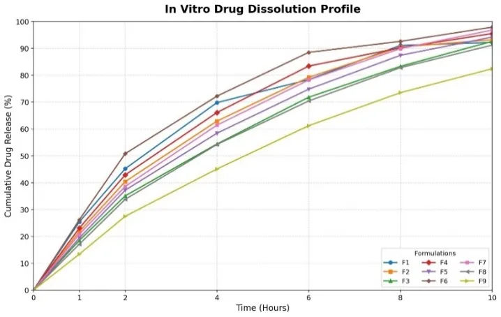
Figure 10: Comparative In-Vitro Cumulative Drug Release Profiles of Formulations F1–F9.
CONCLUSION
The present investigation was undertaken to formulate and characterize a gastroretentive floating mucoadhesive in-situ gelling system containing Litchi chinensis extract using sodium alginate and Carbopol 934.
Nine formulations (F1–F9) were prepared by varying the concentrations of sodium alginate and Carbopol 934 to investigate their influence on the physicochemical and performance characteristics of the prepared formulations.
The formulations were evaluated for physical appearance, pH, viscosity, gel strength, gelation time, floating behavior, ex-vivo mucoadhesive strength, drug content and in-vitro drug release.
The prepared formulations exhibited a clear, pale-yellow appearance, with pH values ranging from 4.12 to 4.71. The viscosity values ranged from 328 to 672 cP, indicating differences in the flow characteristics of the formulations with varying polymer concentrations.
The gelation time ranged from 32.8 to 43.2 seconds, while the gel strength measurements ranged from 28.4 to 48.8 seconds. All formulations demonstrated a reported floating duration exceeding 12 hours, indicating prolonged in-vitro buoyancy under the experimental conditions.
The ex-vivo mucoadhesive strength ranged from 8.4 to 15.6 g, and the drug content ranged from 84.56% to 93.23%. Among the tested formulations, F6 demonstrated the highest reported mucoadhesive strength and drug content.
The in-vitro dissolution study conducted over 10 hours demonstrated differences in the release profiles of the nine formulations. F6 exhibited the highest reported cumulative drug release of 97.90% at the end of the study, whereas F9 exhibited 82.40%.
Based on the reported physicochemical, floating, mucoadhesive and dissolution characteristics, formulation F6 appeared to provide a comparatively favorable balance of the evaluated parameters
REFERENCES

Salma Sulthana, Dr. Konda Shravan Kumar, Formulation of a Gastroretentive in Situ Gelling System Containing Litchi Chinensis Extract for Sustained Antiulcer Activity, Int. J. of Pharm. Sci., 2026, Vol 4, Issue 11, 2499-2521. https://doi.org/10.5281/zenodo.24279523
 10.5281/zenodo.24279523