

We use cookies to ensure our website works properly and to personalise your experience. Cookies policy
Department of pharmacology, Ch. Devi Lal College of Pharmacy, Jagadhari, Yamunanagar, 135003
Vascular dementia (VaD) is the most common type of neurocognitive disorder in the world that is mostly the result of cerebrovascular disease and neurodegenerative pathology. Recent studies highlight the multifaceted and bidirectional relationship between immune mechanisms and vascular dysfunction, dubbed vascular-immune crosstalk, in the progression of VaD. Vascular dysfunction, including chronic cerebral hypoperfusion, endothelial dysfunction, cerebral small vessel disease, and oxidative stress, all play an important role in the disruption of the blood-brain barrier (BBB) and the neurovascular unit. As a result, this damage to the blood vessel leads to chronic neuroinflammation, which involves the chronic maladaptive activation of microglia and astrocytes. The leaky BBB also enables peripheral immune cells to enter the brain parenchyma and further amplifies inflammation. Neurotoxic cytokines, chemokines and ROS are released continuously
1.1 DEMENTIA AS A GLOBAL BURDEN
1.1.1 DEFINTION
The characteristic of dementia, a group of symptoms caused by certain brain illnesses, is a persistent, gained impairment of two or more cognitive abilities that is significant enough to affect day-to-day activities and quality of life. Dementia can result from more than 55 illnesses, some of which do not progress.[1,2,3] Medical conditions that originate in other parts of the body may be the reason. For instance, a person’s ability to think may be impavted by a blood clot that forms in the heart, travels to the brain, and limits blood flow to a part of the brain due to an irregular heartbeat. When the brain is affected by two or more common ageing conditions, such as Alzheimer’s and stroke, dementia frequently results. The most common symptom that the affected person, their family, or others first notice is short-term memory problems.[4]
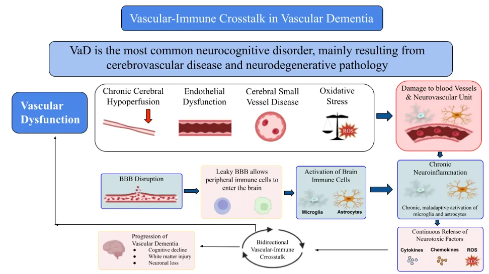
FIGURE 1 Mechanistic Framework of Biderctional Vascular-Immune Crosstalk in Vascular Dementia (VaD)
1.2 PREVALENCE
By 2050, the proportion of dementia patients residing in low- and middle-income nations will increase from 60% to 70%. China and India, together with their neighbours in South Asia and the western Pacific, have the fastest rates of population increase among the elderly. Over 10 million new cases of dementia are reported each year globally, or one new case every 3.2 seconds. With a goal of 100 countries by 2030, the Rehabilitation of WHO’s 2030 project has supported initiatives to enhance rehabilitation in more than 80 countries since 2017. The World Alzheimer Report of 2024 explores attitudes about dementia through a global poll of more than 40,000 people in 166 nations and territories and 24 expert articles, looking at how society views and understands the condition and the stigma that still surrounds it. [5]
1.3 SOCIOECONOMIC IMPACT
Alzheimer’s disease and associated dementia affect over 57 million people globally. By 2050, this population is predicted to surpass 150 million, with the Middle East, North Africa, and eastern sub-Saharan Africa experiencing the greatest growth. Dementia is becoming an increasingly serious issue in many nations as the world’s population ages. Direct and indirect costs are the two categories of costs associated with dementia. Physician visits, hospital stays, prescription medications, paid home care, and nursing home care are examples of direct expenditures. One example of an indirect cost is the lost productivity of carers, which is operationalised as their expected lost wages. [6] One of the main causes of mortality and disability in the world is dementia. The extensive consequences of dementia and its primary direct and indirect economic components are demonstrated by estimating the overall costs to society. In 2019, the projected yearly cost of dementia to society was $1313.4 billion, or $23,796 per person, for 55.2 million dementia sufferers. Direct medical costs accounted for US $213.2 billion (16%), direct social sector spending (including long-term care) for US $448.7 billion (34%), and informal care for US $651.4 billion (50%). Approximately half of the world's expenses are related to informal care. Enormous costs as per dementia across the world have a detrimental effect on both families and care systems. [7]
1.4 TYPES OF DEMENTIA
TABLE 1 Comparative Analysis of Neuropathology, Clinical Features, Progression Trajectories, and Diagnostic Criteria Across Major Dementia Subtypes.
|
Dementia |
Neuropathology & Regions |
Motor Signs & Neuropsychiatric Symptoms |
Progression Trajectory |
Diagnosis |
Reference |
|
Alzheimer’s Disease |
Neurofibrillary Tangles-Transentorhinal cortex, Abnormal Phosphorylated tau-perikaryal cytoplasm |
Parkinsonian gait, tremor, stiffness, and bradykinesia. Prefrontal syndromes, hyperactivity/agitation, depression, psychosis, apathy, and sleep disturbances. |
Sneaky beginning and steady, gradual fall. |
Variations in salivary Aβ-42, ptau, and ptau, t-tau. Ratio levels. |
[8, 9, 10, 11, 12] |
|
Vascular Dementia |
Frontal-subcortical systems are often affected. |
depression, and agitation, cognitive impairment, eating disorders, apathy, irritability/lability, sleep disturbance and eating disorders. |
VaD, it remains underrecognized and under researched compared to other forms of dementia, still frequently in steps |
Delirium, depression, cognitive screening test, brain imaging, neuro-psychological evaluation |
[13, 14, 15, 16] |
|
Lewy Body Dementia |
Depigmentation of the substantia nigra, locus coeruleus, cingulate, entorhinal, inferior parietal, superior temporal, and middle frontal |
Apathy, dysphoria, anxiety, hallucinations, and abnormal motor behaviours |
Short-interval progression, which is more statistically efficient than single-domain measurements. |
Rapid eye movement sleep behaviour disorder, parkinsonism, visual hallucinations, and varying cognitive performance. |
[17, 18, 19, 20, 21] |
1.5 EMERGING ROLE OF VASCULAR AND IMMUNE MECHANISM
AMYLOID CASCADE HYPOTHESIS: Aβ42 is an AD (Alzheimer’s disease biomarker, as Aβ42 decreases in cerebrospinal fluid (CSF), and it is the earliest to be detected as an increase in Aβ accumulation in the brain, that diagnosed or occurs many years before clinical diagnosis of dementia. In the default mode network, the cortical Aβ accumulation spreads Principally, embraces: orbitofrontal cortex (OFC), posterior cingulate (PC), and precuneus, to a collection of lower-order sensory-motor regions, such as lingual gyrus, precentral, post-central, peri-central, and peri-calcarine. The two primary pathology features of Alzheimer's disease are SPs (senile plaques) and NFTs (neurofibrillary tangles). In the signal transduction process, there are two products of amyloid beta (Aβ) works for cleavage I.e., APP (amyloid precursor protein), and glycoprotein. Senile plaque develops as a result of derangement of Aβ brain levels and also Aβ deposition; both of these cause Alzheimer’s disease by cognitive disabilities. Also, when there is a mutation of APP, amyloid presenilin 1 & 2; all these also lead to accumulation of beta amyloid (Aβ). All of these result in the amyloid cascade hypothesis [22,23,24].
FAILURE OF ANTI-AMYLOID THERAPIES: Despite their groundbreaking ability to modify pathology, anti-amyloid beta monoclonal antibodies exhibit several critical limitations. As we see, the main concern is the instability in clinical efficacy data all across large-scale trials, as demonstrated by the variation in positive and negative outcomes as observed in parallel Phase III studies (e.g., aducanumab’s EMERGE and ENGAGE trials). In addition, severe safety concerns involve strict dosing reductions in high-risk subgroups, particularly ApoE4-positive genotypes, which can limit the maximum clinical effect of treatment. Also, past target-specific interventions, such as secretase inhibitors, have had limited therapeutic ranges and severe adverse effects that led to premature trial closing. Critically, a significant gap remains with respect to the translation of biomarker clearance into visible patient care; the Alzheimer’s field still lacks validated, quantitative guidelines to define whether statistically considerable plaque reduction truly matches a “clinically meaningful” improvement in daily life for patient and family. [25-28]
1.6 VASCULAR-IMMUNE CONCEPT
According to available data, dementia is a mixed illness involving intricate interactions between immune and vascular systems. Cerebrovascular abnormalities, including chronic cerebral hypoperfusion, endothelial dysfunction, cerebrovascular small vessel diseases, and BBB disruption, have appeared as important contributors to cognitive decline. Impaired cerebral blood flow reduces the supply of nutrients and oxygen to brain regions, leading to gradual cognitive impairment, white matter injury, and neuronal malfunction. However, there is mounting evidence that the pathogenesis of dementia is significantly influenced by neuroinflammation. When microglia and astrocytes are activated, pro-inflammatory mediators such as TNF-α, IL-1β, and IL-6 are produced, which contribute to dysfunction in synapses and injury in neurons, i.e., neuronal injury. With all the above, vascular dysfunction and neuroinflammation are closely similar. When the blood-brain barrier is weakened, immune cells and peripheral inflammatory mediators can reach the brain more readily, while chronic inflammation further aggravates endothelial injury and cerebrovascular dysfunction. This bidirectional interaction, often referred to as vascular-immune crosstalk, is increasingly recognised as a key mechanism underlying growth and advancement in vascular dementia and other forms of cognitive impairment.
2. OVERVIEW OF VASCULAR DEMENTIA
2.1 DEFINITION AND CLASSIFICATION
DEFINITION
One of the most prevalent types of dementia, vascular dementia (VaD) is brought on by vascular brain injury, which is mostly caused by damage to the brain parenchyma and is secondary to ischaemia, infarction, or haemorrhage. VaD is acknowledged as a neurocognitive condition that includes locomotor abnormalities and behavioural symptoms including hostility, agitation, dysarthria, and Parkinson’s disease. The etiology of vascular dementia is distinct and involves both alterations in the brain and a range of vascular processes. Patients with VaD symptoms have been shown to have peripheral pro-oxidant indicators and poor levels of antioxidant capacity. There is mounting evidence that cerebrovascular disease, in conjunction with neurodegenerative pathology, is the major etiology of dementia. The most common underlying cause is chronic age-related dysregulation of cerebral blood flow (CBF), however, other variables, including oxidative stress, inflammation, and cardiovascular failure, also play a significant role. The cognitive impairment linked to this illness is made worse by oxidative stress, which encourages vascular and neural damage. Reactive oxygen species (ROS) may harm lipids, proteins, and DNA in cells, disrupting neural signalling and encouraging cell death. [35] Breakdown of the cortical-subcortical circuit frequently results in deficiencies in frontal-executive function, complicated attention, and information processing. Cerebral small-vessel (cSVD) is the most prevalent illness leading to VD, which is linked to a variety of degenerative disorders in the cerebrovascular system of the brain.[36]
Numerous neurodegenerative diseases, cerebrovascular diseases, traumatic brain injuries, substance abuse (such as alcohol), and other specific causes can cause dementia; mixed diseases can occur when multiple factors contribute. On the scale of cognitive and motor illnesses, mild cognitive impairment (MCI), also called mild neurological disorder, is a state less severe than dementia.[37]
CLASSIFICATION
Vascular dementia is classified into three main types, i.e., multi-infarct dementia, Sub-cortical ischemic VD, and strategic-infarct dementia.
MULTI-INFARCT DEMENTIA: is stated as Vascular pathology is currently identified as the primary cause of dementia in approximately 8-10% of all cases of dementia, and between 25 and 80 percent of patients with Alzheimer’s disease exhibit vascular lesions that might be related to their dementia. Prior to the identification of dementia subtypes due to single infarcts or generic arteriopathy, MID was adopted as a nearly equivalent term alongside VD.[38] Large-vascular disease and small-vascular disease are the two main groups into which MID etiology falls. The frontal lobe, the hippocampus, and basal forebrain are among the key regions affected by a major stroke in big-vessel disease. The resulting deficiencies could or might not meet dementia diagnosis criteria. The damage caused by small-vessel disease is typically so little that it only manifests as a sequence of tiny changes. However, mentation gradually decreases over time as more tiny vessels become occluded. The primary changes linked to ageing include a decrease in endothelial mitochondria, a decrease in cerebral blood flow, a discernible thickening of the vascular basement membrane, and an increase in degenerative pericytes.[39] Managing executive dysfunction that results in a loss of cognitive independence is indicative of an abrupt beginning of dementia. Neuroimaging shows significant cerebrovascular lesions. There exists a clear temporal relationship among.[40]
SUBCORTICAL ISCHEMIC VaD: The cognitive deficits linked to SIVD frequently exhibit a gradual decline and subtle beginning, resembling the progression of AD. In contrast, a current amyloid imaging investigation has revealed the existence of a true SIVD, which means there is no indication of amyloid plaque formation in the brain, but there are notable subcortical white matter ischemia alterations. In addition, pure SIVD is more common than previously believed. It is not the same as AD or mixed dementia, which show fibrillar types of amyloid accumulation in the brain.[41] Ischemic damage, which comprises both full infarction (lacunar infarcts and microinfarcts) and incomplete infarction of deep cerebral white matter, is the etiology of dementia in SIVD. Small cavitated ischemic infarcts with a diameter of less than 15 mm are known as lacunar infarcts or lacunes. The basal ganglia, internal capsule, thalamus, pons, corona radiata, and centrum semiovale are the usual locations for them. Non-confluent regions of ischemic white-matter alterations may coincide with white-matter lacunes. Microinfarcts occur in cortical and subcortical regions and are primarily noncavitated. They can be as small as a few microns or as small as a tenth of the size of lacunes.[42]
STRATEGIC INFFRACT DEMENTIA: With an ischemic vascular lesion, limbic, associative, and paralimbic circuitry-specific brain regions essential for cognition and behaviour-are affected by strategic infarct (SI) stroke. The most commonly impacted areas are the caudate nuclei, medial frontal lobe, inferomedial temporal lobe, left angular gyrus, left capsular genu, and thalami. A fascinating issue in clinical neurology is the link between brain anatomy and function, which may be examined from cognitive disability and primary neurological damage. These are mostly associated with vascular cognitive impairment and dementia, which can occur gradually or rapidly. Patients may show different kind and levels of impairment in the different cognitive domains depending on the size, location, and severity of the lesion. The paramedian nuclei of the thalamus may be involved in anterograde and retrograde memory impairment, as well as inattention and amotivation.[43]
2.2 EPIDEMIOLOGY
One of the most prevalent illnesses, dementia has a high rate of morbidity, death, and decreased quality of life, particularly among older people. Notably, as the globe moves into an aging society, the number of people affected by dementia or related cognitive decline is predicted to increase to 115 million by 2050. Drawing on a large study population of numerous national health databases, research into young-onset dementia pointed to a pattern of dementia appearing at increasingly younger ages. About two-thirds of dementia patients now reside in low- and middle-income nations, where the average national spending on dementia accounts for 0.45% of GDP. [44] The percentage of VaD among all dementia cases varied significantly between studies, ranging from 10.4% in Norway to 46.7% in rural Japan (1997), according to nine recent population-based studies with sample sizes ranging from 927 to 446,814 participants. Intermediate values included 13.9% in Korea, 17.9% in the UK Biobank, 26.1% in China, and 20.3% in Hong Kong. [45] India’s population is rapidly changing due to an ageing population, which is increasing the country’s dementia burden. The national Longitudinal Ageing Study in India (LASI) estimates that 7.4% of Indians over 60 have dementia, which translates to around 8.8 million afflicted people. [46] In 2020, around 5.8 million Americans has dementia; by 2060, that number is predicted to rise to 13.9 million, or 3.3% of the total population. Centers for Medicare & Medicaid fee-for-service claims data from 2011 to 1013 were analysed to determine the prevalence of vascular dementia based on physician diagnosis. The prevalence of vascular dementia was estimated to be 1.6% across many age categories, with an overall incidence rate of 4.4 per 1000. A symptomatic stroke was temporally linked to 59% of incident vascular dementia cases in the Rochester Epidemiology Project; the remaining cases were linked to bilateral infarcts seen on imaging. [47] intake; deficiencies of folate, vitamin B12, and vitamin.
2.3 RISK FACTORS
TABLE 2. Metabolic, Physiological, and Environmental Risk Factors Contributing to Homocysteine Dysregultion, Vascular Injury, and Cognitive Decline.
|
Category |
Key factors / Findings |
References |
|
Major Metabolic and Lifestyle Drivers |
Hypertension; elevated systolic (134.1 ± 17.0mmHg) and diastolic blood pressure (82.1 ± 11.0mmHg); diabetes (23.1%); increased BMI (≥25 kg/m2); hyperhomocysteinemia (Hhcy); high methionine intake; deficiencies of folate, vitamin B12, and vitamin B6 leading to impaired homocysteine metabolism. Hypertension and Hhcy significantly increase the risk of vascular dementia and cognitive decline. |
[49,50] |
|
Primary Physiological Drivers |
Oxidative stress; vascular inflammation; endothelial dysfunction; blood-brain barrier (BBB) permeability; cerebral blood flow abnormalities; cerebral amyloid angiopathy (CAA); amyloid-β (Aβ) accumulation; tau hyperphosphorylation; neuroinflammation; microhemorrhages; cognitive impairment. Elevated plasma homocysteine increases dementia risk by approximately 9% and Alzheimer’s disease risk by approximately 12% for every 5 μmol/L increase. |
[50] |
|
Metabolic Pathway Co-factors |
Homocysteine metabolism regulated by folate, vitamin B12, vitamin B6, and methionine. Deficiency of B vitamin or excess methionine disrupts homocysteine metabolism, resulting in homocysteinemia, which contributes to oxidative stress, vascular injury, and dementia pathogenesis. |
[50] |
|
Environmental and Biological Co-factors |
Air pollutants including fine particulate matter (PM2.5), particulate matter (PM10), nitrogen dioxide (NO2), nitrogen oxide (Nox), ozone (O3), carbon monoxide (CO), sulphur dioxide (SO2); aluminium; silicon; solvents; pesticides; environment tobacco smoke; extremely low-frequency magnetic fields (ELF-MF); chronic community noise (>25dB); night-shift work; low neighbourhood socioeconomic status (SES); reduced neighbourhood greenness; proximately to roads; rural residence. These environmental exposures have been associated with an increased risk of vascular dementia. |
[48] |
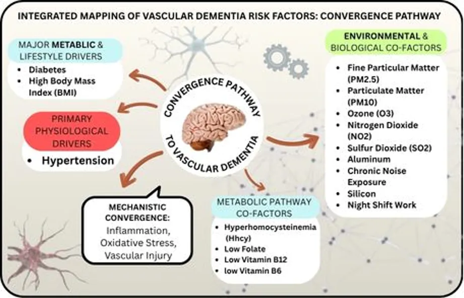
FIGURE 2. Integrated mapping of vascular dementia risk factors detailing the convergence pathways across metabolic, environmental, physiological, and mechanistic drivers leading to cognitive impairment.
3. VASCULAR MECHANISM IN DEMENTIA
The brain uses around 1/4 of the oxygen and glucose used by the entire body. In a healthy adult, the typical whole cerebral blood flow (CBF) is around 57 ml/100gr/min, or 800 ml/min, or almost 15% of the total basal cardiac output. Every time the consumption for energy rises, the CBF must also rise. In accordance to the dynamic shifts in brain activity, CBF and energy consumption fluctuate not just between regions but also over time. In order to fulfil the energy requirement, each local spike in functional activity at physiological circumstances is linked to a local rise in CBF. Therefore, under physiological settings, each local spike in functional activity is linked to a local rise in CBF to fulfil the energy requirement (functional hyperanemia).
Thus, it appears that CCH (Chronic Cerebral Hypoperfusion) also refers to situations of variable functional hyperanemia and insufficient delivery of nutrients (basically, oxygen and glucose) to fulfil the energy needs of the tissue.[51] Neurodegeneration and different levels of cognitive impairment that fall under the VCI spectrum are also linked to CCH. In addition to being able to predict which people will moderate cognitive impairment (MCI) may eventually acquire a severe form of dementia, lowers reduced cerebral perfusion has been linked to the severity of dementia. Numerous pathways that are persistently active in the brain under conditions of CCH lead to damage accumulation and thwart healing efforts. Sustained energy imbalance, oxidative stress, inflammation, endoplasmic reticulum (ER) stress, and mitochondrial failure are some of the pathways.[52,53]
CCH does not occur in isolation; it is associated with other vascular risk factors, such as hypertension. Growing evidence indicates that there is a reciprocal relationship between cerebral hypoperfusion and hypertension, which contributes to the onset of memory deficits. The interaction between Ang II and AT1R can lead to changes in vascular structure, enhance vascular inflammation, and cause oxidative stress damage, ultimately resulting in brain injury.[54] In addition, there are many other reasons or causes of CCH like; shock/hypovolemia, carotid artery stenosis, autonomic neuropathy etc.
From the heart to the tiniest capillaries, the whole circulatory system is lined by vascular endothelial cells. Fluid filtration, which occurs in the kidney’s glomeruli, blood vessel tone, haemostasis, neutrophil recruitment, and hormone trafficking are among their functions. Additionally, endothelial cells (ECs) are essential for controlling blood flow. The layer of cells that lines the blood luminal surface of veins is called endothelium.[55] An active organ that is essential to preserving vascular homeostasis is the endothelium. It generates a number of factors that control artery wall inflammation, smooth muscle cell proliferation, coagulation, vascular tone, and the adherence of circulating inflammatory cells to the endothelium. Managing the delicate equilibrium between vasoconstriction and vasodilation is one of the endothelium’s most crucial roles. Nitric oxide (NO) and prostacyclin are the primary mediators of vasodilation, whereas endothelin-1 (ET-1), angiotensin II, and thromboxane A2 sustain vasoconstriction.
In addition to being the body’s most powerful endogenous vasodilator, nitric oxide also prevents leukocyte adhesion, oxidative stress, inflammation, platelet aggregation, and smooth muscle cell proliferation. L-arginine is converted to nitric oxide by activating endothelial NO synthase (eNOS) in response to chemicals that act on certain endothelium chemoreceptors or mechanical forces that cause shear stress on mechanoreceptors. ED is caused by loss of endothelial integrity after inhibition of the release of protective chemicals.[56]
Endothelial dysfunction is largely caused by oxidative stress and uncoupling of eNOS, the enzyme responsible for NO production. Excess reactive oxygen species (ROS) may scavenge NO, which is mostly formed in the blood vessel wall by eNOS. Therefore, the equilibrium between ROS in the vascular wall and NO synthesis determines the availability of NO to dilate blood vessels. In a physiological condition, vessels are typically kept dilated and quiescent by NO. Excess ROS, mostly produced in blood vessels by vascular NADPH oxidase, quench NO under pathological conditions. A healthy endothelium requires adequate NO. Excess ROS reduces NO availability, leading to endothelial dysfunction. When eNOS becomes uncoupled, it produces superoxide instead of NO, causing a NO/ROS imbalance that contributes to vascular disease and endothelial dysfunction. According to preclinical data, endothelial NO levels are influenced by a number of variables, such as eNOS activity and coupling, which dictate whether NO or superoxide and peroxynitrite (ONOO) are the main products. [57] recently shown that inhibiting the production of nitric oxide (NO) causes local arterial stiffness, indicating that NO produced from the endothelium plays a role in controlling the elasticity of major arteries. Reduced aortic compliance directly leads to arterial stiffness. Increased arterial stiffness also has an indirect impact, speeding up pressure waves as they pass through the walls of the aorta and other large arteries. Consequently, an increased pulse wave velocity (PWV) is a sign of arterial stiffness. The well-established technique of measuring PWV using carotid-femoral PWV, initially published by Young in 1804 and by Bramwell & Hill in 1922, remains the gold standard for evaluating central arterial stiffness. Epidemiological studies have demonstrated the prognostic utility of aortic stiffness for cardiovascular events using carotid-femoral PWV. By using ultrasonography to examine the brachial artery, endothelium NO-dependent and independent vasodilatation may be evaluated, and changes in vessel diameter can be used to gauge endothelial function. It is unclear if endothelial dysfunction is likewise linked to vascular stiffness in RHTN patients, despite the fact that both methods have been applied in numerous research including hypertension patients. The goal of the current investigation was to examine and link arterial wall stiffness measured by PWV and endothelial function measured by flow-mediated dilation (FMD) in both normal healthy participants and resistant, well-controlled hypertension patients. [58]
3.3 BLOOD-BRAIN BARRIER DYSFUNCTION
All animals with a fully formed central nervous system have the blood-brain barrier (BBB). The blood-brain barrier is made up of endothelial cells that form the capillary walls in the brains and spinal cords of animals, including humans. The entire surface area of these microvessels is by far the largest interface for blood-brain interaction. The average adult human brain has a total exchange area of 12 to 18 m², depending on the anatomical location. This surface area varies from 150 to 200 cm²/g of tissue. [59] The blood-brain barrier (BBB) is a highly specialized structure—both physical and biochemical—that controls the movement of substances between the brain and the rest of the body's circulatory system. The long-term regulation of the ionic concentration of the brain's interstitial fluid depends on the BBB. The blood-brain barrier is hypothesised to be disrupted or malfunctioning in many brain diseases, especially neurodegenerative conditions. Several pieces of evidence indicate that VaD is characterized by BBB degradation. Oxidative stress and inflammatory signalling pathways are the primary causes of BBB breakdown. Breakdown of the BBB is mainly driven by reactive oxygen species (ROS) and reactive nitrogen species (RNS) acting within endothelial cells, astrocytes, and pericytes, while tau proteins, cytokines, and amyloid β (Aβ) promote inflammation. [60]
Brain tissue oedema, neuroinflammation, and cerebral microvascular damage can result from BBB breakdown in SVD, which can cause plasma or cellular components to seep out of blood vessels. Increased BBB permeability is discovered in normal-looking white matter close to the WMH, suggesting that BBB damage may play a role in the etiopathogenesis of WMH. [61]
3.4 SMALL VESSEL
Cerebral small vessel disease (CSVD) is a chronic, systemic disorder of the brain’s small perforating arterioles, capillaries, and likely venules. While individual microvascular lesions can remain clinically silent, the cumulative burden of CSVD is a primary driver of major neurological disabilities in older populations. Furthermore, it stands as the most common cause of pure vascular dementia. By exacerbating underlying neurodegeneration, CSVD contributes to roughly 50% of all dementia cases worldwide Beyond stroke and dementia, a high burden of small vessel lesions is strongly linked to a variable clinical spectrum of physical and psychological symptoms, including stepwise cognitive impairment, mobility and gait disturbances, late-life depression, and apathy. [62] The term Recent Small Subcortical Infarct (RSSI) refers strictly to an acute, symptomatic or asymptomatic ischemic event that occurs in the territory of a single deep penetrating arteriole. In contrast, a lacune (derived from the Latin lacuna, meaning pond, pit, or cavity) represents the permanent, healed stage of a prior subcortical ischemic infarction or localized microhemorrhage. While acute tissue damage can appear slightly larger due to local inflammatory edema, chronic lacunes stabilize as small, round or ovoid fluid-filled cavities within the deep subcortical grey or white matter structures [63]
3.5 OXIDATIVE STRESS
Chronic cerebral hypoperfusion caused by cerebral vascular injury (such as ischemic events or cerebral small artery disease) is the main cause of VaD in pathological conditions. This leads to mitochondrial dysfunction and an excessive accumulation of reactive oxygen species (ROS). Mitochondrial dysfunction impedes the electron transport chain (ETC), leading to reduced ATP synthesis and elevated reactive oxygen species (ROS) generation. Furthermore, it may lead to an increase in reactive oxygen species (ROS), particularly mitochondrial ROS (mtROS), which can initiate a detrimental cycle of oxidative stress. Reactive oxygen species (ROS) induce neuroinflammation by causing oxidative damage, increasing vascular endothelial permeability, and disrupting the blood-brain barrier (BBB). This enables toxins and inflammatory mediators to pass into the brain parenchyma. Neurovascular unit (NVU) dysfunction is directly caused by the production of reactive oxygen species (ROS). Excessive ROS production leads to activation of microglia and astrocytes, which release pro-inflammatory cytokines like TNF-α and IL-1β.
Oxidative stress is a pathological condition that is caused by the imbalance between the body's antioxidant system and ROS, resulting in the excessive accumulation of reactive oxygen species (ROS). By oxidatively damaging cellular macromolecules, including lipids, proteins, and DNA, this accumulation interferes with normal cell signal transmission mechanisms.
Reactive oxygen species (ROS) are a group of very reactive oxygen derivatives, such as superoxide anion (O2−), hydrogen peroxide (H2O2), and hydroxyl radical (⋅OH). ROS can be produced by both endogenous (leakage from the mitochondrial electron transport chain, the NADPH oxidase system, and other enzyme systems) and exogenous (radiation and environmental pollution) sources.6. The mitochondria are the primary site of reactive oxygen species (ROS) production. Under normal physiological conditions, mitochondria create adenosine triphosphate (ATP) by oxidative phosphorylation, and 1% to 2% of oxygen is partly transformed to superoxide anion (O₂⁻). However, when cells experience ischaemia, hypoxia, or mitochondrial failure, the electron transport chain (ETC) is disrupted, leading to a significant increase in O₂⁻ production. O₂⁻ (superoxide ion) is further transformeed into hydrogen peroxide (H2O2) by a dismutation process. H2O2 next goes through a Fenton reaction with ferrous ions (Fe2+) to produce the more dangerous hydroxyl radical (·OH). [64]
Here, oxidative stress due to accumulation of ROS has been identified as a critical upstream mediator of lipid peroxidation, and consequently neurovascular pathology, in the case of vascular dementia (VaD). The electron leakage from complexes I and III of the electron transport chain in an ischemic cerebrovascular environment leads to a high rate of production of the superoxide O2 radical. This superoxide may spontaneously or enzymatically transform into hydrogen peroxide (H2O2), a major ROS that can directly oxidize components of cellular membranes such as lipids. At the same time, these high levels of ROS convert healthy circulating LDL particles into harmful oxidized LDL (ox-LDL). Ox-LDL compromises blood-brain barrier (BBB) integrity and enhances leukocyte adhesion, which allows the migration of monocytes to the tunica intima, where they become foam cells. This lipid-peroxidation cascade drives the formation, expansion, and eventual rupture of atherosclerotic plaques to a basic level. This creates microinfarcts and focal necrosis that lead to a progressive loss of neurons, which may be reflected biochemically by markers such as malondialdehyde (MDA), and which leads to clinical symptoms of step-wise cognitive deterioration as seen in VaD. [65]
4. IMMUNE MECHANISM IN DEMENTIA
4.1 NEUROINFLAMMATION
Pathological changes in the nervous system that accompany dementia have been better appreciated with a dual-role model, which is sometimes viewed as a ‘Jekyll-and-Hyde’ paradox. [68] In physiological state and in the early stage of neurodegeneration, acute neuro-inflammation is a well-coordinated adaptive defence mechanism. [66,67] This transient immune response is mainly neuroprotective in function and involves a rapid and transient activation of resident microglial and astrocytes in response to early protein misfolding and/or acute triggers. [66,68] The phagocytic properties of these innate cells are advantageous to the clearance of toxic physiological insults, including early soluble amyloid-beta (Aβ) oligomers, hyperphosphorylated tau monomers, metabolic toxins and cellular debris. [66,67,68] The central nervous system (CNS) thus triggers this controlled inflammatory response and the production of anti-inflammatory resolution molecules, which help to minimize secondary tissue injury, encourage cellular repair, and facilitate tissue homeostasis to return to normal levels. [66,67] Thus, this acute phase is typically self-limited and self-resolving if the principal pathogenic triggers are effectively neutralised or eliminated from the brain parenchyma. [66,67]
But when these initial proteotoxic triggers remain unresolved and unchecked over time a critical maladaptive shift occurs. [66,69] This breakdown in immune resolution precipitates the onset of chronic neuroinflammation, a low-grade, chronic and sterile inflammatory state that occurs from persistent exposure to DAMPs such as dense and insoluble amyloid plaques and neurofibrillary tangles. [66,67] Systemic “inflammation” (chronic elevation of peripheral pro-inflammatory mediators in a state of ageing) is often the trigger and is a major cause of the impaired capacity of the immune system to reset. [66,67,69] Within this persistently hostile environment, the idea of microglial “priming” is at the core of the degenerative cascade. [70] Conditions of acute systemic insults (e.g., peripheral infections, focal trauma or chronic systemic vascular disease) lead to an excessive cytokine response, which is highly neurotoxic within the CNS, due to underlying neurodegenerative pathology, and this response can destroy local neural networks. [69,70]
Chronic changes over time create a vicious circle of permanent phenotypic reprogramming of microglia and astrocytes to a consistent pro-inflammatory state. [66,67] These chronically activated glia produce high levels of cytokines that are neurotoxic such as interleukin-1 beta (IL-1β), interleukin-6 (IL-6), and tumor necrosis factor alpha (TNF-α). [66,71] This ongoing chemical exposure has a clear downstream influences on the ongoing dementia trajectory. [66,69] It is a chronic neuroinflammatory microenvironment that leads to excessive and improper removal of synapses, promotes progressive axonal degeneration and induces NMDA-mediated excitotoxicity in vulnerable neuronal pathways. [69,71] At the same time, a persistent inflammatory stimulus leads to a permanent permeabilization of the blood-brain barrier (BBB). [69] The unobstructed passage of peripheral immune cells thus brings about a breakdown in the structure that allows for the free movement of peripheral immune cells and halts the neurodegenerative process, thereby accelerating the cognitive decline seen in dementia. [67,69,71]
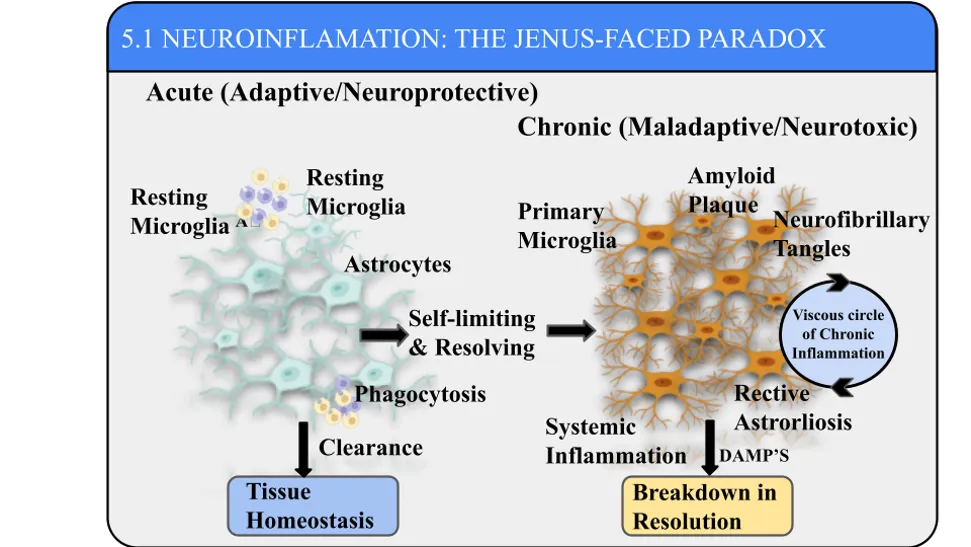
Figure 3. The Janus-Faced Paradox of Neuroinflammation: Overview comparing the protective vs. Neurotoxic pathways of neuroinfammation. Acute response (left) utilizes resting microglia and astrocytes to perform phagocytosis and clear debris, maintaining tissue homeostasis. Chronic response (right) is driven by persistent factors (amyloid plaques, neurofibrillary tangles, DAMPs) and rective astrogliosis, creating a vicious inflammatory cycle that leads to a breakdown in resolution.
4.2 MICROGLIAL ACTIVATION
Microglial activation is considered the main cellular lesion of the central nervous system (CNS) chronic inflammation observed in the pathological architecture of dementia. [72] In normal conditions, resident microglia are highly motile and exhibit a surveillance phenotype to scan the neural microenvironment and ensure maintenance of the integrity of synapses and elimination of metabolic waste. [72,73] The persistant accumulation of damage-associated molecular patterns (DAMPs), namely oligomeric amyloid-beta (Aβ) and hyper-phosphorylated tau, however, activated the microglial pattern recognition receptors (PRRs), including Tool-like receptors (TLRs) and the NLRP3 inflammasome complex. [73,74] This binding induces a dramatic switch in the phenotype, from a homeostatic microglia to a state of high reactivity and neurotoxic phenotype. [72,74] Chronically activated, these cells become extensively morphologically remodeled and transcriptionally reprogrammed, and serve as long-term sources of chronic neuroinflammation. [74,75] Massive and dysregulated production and release of an important triad of pro-inflammatory cytokines (tumor necrosis factor-alpha (TNF-α), interleukin-1β (IL-6)) is the hallmark of this persistent activation state. [72,75]
Of this inflammatory triad, IL-1β is an early orchestrator in an upstream manner, which hastens the neurodegenerative cascade. [76] Maturation and release of IL-1β is dramatically reliant on the activation of the molecular platform called the NLRP3 inflammasome, which is chronically hyper-activated in the brains of dementia patients. [72,76] Soon after release into the parenchyma, IL-1β triggers potent autocrine amplification, also binding back to the microglial receptors to induce additional production of cytokines, thereby leading to the activation of neighbouring astrocytes in a neurotoxic state. [75,76] This chronic increase of IL-1β directly disrupts long-term potentiation (LTP) in the hippocampal network and promotes the hyperphosphorylation of tau protein inside the cell, accelerating the onset of the development of neurofibrillary tangles. [76,77] In addition, IL-1β functions together with elevated levels of IL-6, a cytokine which activates a vast downstream pathway through the Janus kinase/signal transducer and activator of transcription (JAK/STAT) pathway. [75,77] Although transient IL-6 signalling is necessary for neurodevelopment, chronic overproduction of IL-6 in dementia leads to changes in gene transcription in neurons, marked axonal dystrophy, and a non-resolving inflammatory background throughout localized neurovascular units. [72,77]
At the same time, hypersecretion of TNF-α is a potent direct mediator of synaptic dysfunction and neuronal apoptosis. [73,75] Chronically activated microglia release large amounts of soluble TNF-α, which binds with high affinity to neuronal tumor necrosis factor receptor 1 (TNFR1), activating pro-apoptotic cascades like the caspase-3 and mitogen-activated protein kinase (MAPK) pathways. [73,78] Apart from the direct neurotoxicity of the pathogen induced by its direct effects on the brain, the high level of TNF-α significantly affects synaptic transmission by reducing the number of protective AMPA receptors by promoting their rapid endocytosis and causing glutamate excitotoxicity by downregulating the expression of the glutamate transporters in the astroglia. [75,78] This localized chemical stress is compounded by “priming’’ or hypersensitization in the microglial cells, which occurs when the underlying proteotoxic pathology makes the cell easily activated. [76] The primed brain encounters secondary systemic insults, and this microglial/cytokines axis responds with an unchecked hyper-inflammatory response. [76,78] In the end, the constant interplay of TNF-α, IL-1β and IL-6 turns the microglial response into a global neurotoxic engine, which leads to extensive pruning of synapses, irreversible permeabilization of the blood-brain barrier and irreversible loss of cognitive function in dementia. [72,75,78]
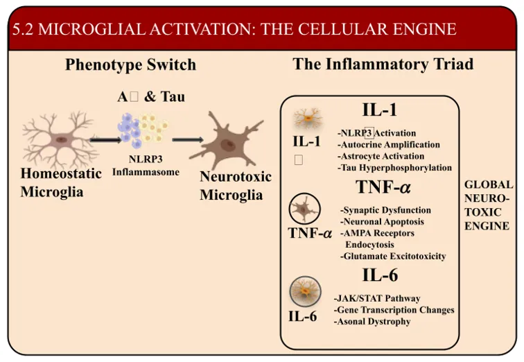
FIGURE 4. Microglial Activation and the Inflammatory Traid: Overview of the phenotypic transition of homeostatic micrglia to a neurotoxic state driven by Aβ, Tau, and the NLRP3 inflammasome. The resulting cascade releases an inflammatory traid – IL-1, TNF-α, and IL-6 – which collectively function as a global neurotoxic engine through synaptic dysfunction, excitotoxicity, and altered gene expression.
4.3 ASTROCYTE ACTIVATION
In addition to microglial activation, there is a critical transition point in the neuroinflammatory process of dementia whereby resident macroglia, also called astrocytes, undergo structural and functional remodeling, called reactive astrogliosis [79]. Protoplasmatic astrocytes participate in maintaining metabolic homeostasis, controlling the local blood flow, stabilizing the concentration of extracellular potassium ions, and recycling neurotransmitters such as glutamate at the tripartite synapse [79, 80] and play an indispensable role in a healthy central nervous system (CNS). But in the course of neurodegenerative proteinopathies, astrocytes undergo a dramatic morphological and biochemical change in reaction to chronic stress of the cell and the presence of danger signals in the environment [80, 81]. This reactive astrogliosis is characterized by astrogliosis with prominent hypertrophy of the major cellular processes of the astrocytes, with a marked upregulation of the transcription of intermediate filament proteins, especially glial fibrillary acidic protein (GFAP) and vimentin [81, 82]. The initial, acute activation of astrocytes in the injured brain attempts to compartmentalize the damaged neural tissue by forming a scar, but over time the persistent activation of astrocytes in the aging brain becomes maladaptive and neurotoxic, promoting the progressive deterioration of cognition [79, 82].
These reactive astrocytes have an operational phenotype that is strongly influenced by the adjacent chronically activated microglia [80, 83]. Signals from microglia are the major upstream regulators that trigger the neurotoxic astrocyte configuration and result in a virtual shutdown of the typical neurotrophic properties of the astrocyte [81, 83]. Reactive astrocytes change from a supportive role to a secretory function and continue to secrete a broad range of inflammatory mediators, such as chemokines CCL2 and CXCL10, reactive oxygen species (ROS) and matrix metalloproteinases (MMPs) [82, 83]. Importantly, this chronic inflammatory mediator release leads to dysregulation of the highly regulated expression of glutamate transporters on astrocytes, such as glutamate transporter-1 (GLT-1/EAAT2) [80, 84]. Following the downregulation of these clearance transporters, there is an excessive build-up of extracellular glutamate in the synaptic cleft, causing a persistent, low level of activation of NMDA receptors on neighbouring neurons and resulting at the end of the day in a catastrophic loss of synapses and pruning of the dendritic spines [79, 84].
In addition, chronic secretion of inflammatory mediators from astrocytes has a profound effect on the integrity of the neurovascular unit [82, 85]. The chronic astrocytic stress disrupts the local expression of AQP4 water channels at astrocytic end-feet and this loss of function is a key feature that severely affects the glymphatic clearance mechanism from the brain, leading to the subsequent increase in the accumulation of toxic protein aggregates in the parenchyma [81, 85]. At the same time, continuing release of pro-inflammatory factors and MMPs destroys the tight junction proteins of vascular endothelial cells, permanently altering the permeability of the blood-brain barrier (BBB)[82, 85]. So, this localized barrier dysfunction allows peripheral immune cells to enter and enhance polarization of the astrocytes to a chronic and self-sustaining inflammatory state [79, 85].
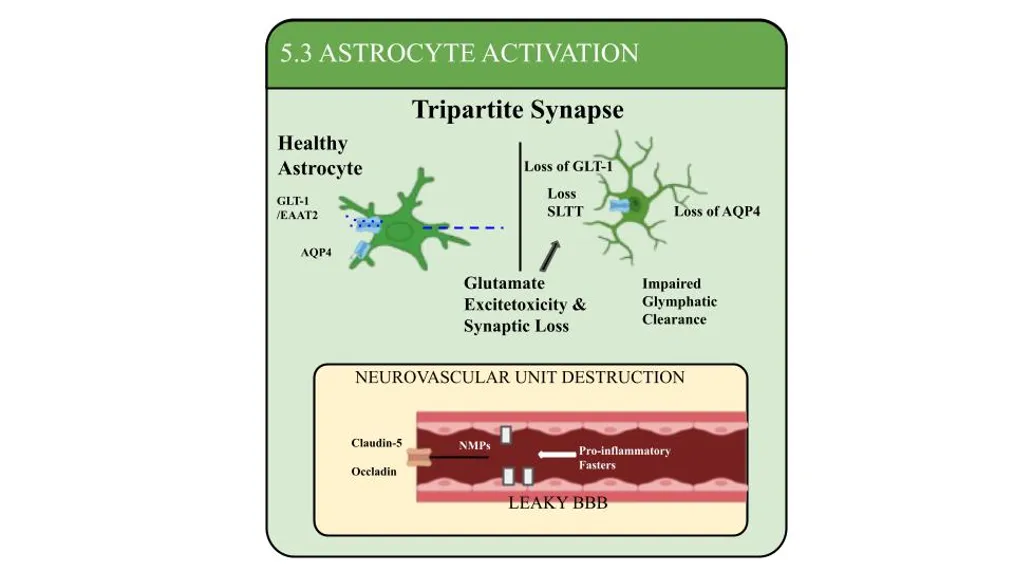
FIGURE 5. Astrocyte Dysfunction in Synaptic and Vascular Homeostasis: Schematic illustrating how reactive astrocyte transformation leads to loss of key transporters (GLT-1/EAAT2 and AQP4), precipitating glutamate excitotoxicity, synaptic loss, and impaired glymphatic clearance. This dysfunction cascades into neurovascular unit destruction, charaterstized by tight junction disruption (Claudin-5, Occludin) and blood-brain barrier hyperpermeability.
4.4 PERIPHERAL IMMUNE INFILTRATION
The last stage of neuroinflammatory cascade progression in dementia pathogenesis is the recruitment of peripheral immune cells [86] which are actively recruited. The blood brain barrier (BBB) is a highly selective physical and metabolic barrier made up of specialized brain capillary endothelial cells, pericytes and astrocytic end-feet that under physiological conditions tightly restricts leakage of leukocytes into the immune-privileged brain parenchyma [86, 87]. But long-term, continuous exposure to pro-inflammatory cytokines and matrix metalloproteinases, or MMPs, such as TNF-α and IL-1β, from microglia and astrocytes, leads to a reduction in important endothelial tight junction proteins like claudin-5 and occludin [87, 88]. Such a “leaky” BBB leads to significant increases in adhesion molecule expression on endothelial cells, such as Intercellular Adhesion Molecule-1 (ICAM-1) and Vascular Cell Adhesion Molecule-1 (VCAM-1) [3, 5]. They are molecular entry gates that allow circulating peripheral immune populations to tether, roll, and diapedese across the damaged endothelial wall [86, 89].
Infiltrating peripheral populations, migration of hematogenous macrophages, which cross the permeabilized BBB, greatly alters the local inflammatory balance [90]. These peripheral monocyte-derived macrophages are known to easily enter the cortical and hippocampal regions guided by the chemokine gradient set up by reactive glia, which include CCL2 and CCL5 [89, 90]. In contrast to resident microglia, these newly recruited peripheral macrophages have very loose regulatory homeostatic checkpoints and upon contact with parenchyma amyloid-beta (Aβ) and tau aggregates, display a highly unconstrained pro-inflammatory effector phenotype [90, 91]. These peripheral macrophages, rather than driving efficient resolution or long-term protective clearance, produce a second, massive release of reactive oxygen species (ROS), nitric oxide and other proteases [86, 91]. This large chemical influx permanently disrupts any remaining neuroprotective mechanism (self-regulatory) in these localized neurovascular units and creates a vicious cycle of neurotoxicity and physical cell death that, is highly destructive [89, 91].
At the same time, the active infiltration of adaptive immune cells, namely T-lymphocytes, changes the neurodegenerative niche in a fundamental way [87, 92]. CD4 helper T cells and CD8 cytotoxic T cells selectively enter the brain parenchyma via chronically reactive endothelial cells and antigen presenting microglia [88, 92]. After entering the CNS, these T-cells come into contact with their cognate antigens that are displayed on the major histocompatibility complex (MHC) molecules of activated glia [87, 92]. The antigen-driven reactivation results in the release of the interferon gamma (IFN-ɣ) and the interleukin 17 (IL-17) from the local microenvironment, directly polarizing resident microglia into a highly neurotoxic and irreversible state [86, 92]. In addition, infiltrating CD8 T-cells have the ability to directly engage stressed, MHC-I expressing cortical neurons to induce aberrant antigen-specific perforin/granzyme mediated cell death [91, 92]. At the end, the structural defect of the BBB and subsequent recruitment of T-cells and peripheral macrophages turns dementia into an invasive and systemic-central immune disease, resulting in an irreversible cycle of pruning synapses, losing axons, and cognitive damage [86, 90, 92].
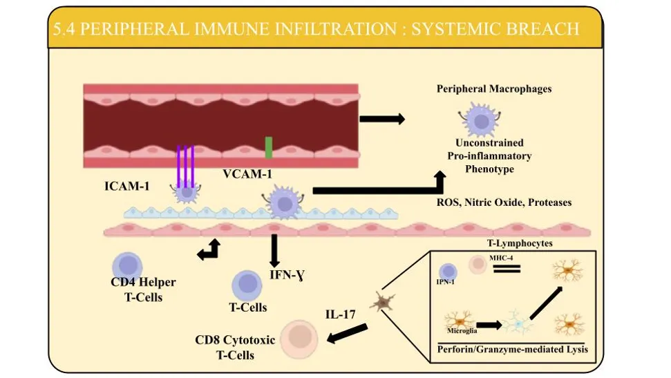
FIGURE 6. Systemic Immune Infilteration in Neuroinflammation: Schematic illustration showing ICAM-1 and VCAM-1 mediated extravasation of peripheral immune cells into the CNS. Infilterating macrophages exhibit an unconstrained pro-inflammatory phenotype releasing ROS and proteases, while CD4+ and CD8= T-lympho’ release IFN-ɣ and IL-17, triggering sustained microglial activation and perforin/granzyme-mediated cell lysis.
4.5 CYTOKINES AND CHEMOKINES
The final result of glial activation and infiltration of peripheral immune cells is the creation of an extremely neurotoxic extracellular environment characterized by overlapping, chronically elevated cytokines and chemokines. [93] The constant, dysregulated levels of these soluble signalling molecules directly promote the progressive collapse of the neuronal network and synapse via the cortical and hippocampal niche.[93,94] Cytokine-induced neurotoxicity appears to occur through mechanisms of chronic overactivation of the neuronal surface receptors that interfere with important intracellular signalling cascades.[94,95] In particular, chronic exposure to pro-inflammatory cytokines leads to impaired CREB activation via the cAMP pathway, a master key regulating neuroplasticity, and it also inhibits the expression of BDNF [95., 96] Absence of such neurotrophic support causes severe loss of dendritic spines and structural atrophy in vulnerable populations of pyramidal neurons and halts long-term potentiation (LTP) necessary for long-term memory [93, 96].
At the same time, the common release of chemokines leads to the initiation of an inflammatory signal that is now extended to an aggressive targeted neurodegenerative process [97]. Long-lasting activation of microglia and astrocytes leads to the release of large amounts of homeostatic and inflammatory chemokines such as CCL2, CXCL10, and CX3CL1 [96, 97]. The fractalkine chemokine receptor axis (CX3CL1–CX3CR1) is normally responsible for stable non-destructive communication among microglia and neurons, but chronic disruption of this axis in dementia leads to microglia to achieve aberrant hyper-aggressive synaptic pruning through the complement cascade [97, 98]. At the same time, both CCL2 and CXCL10 are present in high levels within the parenchymal cells and their corresponding neuronal receptors (CCR2 and CXCR3) lead to downstream dysregulation of intracellular calcium (Ca2+) [94, 98]. The influx of this intracellular calcium leads to an overload of neuronal mitochondria, causing a dramatic depolarization of the mitochondrial membrane and a large production of Reactive Oxygen Species (ROS) [93, 98]. So, this metabolic failure directly triggers the cytochrome c release that promotes caspase-3 activation and programmed the neuronal apoptosis in vulnerable brain regions [96, 98].
Moreover, the synergistic cross-talk between cytokines and chemokines is a crucial factor in promoting downstream neurodegenerative pathologies, and inflammation is directly linked to the physical features of dementia [94, 99]. The transcription of beta-site amyloid precursor protein cleaving enzyme 1 (BACE1) is directly increased by high levels of TNF-α and IL-1β, which can significantly boost the production and aggregation of neurotoxic amyloid-beta (Aβ) peptides. [95,99] At the same time, neurons activate the intracellular signalling pathways induced by chemokines, which include glycogen synthase kinase-3 β (GSK-3β) and mitogen-activated which promote pathological hyper-phosphorylation of microtubule-associated tau protein, leading to destabilisation of axonal microtubules and promoting assembly of neurofibrillary tangles. [93,99] In the end, the chronic inflammatory cytokine/chemokine system becomes a direct, all-encompassing neurotoxic powerhouse, impacting the architecture of the brain and setting the stage for cognitive decline. [94,97,99]
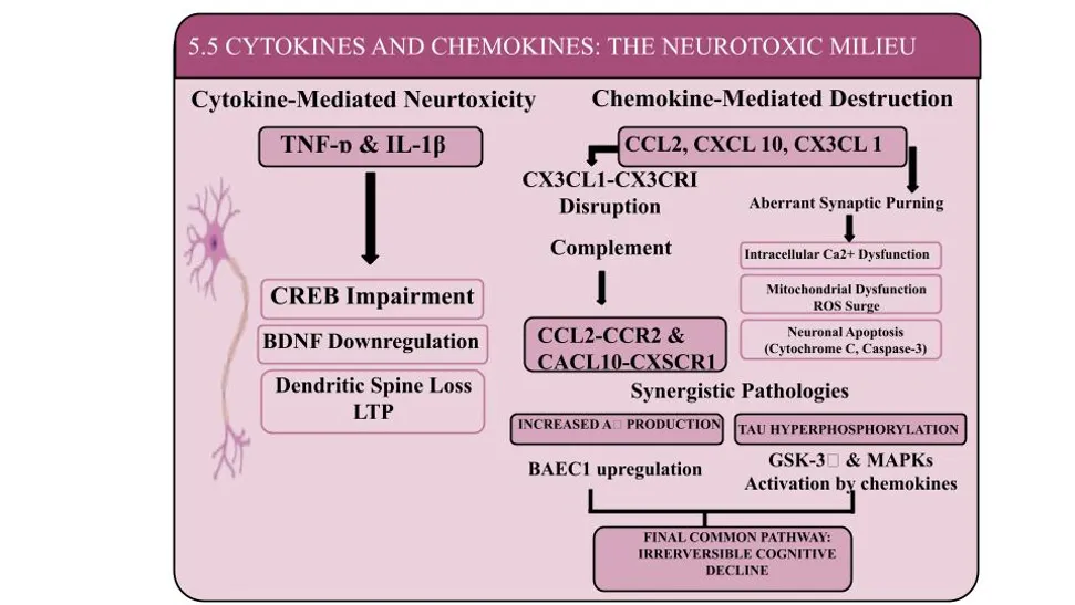
FIGURE 7. Inflammatory Signaling Pathways in Neurotoxicity and Cognitive Decline: Overview of cytokine (TNF-α, IL-1β) and cytokines impairing synaptic plasticity, BDNF levels, and LTP. Dysregulated chemokine pathways trigger complement activation, aberrant synaptic pruning, mitochondrial ROS production, and apoptosis, while upregulating BACE1 and GSK-3βMAPK signaling to accelerate Aβ accumulation, Tau hyperphosphorylation, and progressive cognitive decline.
5. VASCULAR IMMUNE CROSSTALK IN VaD
5.1 INTRACTION BETWEEN VASCULAR INJURY AND INFLAMMATION
Neuronal damage brought on by cerebral ischemia eventually leads to serious neurological conditions including vascular dementia and even patient death. After ischemia damage, microglia will degrade the activities of mitochondria and promote inflammation through pro-inflammatory cytokines and chemokines. This will activate apoptotic pathways and change the function of neurons in the contralateral hemisphere. [100]
One of the main causes of the cognitive deterioration in dementia is neuroinflammation. When microglia are activated, they produce chemokines like CCR3, CCR5, and CCL12 as well as pro-inflammatory cytokines including TNF-α, IL-1 and IL-6. As the brain ages, chronic inflammation can damage the blood-brain barrier (BBB) and make it leaky. Inflammatory cells can then penetrate the BBB and enter the brain. More inflammatory reactions are released by the microglia as a result of this breach, which leads to tau protein hyperphosphorylation and NFT development. [101] NFT production produces exosomes that trigger NLRP3 complexes, cytokine release, and pro-inflammatory reactions. The IL-1β produced by the activated NLRP3 complex interacts to the IL-1β receptor, starting dementia and neuroinflammatory pathways. [102] The neuronal proteins CREB and BDNF can change as a result of the inflammatory mediators effects on IL-1β, TNF-α, and mitogen-activated protein kinase (MAPKs). Dementia and cognitive impairment are thus caused by changes in these neurochemicals. [103] Similar inflammatory mediators are involved in their downstream inflammatory pathways. Protein aggregation, mutation, or any other reason can first activate microglial cells and astrocytes, which then produce pro-inflammatory cytokines that can induce synaptic dysfunction, neuronal degeneration, and reduced cognitive function. [100] The complex structure known as the neurovascular unit is made up of glial and neuronal components from the central nervous system (CNS), endothelial cells, and pericytes. Vascular pathology’s underlying processes and how they contribute to neurodegenerative illnesses. Most neurodegenerative illnesses include neuroinflammation and reactive gliosis, and new research indicates that immune-mediated disruption of the blood-brain barrier may play a role in including reactive gliosis and neuronal dysfunction. Therefore, neuroinflammation brought on by vascular injury will result in more microvascular damage and breakdown of the blood-brain barrier. [104]
5.2 OXIDATIVE STRESS AS A LINK
Oxidative stress and vascular dysfunction are linked by inflammation. It has been suggested that oxidative stress may be a major factor in the development of VaD. In fact, free radicals, like reactive oxygen species (ROS), can react with substances such as proteins and lipids, leading to damage to the brain. This underscores the neurotoxicity of the amyloid and suggests that oxidative stress may be a cause and consequence of Aβ accumulation by increasing the Aβ precursor protein (APP). Additionally, the pathophysiology of VaD demonstrates that an increase in reactive oxygen species (ROS) is caused by the dissociation between endothelial nitric oxide synthase (eNOS) and the overactivity of NADPH oxidase. . Protein damage, abnormal folding, amyloidosis, islet amyloid polypeptide, ER stress, dysmorphic mitochondria, direct oxygen destruction of endothelial cell tight junction, and priming of the local glial cells are all consequences of ROS-mediated DNA base modification. [107] All of these lower activation threshold leads to an excessive and uncontrolled generation of inflammatory mediators and mitochondrial ROS. All of this results in systemic-central neurodegeneration that is persistent.
5.3 NEUROVASCULAR UNIT DYSFUNCTION
The BBB also serves as a component of a neurovascular unit (NVU), which is made up of neurons, astrocytes, and microglia in addition to mural cells, specialised endothelial cells, and the BBB’s basement membrane, which serves as a regulatory center for preserving cerebral homeostasis. [108] Neurovascular uncoupling, CBF decreases and dysregulation, and BBB disruption, including pericyte loss, are increasingly recognised as early stages in the pathophysiological cascade. Aβ oligomers probably contribute significantly to neurovascular unit dysfunction because they cause vasoconstriction, which lowers CBF. Blood vessels that aren’t working correctly won’t be able to provide vital nutrients like glucose and oxygen to the brain or eliminated waste products from metabolism. Systemic infections and inflammation frequently increase BBB permeability, allowing immune mediators and cells to enter the brain. Additionally, capillary leakages of blood-derived products, such as fibrin (ogen), immunoglobulin G (IgG), thrombin, albumin, and hemosiderin, have been found in the cortex and hippocampus of VaD post-mortem brain tissues. [109] All of these directly lead to the demise of the nearby European people.
5.4 COGNITIVE DECLINE MECHANISM
The last clinical sign of synaptic and neuronal integrity is the cognitive loss characteristic of VaD. Cognitive impairment can be attributed to two main mechanisms: through the long-term and dysregulated release of chemokines (CCL2, CXCL10) and cytokines (TNF-α, IL-β, IL-6). Irreversible neuronal death is caused by glutamate-mediated excitotoxicity, pro-apoptotic signalling via caspase-3, and metabolic fatigue brought on by hypoperfusion.
CONCLUSION
Vascular dementia (VaD) is a complex, multi-factorial neurocognitive disorder that is associated with complex pathophysiological mechanism that go well beyond conventional ischemic cerebral injury. There is growing evidence for the pivotal contribution of vascular-immune crosstalk, a feed-forward loop between cerebral hypoperfusion, endothelial dysfunction, blood-brain barrier (BBB) dysfunction, and chronic neuroinfammation. Changing dynamics of the neurovascular unit (NVU) and chronic glial activation lead to a feedback loop of neurotoxicity with increased levels of pro-inflammatory cytokines, matrix metalloproteinases and reactive oxygen species (ROS). Eventually, these molecular cascades lead to pruning of synapses, loss of structure in the axons, and a gradual deterioration of cognitive function. While significant advances have been made in identification of important signalling pathways such as NF-κB, NLRP3, MAPK, Nrf2/HO-1, the challenge of moving these mechanistic insights into effective clinical interventions is still significant. Symptomatic treatment with cholinestrase inhibitors, NMDA receptor antagonists and intensive secondary cardiovascular risk factors managementare the mainstays of current management strategies. But therapeutic agents are still missing that can stop or repair any damage to the NVU and immune system. But therapeutic agents are still missing that can stop or repair any damage to the NVU and immune system. In the near future, the clinical paradigm needs to evolve into target-oriented, multi-mechanistic therapeutics. Targeted BBB restoration, accurate microglial immunomodulation, cellular repair using stem cells, and effective drug delivery systems, such as nanocarriers, are promising directions in development. The efforts to close the gap between the preclinical observations and the ultimately succesful translation to patients with VaD will need to be accompanied by strong, long-term clinical trials, based on standardised biomarkers, for early detection, accurate disease staging, and tailored therapeutic options for VaD patients.
REFERENCES


Nikhil Goyal, Shalu Tomar, Saloni Sharma, Anurag Bhargava, Vascular–Immune Crosstalk in Vascular Dementia: Role of Neurovascular Dysfunction and Neuroinflammation, Int. J. of Pharm. Sci., 2026, Vol 4, Issue 8, 2879-2906. https://doi.org/10.5281/zenodo.22023140
 10.5281/zenodo.22023140