

We use cookies to ensure our website works properly and to personalise your experience. Cookies policy
Department of Pharmaceutics, Prasanna College of Pharmacy, Laila, Belthangady, Karnataka, India - 574214.
Transdermal drug delivery has gained significant importance as a promising approach for achieving controlled and sustained delivery of therapeutic agents across the skin. Compared with conventional routes of administration, transdermal drug delivery offers several advantages, including avoidance of hepatic first-pass metabolism, reduced gastrointestinal adverse effects, improved patient adherence, and more consistent systemic drug concentrations. However, effective drug transport across the skin remains challenging because the stratum corneum acts as the primary barrier to drug permeation. This limitation is particularly significant for drugs with high molecular weight, low lipid solubility, or unfavourable partitioning properties. Drug transport across the skin occurs through intercellular, intracellular, and appendageal pathways and is influenced by the physicochemical properties of the drug and the physiological characteristics of the skin. Recent advances in pharmaceutical formulation technology have led to the development of novel vesicular carriers to overcome some of the limitations associated with conventional transdermal drug delivery systems. These vesicular systems can enhance drug solubility and skin permeation, protect therapeutic agents from degradation, and facilitate controlled drug release. Among the widely investigated vesicular carriers are liposomes, niosomes, transfersomes, and ethosomes. Liposomes are phospholipid-based vesicles capable of encapsulating both hydrophilic and lipophilic drugs, whereas niosomes are composed primarily of non-ionic surfactants and may offer improved stability and formulation flexibility. Transfersomes are highly deformable vesicles containing edge activators that facilitate their passage through narrow skin pathways. Ethosomes contain relatively high concentrations of ethanol, which can alter skin lipid organization and enhance drug penetration. Various techniques, including thin-film hydration, reverse-phase evaporation, ethanol injection, and sonication, are employed in the preparation of these vesicular systems.
Transdermal drug delivery systems (TDDS) are advanced drug delivery approaches in which medicated patches are applied to the skin to deliver drugs across the skin, generally into the systemic circulation, at a predetermined and controlled rate. These systems can provide sustained drug release over an extended period, helping maintain drug concentrations within the therapeutic range and minimizing fluctuations in plasma drug levels.1
A drug delivery system (DDS) employs various technologies to deliver active pharmaceutical ingredients into the body in a controlled manner, thereby maximizing therapeutic efficacy while minimizing adverse effects.2
Transdermal drug delivery is an important non-invasive method of drug administration. It bypasses the gastrointestinal tract and avoids exposure to gastrointestinal conditions that may degrade certain drugs. It also bypasses hepatic first-pass metabolism and eliminates the discomfort associated with injectable (parenteral) therapy. Furthermore, it can be a suitable route for systemic drug delivery in paediatric and geriatric patients and individuals with dysphagia.3
The field of drug delivery has undergone significant advancements with the emergence of novel drug delivery systems (NDDS), which aim to improve drug targeting, enhance therapeutic efficacy, reduce adverse effects, and improve patient compliance. Transdermal drug delivery is one such approach that offers several advantages when suitable drugs are formulated appropriately.4
The skin is the body's largest organ and serves as a protective barrier against the external environment. The skin proper consists of two principal layers: the epidermis and the dermis, while the hypodermis (subcutaneous tissue) lies beneath them. The stratum corneum, which is the outermost layer of the epidermis, acts as the primary barrier to drug permeation and plays a crucial role in transdermal drug delivery.5
The effectiveness of transdermal drug delivery systems is limited by the barrier function of the stratum corneum, which generally favours the permeation of relatively small molecules with suitable physicochemical properties. Conventional transdermal delivery is particularly challenging for drugs with molecular weights above approximately 500 Da, although this is a general guideline rather than an absolute limit. Despite these limitations, TDDS offers several therapeutic advantages, including sustained and controlled drug release, maintenance of relatively stable plasma drug concentrations, reduced exposure to gastrointestinal adverse effects, improved patient adherence, and avoidance of hepatic first-pass metabolism. These advantages make TDDS a valuable alternative for suitable drugs, particularly those that undergo significant gastrointestinal degradation or hepatic first-pass metabolism.6
Various technologies and approaches have been explored to overcome the skin barrier, including iontophoresis, electroporation, chemical permeation enhancers, microemulsions, sonophoresis, and vesicular carriers such as liposomes, niosomes, ethosomes, and transfersomes. These approaches may enhance drug permeation through different mechanisms. Among them, vesicular carrier systems have attracted considerable research interest because of their potential to improve drug interaction with the skin, enhance permeation, and facilitate controlled drug delivery.7
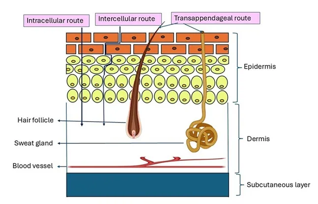
Figure 01: Schematic Representation of Skin Permeation
Pathways in Transdermal Drug Delivery
The skin consists of the epidermis and dermis, with the hypodermis (subcutaneous tissue) situated beneath the skin proper. The stratum corneum, which forms the outermost layer of the epidermis, is the principal barrier to drug permeation. Skin appendages, including hair follicles and sweat gland ducts, also contribute to drug transport by providing alternative pathways through the skin barrier.8
The barrier function of the skin is primarily attributed to the stratum corneum. This layer consists of keratin-filled corneocytes embedded in a highly organized lipid matrix. This structure minimizes water loss and restricts the penetration of many substances, including drugs. Consequently, effective transdermal delivery depends on the physicochemical properties of the drug, including its molecular size, lipid solubility, and ability to partition into and diffuse through the skin.9
Novel vesicular systems, including liposomes, niosomes, transfersomes, and ethosomes, have been investigated for their potential to enhance drug permeation across the stratum corneum. Depending on their composition and properties, these systems may interact with skin lipids, improve drug partitioning into the skin, and facilitate drug transport across the skin barrier. However, their effectiveness depends on the formulation, drug properties, and mechanism of interaction with the skin.10
Drug permeation through intact skin occurs mainly through two pathways: the transepidermal pathway and the transappendageal pathway. The transepidermal pathway is further divided into the transcellular and intercellular routes. The route followed by a drug depends on its physicochemical properties and its interactions with the skin barrier.11
1. Transcellular Route
In the transcellular route, drug molecules pass through the corneocytes and the lipid domains between them. As the drug encounters alternating aqueous and lipid environments, its ability to cross the skin depends on its capacity to partition between and diffuse through these different regions.
2. Intercellular Route
In the intercellular route, drug molecules pass through the lipid matrix surrounding the corneocytes. This pathway is particularly important for the permeation of many drugs because the intercellular lipids provide a tortuous pathway through the stratum corneum. Drug permeation depends on the ability of the molecules to partition into and diffuse through this lipid matrix.12
3. Transappendageal Route (Shunt Route)
The transappendageal route, also known as the shunt route, involves drug permeation through skin appendages, including hair follicles and sweat gland ducts. These structures provide alternative pathways through the stratum corneum. The extent of drug transport through this route may be influenced by the density and distribution of skin appendages, the dimensions of their openings, and the physicochemical properties of the drug.13
NOVEL VESICULAR DRUG DELIVERY SYSTEMS
ETHOSOMES
Ethosomes were developed by Touitou et al. in 1997 as novel lipid-based carriers composed primarily of ethanol, phospholipids, and water. They are soft, flexible vesicular carriers designed to enhance drug penetration through the skin and improve transdermal drug delivery.14
Drug Permeation Mechanism of Ethosomes
The exact mechanism by which ethosomes enhance drug permeation through the skin is not completely understood. However, their effect is generally attributed to the combined actions of ethanol and the flexible phospholipid vesicles.
i. Ethanol Effect
Ethanol acts as a skin penetration enhancer by interacting with the intercellular lipids of the stratum corneum. This interaction increases lipid fluidity and alters the organization of the lipid bilayers, thereby reducing the barrier resistance of the skin and facilitating drug permeation.
ii. Ethosome Effect
The presence of ethanol enhances skin permeability, while the flexible phospholipid vesicles may facilitate the transport of the encapsulated drug into the skin. The vesicles can interact with the skin lipids and may contribute to the gradual release of the drug within the skin layers, improving transdermal drug delivery.15
Ethosomes enhance drug permeation through the combined action of ethanol and phospholipid vesicles. Ethanol modifies the lipid organization of the stratum corneum, while the vesicular system facilitates drug delivery into the skin. Drug transport may occur through the stratum corneum and skin appendages, including hair follicles. These combined effects contribute to improved skin penetration and transdermal drug delivery.16
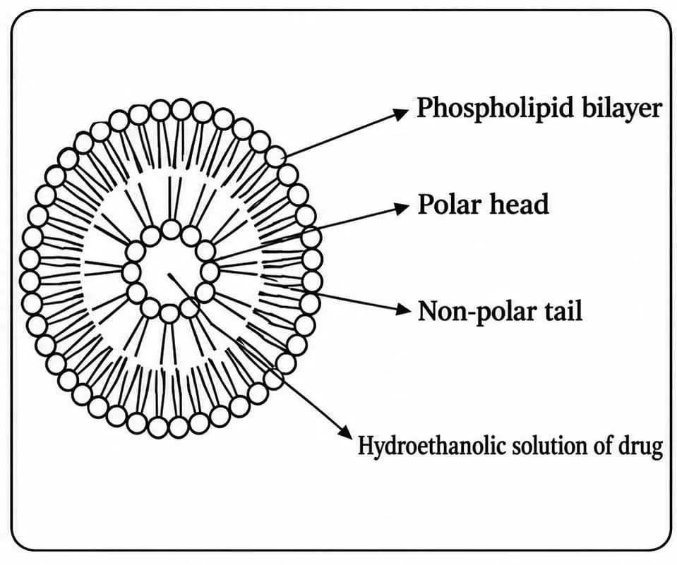
Figure 02: Schematic Representation of Ethosomes
Types of Ethosomal Systems
1. Classical Ethosomes
Classical ethosomes consist of phospholipids, water, and a relatively high concentration of ethanol, which may reach up to 45% w/v. They are characterized by flexible vesicles, a generally negative zeta potential, and the potential for high drug entrapment efficiency. Compared with conventional liposomes, they may provide enhanced skin permeation. Their suitability for drug encapsulation depends on the physicochemical properties of the drug and the formulation composition.18
2. Binary Ethosomes
Binary ethosomes are modified classical ethosomes that contain an additional alcohol, such as propylene glycol (PG) or isopropyl alcohol (IPA). The additional alcohol may modify vesicle characteristics and influence the stability and skin permeation properties of the formulation.19
3. Transethosomes
Transethosomes are modified ethosomal vesicles that combine the characteristics of ethosomes and transfersomes. In addition to the basic ethosomal components, they contain an edge activator, usually a surfactant, or another suitable penetration-enhancing component. These ingredients may improve vesicle flexibility and enhance drug permeation through the skin.20
Methods of Preparation of Ethosomes
• Cold Method
The cold method is one of the commonly used techniques for preparing ethosomes. In this method, phospholipids, the drug, and other lipid components are dissolved in ethanol in a covered vessel at room temperature under continuous, vigorous stirring. The resulting mixture is then heated to approximately 30°C using a water bath.
Separately, water is heated to the same temperature and gradually added to the ethanolic mixture with continuous stirring for approximately 5 minutes in a covered vessel. If required, the vesicle size can be further reduced by sonication or extrusion. The final formulation is stored under refrigerated conditions to help maintain its stability.21
• Hot Method
In the hot method, phospholipids are dispersed in water and heated in a water bath at approximately 40°C until a colloidal dispersion is formed. In a separate vessel, ethanol and propylene glycol are mixed and heated to the same temperature.
The ethanolic phase is then added to the aqueous phase with continuous stirring. The drug is incorporated into the appropriate phase depending on its hydrophilic or lipophilic nature. If necessary, the vesicle size can be reduced further by probe sonication or extrusion. 15
LIPOSOMES
Liposomes were first described by Bangham and colleagues in the 1960s during studies investigating phospholipids and blood clotting.22
Liposomes are small, spherical vesicles composed primarily of lipids arranged in one or more phospholipid bilayers. 23
Liposomes are primarily composed of phospholipids, which are amphiphilic molecules consisting of a hydrophilic head group and two hydrophobic fatty acid chains. When dispersed in an aqueous medium, phospholipids spontaneously arrange themselves into bilayer membranes because of their amphipathic nature. The hydrophilic head groups interact with the surrounding aqueous environment, whereas the hydrophobic fatty acid chains associate with one another. This arrangement forms a phospholipid bilayer with hydrophobic regions oriented inward and hydrophilic surfaces facing the aqueous medium. This structure enables liposomes to encapsulate drugs with different physicochemical properties. 24-25
Mechanism of Action of Liposomes
Liposomes are spherical vesicles composed of one or more phospholipid bilayers enclosing an aqueous core. This structure enables them to encapsulate both hydrophilic and hydrophobic therapeutic agents. Hydrophilic drugs are generally entrapped within the aqueous core, whereas hydrophobic drugs are incorporated into the lipid bilayer. Drug localization depends primarily on the physicochemical properties of the drug and the lipid composition of the vesicle. Liposomes can interact with cellular membranes and facilitate drug delivery through mechanisms such as membrane fusion, endocytosis, and other membrane-associated interactions.
Mechanism of Liposome - Mediated Drug Delivery
i. Adsorption
The initial step involves the attachment of liposomes to the cell surface through interactions with the plasma membrane. This promotes close contact between the liposomal carrier and the target cell.
ii. Endocytosis
Following adsorption, liposomes may be internalized by cells through endocytosis. This process involves the engulfment of the vesicles into intracellular compartments, from which the encapsulated drug may subsequently be released.
iii. Membrane Fusion
Liposomes may fuse directly with cellular membranes through the mixing of phospholipids in the liposomal and cellular bilayers. This process can facilitate the transfer of liposomal contents into the cell.
iv. Lipid Exchange
Lipids may be transferred between liposomal and cellular membranes through membrane interactions and, in some cases, lipid-transfer proteins. Such exchange may alter membrane composition and influence drug delivery.26
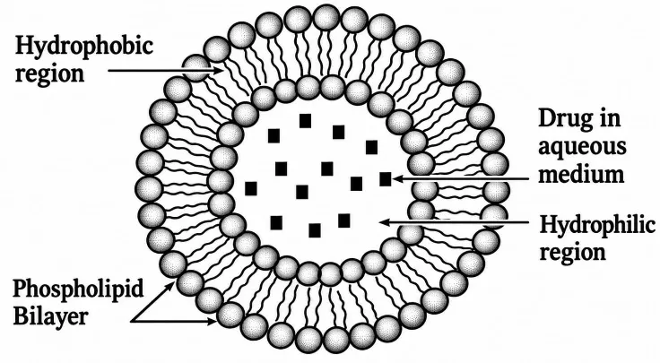
Figure 03: Schematic Representation of Liposomes
Types of Liposomes
a. Multilamellar Vesicles (MLVs)
Multilamellar vesicles consist of multiple concentric phospholipid bilayers separated by aqueous layers, giving them an onion-like structure. The bilayers are arranged one inside another, with aqueous compartments between successive layers.
b. Unilamellar Vesicles (ULVs)
Unilamellar vesicles consist of a single phospholipid bilayer enclosing an aqueous core. Based on their size, they are further classified into the following types:
i. Small Unilamellar Vesicles (SUVs)
Small unilamellar vesicles consist of a single phospholipid bilayer surrounding an aqueous compartment and have a relatively small vesicle diameter.
ii. Large Unilamellar Vesicles (LUVs)
Large unilamellar vesicles also consist of a single phospholipid bilayer enclosing an aqueous core, but they have a larger diameter than SUVs. Consequently, they can generally encapsulate a greater volume of aqueous solution. 28-30
Methods of Preparation
• Thin-Film Hydration (TFH) Method (Bangham Method)
The thin-film hydration (TFH) method, also known as the Bangham method, is one of the oldest and most widely used methods for preparing multilamellar vesicles (MLVs).
In this method, phospholipids are first dissolved in an organic solvent, such as chloroform, dichloromethane, ethanol, or a chloroform - methanol mixture, to obtain a uniform lipid solution. The organic solvent is then removed under reduced pressure at 45 - 60°C using a rotary evaporator. For small volumes of less than 1 mL, the solvent may also be removed by passing a stream of dry nitrogen or argon gas.
After solvent evaporation, a thin, dry lipid film forms on the inner wall of the flask. The film is then hydrated with an aqueous medium, such as distilled water or phosphate-buffered saline (pH 7.4). Hydration is carried out for 1 - 2 hours at 60 - 70°C, or at a temperature above the phase-transition temperature of the phospholipids. Gentle stirring helps detach the lipid layers from the flask wall and promotes hydration.
During hydration, the lipid bilayers swell and self-assemble into multilamellar vesicles. The resulting liposomal suspension may be stored overnight at 4°C to allow further vesicle formation. The final suspension generally contains vesicles with varying sizes and numbers of bilayers.31
• Reverse - Phase Evaporation Method (REV)
The reverse-phase evaporation (REV) method was first described by Szoka and Papahadjopoulos. In this method, phospholipids are dissolved in an organic solvent, while the drug intended for encapsulation is dissolved in an appropriate aqueous medium. The aqueous phase is then added to the organic phase, and the mixture is sonicated to form a water-in-oil (W/O) emulsion containing inverted micellar structures.
The organic solvent is gradually removed under reduced pressure using a rotary evaporator, resulting in the formation of a viscous gel-like phase. As solvent removal continues, the gel structure collapses, and the inverted micelles reorganize and fuse. The phospholipids then form bilayer membranes around aqueous compartments, resulting in the formation of liposomes. This method can facilitate the encapsulation of hydrophilic drugs within the aqueous compartments.32
NIOSOMES
Niosomes are microscopic vesicular drug delivery systems composed of one or more bilayers of non-ionic surfactants, usually in combination with cholesterol. They are formed through the self-assembly of these components in an aqueous medium. Niosomes are widely investigated as carriers for controlled, sustained, and targeted drug delivery because of their potential biocompatibility and ability to encapsulate different types of drugs.33
Mechanism of Penetration of Niosomes
When applied topically, niosomes may prolong drug retention within the stratum corneum and epidermis, potentially reducing the rate of systemic drug absorption. They may also influence the barrier properties of the stratum corneum by reducing transepidermal water loss and replenishing skin lipids, thereby improving skin hydration and smoothness. Niosomes may interact with the skin surface and, under suitable conditions, penetrate into the skin. Their interaction with the skin may promote the formation of smaller vesicular structures through a process described as niosome re-formation. Furthermore, the adsorption and fusion of niosomes at the skin surface may increase the thermodynamic activity gradient of the drug, promoting the permeation of lipophilic drugs. The surfactants present in niosomes may also act as penetration enhancers by modifying the skin barrier and facilitating drug permeation.34
Niosomes can interact with cells through several mechanisms. One mechanism involves adsorption onto the cell surface with minimal or no internalization of the vesicular components. This adsorption may occur through physical attractive forces between the niosomes and the cell membrane or through specific receptor - ligand interactions involving components on the vesicle surface. Such interactions may facilitate the transfer of the encapsulated drug to the skin or cellular membrane. Niosomes may also fuse with the cell membrane, resulting in the mixing of their membrane components and the release of their contents. Alternatively, cells may internalize niosomes through endocytosis. Following internalization, lysosomal enzymes may degrade the niosomal membrane, facilitating the release of the entrapped drug into the intracellular environment.35
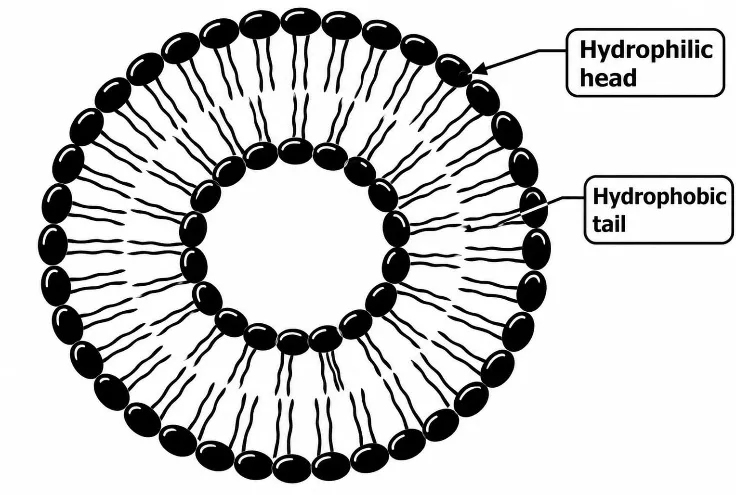
Figure 04: Schematic Representation of Niosomes
Types of Niosomes
Niosomes can be classified according to their size, number of lamellar layers, structural components, and intended applications.37
a. Multilamellar Vesicles (MLVs)
Multilamellar vesicles (MLVs) consist of multiple concentric bilayers enclosing separate aqueous compartments. They typically range from approximately 0.5 to 10 µm in diameter and are among the commonly studied types of niosomes. Their multilayered structure makes them suitable carriers for lipophilic drugs, which can be incorporated into the surfactant bilayers.38
b. Large Unilamellar Vesicles (LUVs)
Large unilamellar vesicles (LUVs) consist of a single bilayer surrounding a relatively large aqueous compartment. Their high aqueous compartment-to-membrane ratio allows the encapsulation of substantial quantities of hydrophilic bioactive substances with comparatively low membrane-material requirements. Their reported size generally ranges from approximately 100 to 3000 nm. 39
c. Small Unilamellar Vesicles (SUVs)
Small unilamellar vesicles (SUVs) consist of a single bilayer surrounding an aqueous core. They can be prepared from multilamellar vesicles (MLVs) using size-reduction techniques such as sonication, French press extrusion, and other mechanical methods. Electrostatic stabilization of SUVs may be achieved by incorporating dicetyl phosphate into Span 60-based niosomes loaded with 5 (6)-carboxyfluorescein (CF).40
Niosomes may also be classified according to their structural components and intended therapeutic applications.
d. Discomes
Discomes are disc-shaped vesicular structures formed from hexadecyl diglycerol ether (C16), cholesterol, and dicetyl phosphate in the presence of the non-ionic surfactant polyoxyethylene cholesteryl ether (Solulan C24). They typically range from 11 to 60 µm in size and have been investigated as carriers for sustained ocular drug delivery because of their potential for prolonged retention at the site of administration. 37
e. Elastic Niosomes
Elastic niosomes are highly deformable vesicular carriers designed to enhance drug permeation through the skin. Their flexibility may allow them to pass through narrow pathways within the skin barrier that are smaller than their original diameter, facilitating transport across the stratum corneum. They have been investigated for delivering therapeutic agents with different molecular weights. Compared with conventional formulations, elastic niosomes may provide prolonged drug release and improve drug penetration and therapeutic performance. Their movement across the skin is primarily associated with the trans-epidermal hydration gradient rather than simply with the vesicle concentration.
The first detergent-based elastic niosomes were developed by Van den Bergh and consisted of sucrose laurate ester and the micelle-forming surfactant octaoxyethylene laurate ester. When loaded with anti-inflammatory agents such as diclofenac and indomethacin, these deformable vesicles have demonstrated potential for enhanced skin penetration and transdermal drug delivery.41
f. Aspasomes
Aspasomes are vesicular carriers composed of ascorbyl palmitate, cholesterol, and the negatively charged lipid dicetyl phosphate. The lipid components are hydrated with water or an aqueous medium, followed by sonication to obtain a uniformly dispersed vesicular system.42
g. Bola Surfactant - Containing Niosomes
Bola surfactant-containing niosomes are prepared using the bola surfactant omega hexadecylbis-(1-aza-18-crown-6), Span 80, and cholesterol. These components are mixed in a molar ratio of 2:3:1 to form the vesicular system. 43
h. Proniosomes
Proniosomes are dry, non-ionic surfactant-based formulations that form niosomes upon hydration. They offer improved physical stability by reducing aggregation, vesicle fusion, and drug leakage. Their dry formulation also facilitates storage, transportation, and handling. Owing to these advantages and their potential for controlled drug release, proniosomes are promising carriers for transdermal drug delivery. 44
i. Vesicles in Water-in-Oil (v/w/o) Systems
Vesicles in water-in-oil (v/w/o) systems are prepared by incorporating an aqueous niosomal dispersion into an oil phase. These systems can be prepared using niosomes composed of sorbitan monostearate, cholesterol, and Solulan C24 (polyoxyethylene cholesteryl ether). The niosomal dispersion is added to the oil phase at 60 °C. Upon cooling to room temperature, the resulting emulsion undergoes gel formation, producing a vesicle-in-water-in-oil gel (v/w/o gel). 45
Methods of Preparation
• Lipid Film Hydration (Hand Shaking Method)
The lipid film hydration (LFH) method, also known as the hand shaking method (HSM), is one of the commonly used techniques for preparing niosomes. In this method, non-ionic surfactants, cholesterol, and other lipid components are dissolved in a suitable organic solvent. The solvent is then evaporated under reduced pressure using a rotary evaporator to form a thin film on the inner wall of a round-bottom flask.
The dried film is hydrated with an aqueous phase containing the drug at a temperature slightly above the phase-transition temperature of the surfactant. Gentle shaking is performed for a specified period to facilitate hydration. During this process, the film swells, detaches from the +flask wall, and forms niosomal vesicles.
Alternatively, the lipid film may be exposed to a stream of water-saturated nitrogen for approximately 15 minutes before hydration without shaking. The hand shaking method predominantly produces multilamellar vesicles (MLVs), whereas the non-shaking method generally produces large unilamellar vesicles (LUVs). 46
• Reverse Phase Evaporation Technique (REV)
The reverse phase evaporation (REV) technique is used for preparing niosomes and may provide high drug encapsulation efficiency. In this method, cholesterol and a non-ionic surfactant are dissolved in a mixture of chloroform and diethyl ether. An aqueous phase containing the drug is then added to the organic phase, and the resulting mixture is sonicated at 4 - 5 °C to form a water-in-oil emulsion.
A small quantity of phosphate-buffered saline (PBS) is subsequently added, followed by further sonication to improve emulsion stability. The organic solvents are removed under reduced pressure at approximately 40 °C using a rotary evaporator, producing a viscous niosomal suspension. Finally, the suspension is diluted with PBS and heated in a water bath at 60 °C for approximately 10 minutes to facilitate niosome formation. 47
Transfersomes
Transfersomes are specialized vesicular drug delivery systems characterized by highly flexible and self-adaptive membranes. Their exceptional deformability enables them to pass through narrow skin pathways that may be considerably smaller than the vesicles themselves, making them promising carriers for transdermal drug delivery.48
The term Transferosome is a registered trademark associated with the German company IDEA AG. It is derived from the Latin word transferre, meaning “to carry across,” and the Greek word soma, meaning “body.” Transfersome technology was introduced by Çevc and Blume in 1991 and has since attracted considerable research interest and patent activity. Although the technology has been studied for several decades, transfersomes continue to be investigated as drug delivery systems because their clinical application remains limited compared with conventional dosage forms. 49
Transfersomes, also known as ultradeformable vesicles, are designed for transdermal drug delivery. They generally consist of a phospholipid bilayer containing an edge activator and enclosing an aqueous core. Depending on the hydrophilic or lipophilic nature of the drug, the active substance may be encapsulated within the aqueous core or incorporated into the lipid bilayer. 50
Mechanism of Transfersomes
Transfersomes are designed to penetrate the stratum corneum and deliver therapeutic agents into deeper skin layers, including the dermis, with the potential for systemic delivery. Their skin penetration ability largely depends on the deformability of the vesicular membrane, which is influenced by the composition of the formulation. Therefore, formulation studies are required to obtain suitable vesicles with adequate deformability, drug-loading capacity, and stability for the intended therapeutic application. 51
The skin penetration mechanism of transfersomes is primarily associated with the hydration gradient across the skin. When a transfersomal formulation is applied under non-occlusive conditions, water evaporates from the skin surface, creating a gradient between the relatively dry outer skin and the more hydrated deeper layers. Because of their highly deformable bilayers and ability to retain water, transferosomes may move towards the deeper, water-rich layers of the skin.
Although this process is not equivalent to conventional forward osmosis, it is believed to involve transport mechanisms influenced by hydration and osmotic forces. During passage through the stratum corneum, the vesicles may undergo partial dehydration and subsequently rehydrate in deeper skin layers. Thus, the penetration and uptake of transferosomes are associated with the hydration gradient between the stratum corneum, viable epidermis, and surrounding environment. 52
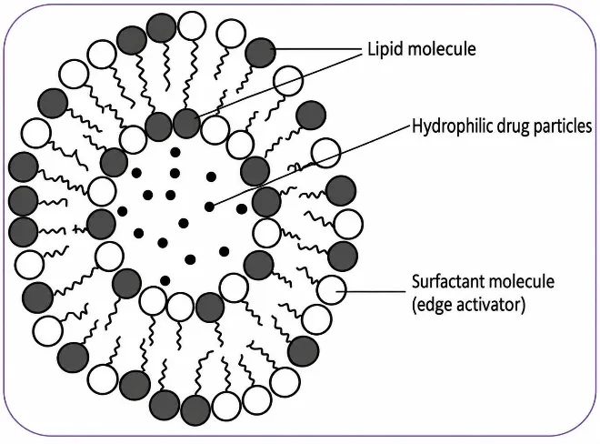
Figure 05: Schematic Representation of Transferosomes
Methods of Preparation
• Thin Film Hydration Technique (Rotary Evaporation - Sonication Method)
The thin film hydration technique, also known as the rotary evaporation - sonication method, is one of the commonly used methods for preparing transferosomes. In this method, phospholipids and an edge activator are dissolved in a suitable volatile organic solvent or solvent mixture, such as chloroform and methanol, inside a round-bottom flask. If the drug is lipophilic, it is incorporated into the lipid phase at this stage.
The organic solvent is evaporated under reduced pressure using a rotary vacuum evaporator at a temperature appropriate for the lipid components, forming a thin, uniform lipid film on the inner wall of the flask. The film is further maintained under vacuum to facilitate the removal of residual organic solvent.
The dried lipid film is then hydrated with an aqueous buffer of suitable pH, such as phosphate buffer at pH 7.4, while rotating the flask at an appropriate temperature for a specified period. Hydrophilic drugs may be incorporated during the hydration step. Hydration causes the lipid film to swell and form vesicles, commonly including multilamellar vesicles.
The resulting dispersion is allowed to hydrate further and is then subjected to sonication using a bath or probe sonicator to reduce vesicle size. Finally, the dispersion may be extruded through polycarbonate membrane filters with pore sizes ranging from 200 to 100 nm to obtain vesicles with a more uniform size distribution. 53
B. Reverse Phase Evaporation Method (REV)
In the reverse phase evaporation method, phospholipids and the edge activator are dissolved in a suitable organic solvent, such as ethanol, diethyl ether, or isopropyl ether, depending on the formulation. The solvent is partially evaporated under reduced pressure using a rotary evaporator, and the lipid components are redissolved to form reverse or inverted micellar structures.
An aqueous drug solution is added gradually while the mixture is sonicated under a nitrogen atmosphere. The remaining organic solvent is then removed by evaporation. During this process, the mixture initially forms a gel-like phase that subsequently collapses to produce a transfersomal suspension containing drug-loaded vesicles.⁵⁴
CONCLUSION
Transdermal drug delivery represents a promising alternative to conventional routes of drug administration because it can provide controlled and sustained drug delivery while avoiding first-pass metabolism. However, the barrier properties of the stratum corneum remain a major challenge to effective drug permeation. Understanding the pathways and mechanisms of drug transport through the skin is therefore essential for developing effective transdermal formulations.
Novel vesicular systems, including liposomes, niosomes, transfersomes, and ethosomes, have demonstrated considerable potential for improving transdermal drug delivery. Their ability to enhance drug solubilization and skin permeation, protect drugs from degradation, and support controlled release makes them valuable carriers for various therapeutic agents. Among these systems, the deformability of transfersomes and the penetration-enhancing properties of ethanol-containing ethosomes are particularly promising for improving drug transport across the stratum corneum.
Despite these advantages, several factors require consideration, including formulation stability, vesicle size, drug entrapment efficiency, skin compatibility, manufacturing reproducibility, and scale-up. Further research involving formulation development, long-term stability studies, safety evaluation, and clinical investigations may facilitate the successful application of novel vesicular carriers in transdermal drug delivery. Overall, vesicular drug delivery systems represent an important and evolving area of pharmaceutical research with the potential to improve the therapeutic performance of transdermal formulations.
REFERENCES

Sindhura Pallavi, Ashitha, Janani N. P., Meghabai P. M., Renuka, Sushmitha, Vesicular Carriers in Transdermal Drug Delivery: Exploring Skin Permeation Mechanisms and Formulation Strategies, Int. J. of Pharm. Sci., 2026, Vol 4, Issue 10, 4247-4259. https://doi.org/10.5281/zenodo.23686090
 10.5281/zenodo.23686090