

We use cookies to ensure our website works properly and to personalise your experience. Cookies policy
Adarsh College of Pharmacy, Near MIDC, Khambale, Vita 415311, Maharashtra, India.
Ocular diseases are a serious health concern worldwide and demand effective drug delivery systems for better treatment results. Traditional eye medicines such as drops and ointments are commonly used, but they often show poor effectiveness because the drug is quickly washed away by tears, drained through the tear ducts, and does not remain long on the eye surface. As a result, only a very small amount of the medicine, usually less than 5%, reaches the inside of the eye. To overcome these drawbacks, advanced drug delivery methods have been developed. Among them, ocular inserts have gained importance because they can provide controlled and long-lasting release of drugs. In recent years, three-dimensional (3D) printing, also called additive manufacturing, has emerged as a promising technique for making customized drug delivery devices. This technology allows accurate control over the shape, internal design, and drug distribution within the device. Therefore, 3D printing offers new opportunities to design patient-specific ocular inserts with improved treatment outcomes and predictable drug release. Different 3D printing methods have been studied for ophthalmic drug delivery, including fused deposition modeling, stereolithography, selective laser sintering, and inkjet printing. This review focuses on the use of 3D printing in the design and preparation of ocular inserts. It explains the barriers in the eye that limit drug delivery, the shortcomings of conventional eye medicines, the types of ocular inserts, and the role of various polymers in their formulation. It also highlights the different 3D printing techniques applied in pharmaceutical manufacturing, their applications in ophthalmic drug delivery, and recent progress in the development of 3D-printed ocular inserts.
Anatomy and Physiological Barriers of the Eye
The eye is a highly specialized sensory organ responsible for vision, and it is safeguarded by several anatomical and physiological barriers that restrict the entry of drugs into ocular tissues. While these protective mechanisms are vital for maintaining ocular homeostasis, they also create significant challenges for effective drug delivery. Figure 1 illustrates the anatomy of the eye. Structurally, the eye is divided into two main parts: the anterior segment and the posterior segment (1). The anterior segment of the eye includes the cornea, conjunctiva, aqueous humor, iris, and lens, while the posterior segment consists of the vitreous humor, retina, choroid, and optic nerve. Most conventional ophthalmic formulations are designed to act on the anterior segment because delivering drugs to the posterior segment through topical administration is particularly challenging. The cornea serves as the main barrier to drug penetration in topical ocular delivery. It is made up of several layers, including the epithelium, Bowman’s membrane, stroma, Descemet’s membrane, and endothelium. The corneal epithelium functions as a lipophilic barrier, restricting the entry of hydrophilic drugs, whereas the stromal layer is relatively hydrophilic and limits the passage of lipophilic compounds (2). This dual barrier significantly reduces drug absorption into intraocular tissues. In addition to corneal barriers, several physiological factors contribute to poor ocular drug bioavailability. Tear turnover and blinking rapidly remove instilled formulations from the ocular surface, leading to short residence time. Furthermore, nasolacrimal drainage can transport a significant portion of the administered drug into the nasal cavity, reducing ocular drug concentration and potentially causing systemic side effects. Other important barriers include the blood–aqueous barrier and the blood–retinal barrier, which control the movement of substances from the bloodstream into ocular tissues. These barriers make it especially difficult to deliver drugs effectively to the deeper parts of the eye. As a result, new and innovative drug delivery approaches are needed to overcome these limitations and achieve better therapeutic outcomes (3).
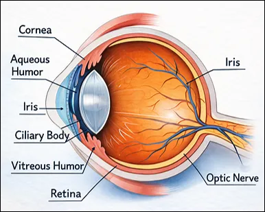
Figure 1: Anatomy of Eye
Ocular Drug Delivery Systems
The field of ocular drug delivery presents significant challenges in pharmaceutical research due to the intricate anatomy and protective barriers of the eye. Traditional ophthalmic dosage forms, such as eye drops, ointments, and suspensions, are commonly implemented in clinical settings because they are user-friendly and easy to apply; however, these methods have notable shortcomings, including low bioavailability, quick elimination before reaching the cornea, and the necessity for frequent administration (1,4). To address these issues, a range of innovative ocular drug delivery systems has been created, such as nanoparticles, liposomes, noisome, dendrimers, in situ gels, ocular implants, contact lenses, microneedles, and ocular inserts (4). These advanced approaches are designed to improve how long drugs remain on the eye’s surface, increase their absorption into ocular tissues, and enable a prolonged and controlled release of the medication (2). Within these systems, nanoparticles and liposomal formulations have been extensively studied for their ability to encapsulate drugs and support their transport across ocular barriers. In situ gelling systems, which change from liquid to gel upon contact with the ocular surface, further improve drug absorption and extend the time the medicine remains in the eye (3,5). Even with these improvements, challenges like stability problems, restricted drug loading capacity, and worries about patient adherence remain. Nonetheless, ocular inserts have surfaced as a notably promising method, providing extended drug delivery, enhanced therapeutic effectiveness, and decreased dosing frequency, establishing them as a beneficial option in contemporary ocular drug delivery techniques (6,7).
Ocular Inserts:
Ocular inserts (Fig.2) are sterile, thin, and flexible drug delivery devices designed to be placed in the conjunctival sac of the eye, typically beneath the lower eyelid. After insertion, these systems gradually release medication over an extended period, maintaining a relatively constant drug concentration within ocular tissues. Compared to conventional ophthalmic dosage forms like eye drops, ocular inserts offer several notable advantages (1,2). They enable controlled and sustained drug delivery, enhance ocular bioavailability, low dosing frequency, and minimize systemic side effects associated with nasolacrimal drainage. Furthermore, they allow more precise dosing and reduce variability linked to patient-administered eye drops (4). Ocular inserts are generally categorized into three main types: insoluble, soluble, and biodegradable. Insoluble inserts remain intact within the conjunctival sac and primarily release the drug through diffusion. Soluble inserts gradually dissolve in tear fluid after delivering the medication, while biodegradable inserts undergo controlled degradation in physiological conditions, eliminating the need for removal. Advancements in polymer science have played a crucial role in improving ocular insert design. Both natural and synthetic polymers are employed to fabricate these devices, allowing precise control over drug release kinetics and mechanical properties (2,4,7).
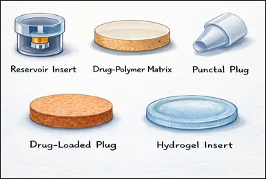
Figure 2: Ocular Inserts
Polymers Used in Ocular Inserts
They significantly influence the device’s mechanical strength, drug release behavior, and biocompatibility, polymers are essential to the formulation of ocular inserts. For the formulation of efficient ophthalmic drug delivery systems, both natural and synthetic polymers have been thoroughly investigated. Because of its superior biocompatibility, biodegradability, and low toxicity, natural polymers such as chitosan, gelatin, alginate, and collagen are used extensively (7). For instance, chitosan has potent mucoadhesive qualities that improve absorption and drug retention on the ocular surface. Alginate is also prized for its ability to form gels and deliver prolonged drug release. However, synthetic polymers such as polylactic acid (PLA), polyethylene glycol (PEG), polycaprolactone (PCL), and polyvinyl alcohol (PVA) are frequently utilized. Because of their excellent stability and strong mechanical properties, these materials are well suited for developing controlled drug delivery systems. They allow precise regulation of drug release patterns as well as predictable control over degradation rates (1,3). The recent advancements have centered on integrating these polymers with advanced manufacturing techniques such as 3D printing. The creation of highly customized ocular inserts with precise mechanical characteristics, well-defined architecture, and regulated drug distribution is made possible by this integration, which eventually improves therapeutic effectiveness and patient compliance (4).
3D Printing Technologies in Pharmaceutical Drug Delivery
The ability of 3D printing technologies to design complex drug delivery systems with precise control over structure, composition, and drug distribution has attracted significant interest in pharmaceutical research. Using these additive manufacturing methods, customized dosage forms can be developed, making them highly suitable for advanced drug delivery applications and personalized medicine (8). Fused deposition modeling (FDM) is one of the most popular methods. This technique creates layered structures by heating and extruding thermoplastic polymer filaments containing drug substances through a nozzle. FDM can be used to fabricate a variety of drug delivery devices because it is reasonably easy, affordable, and adaptable (9) Fig.3 indicates different 3D Printing technologies.
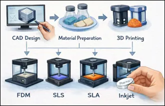
Figure 3: 3D Printing Technologies
Applications of 3D Printing in Eye Inserts
3D printing has surfaced as a potential method for ocular drug delivery, providing the capability to produce highly tailored inserts. This technology enables accurate control over dimensions, shape, and internal design, facilitating the creation of devices that have regulated and anticipated drug release (8,10). Recent studies emphasize the effective creation of 3D-printed ocular inserts for administering various therapeutic agents, such as antibiotics, anti-inflammatory drugs, and medications utilized in glaucoma treatment. These devices can be designed to release medications gradually over extended periods, such as several days or even weeks, which reduces how often they need to be administered and helps improve patient adherence to therapy (10). Moreover, 3D printing allows the creation of personalized ocular inserts, which is particularly beneficial for treating chronic eye issues that necessitate long-term treatment. This method aims to enhance treatment results and overall patient comfort by customizing drug dosage and release properties to meet individual requirements (9).
Obstacles and Future Outlook
Despite the significant promise of 3D printing for ophthalmic drug delivery, several challenges still need to be addressed. A key constraint is the limited access to appropriate pharmaceutical-grade substances that can be efficiently printed without compromising drug stability. In certain instances, the printing process- particularly those that use heat- cause the degradation of delicate drug compounds. Moreover, the lack of clear regulatory guidelines and uniform production protocols creates obstacles for broad acceptance. One more issue is scalability (11). As Compared to conventional manufacturing methods, 3D printing is comparatively slow, complicating large-scale production. Maintaining consistent quality, safety, and efficacy across batches continues to be a vital concern, especially since regulatory frameworks for 3D-printed pharmaceuticals are still evolving. Despite these obstacles, continuous advancements in biomaterials, printing technologies, and digital design are anticipated to address numerous limitations. Through continuous research and technological progress, 3D printing has the potential to transform ophthalmic drug delivery by enabling personalized treatments and supporting the development of advanced ocular therapeutic systems (12,13).
Design Factors for Ocular Inserts
Creating efficient ocular inserts (Fig.4) necessitates a precise equilibrium of formulation and design factors to guarantee safety, therapeutic efficacy, and patient satisfaction. Since these devices are inserted into the conjunctival sac, they should possess appropriate physicochemical and mechanical properties to reduce irritation while ensuring sufficient retention duration. Biocompatibility is a crucial necessity in the design of ocular inserts. The materials employed must be non-toxic, non-irritating, and compatible with ocular tissues to avoid discomfort, excessive tearing, or early removal. This renders the choice of suitable polymers and excipients an essential phase in formulation development (7,14).
The management of drug release behavior is similarly crucial. Ocular inserts are usually created to provide medications in a continual fashion for prolonged periods, ranging from hours to multiple days or even weeks. Factors like polymer composition, device thickness, porosity, drug solubility, and material degradation properties affect drug release profiles. Accomplishing controlled release aids in sustaining steady therapeutic levels and minimizes the frequency of dosing (12). The insert’s physical dimensions, such as its size and shape, significantly influence both patient comfort and the effectiveness of the treatment. Devices need to be small and adaptable enough to fit comfortably in the conjunctival sac without obstructing vision or blinking. Inadequately designed inserts can lead to a foreign body feeling, decreasing patient adherence (11,12).
The mechanical attributes, especially durability and pliability, are essential as well. Inserts must be durable enough to endure handling during administration, yet flexible enough to conform to the curvature of the eye. Consequently, materials that possess appropriate elasticity and tensile strength are favored (15). Ultimately, sterility and stability are crucial for guaranteeing product safety and efficacy. Ocular inserts should be manufactured in sterile environments and maintain their physical and chemical stability during storage. Cutting-edge technologies like 3D printing offer novel opportunities for accurately managing (15,16).
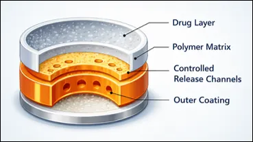
Figure 4: Design Factors for Ocular Inserts
Mechanisms of Drug Release from Ocular Inserts
The polymer composition and structural design of the device play a vital role in the release behavior of the medications that are intended to be delivered via ocular inserts in a controlled and sustained manner. To achieve predictable therapy outcomes, a thorough understanding of these pathways is essential (5). Diffusion-controlled release, in which the drug progressively transitions from the polymer matrix into the tear fluid, is one of the main methods of drug release. The thickness of the insert, the porosity of the polymer, and the drug’s solubility all have an impact on this process. Insoluble polymer-based inserts are frequently linked to such systems. Erosion-controlled release, which entails the slow disintegration of the polymer matrix in the ocular environment, is another crucial mechanism. When the substance deteriorates, the substance that was contained is released as the material breaks down. Because they can safely break down over time, biodegradable polymers like polylactic acid (PLA) and polycaprolactone (PCL) are widely utilized in these systems (4). A third mechanism is osmotic release, in which the osmotic pressure differential between the insert and the surrounding tear fluid drives medication delivery. This strategy may offer a longer-lasting and more reliable release profile. Drug release in sophisticated ocular insert systems is frequently controlled by a mix of processes, such as swelling, polymer breakdown, and diffusion. Drug release patterns can be customized to match individual treatment demands thanks to modern fabrication processes like 3D printing, which further improve control over the interior structure of inserts (16).
Role of 3D Printing in the Development of Ocular Inserts
The use of 3D printing in pharmaceutical research has greatly advanced the design and manufacturing of drug delivery systems, including ocular inserts. Compared to conventional fabrication techniques, this technology provides greater flexibility, precision, and efficiency. Traditional manufacturing methods such as solvent casting, compression molding, and film extrusion have been widely used to produce ocular inserts (6). However, these methods often provide only limited control over the internal architecture of the device. In contrast, 3D printing enables the creation of intricate geometries with well-defined architectural features, allowing better regulation of drug release behavior (9). A key advantage of 3D printing is its ability to enable personalized medicine. With the help of computer-aided design, ocular inserts can be tailored to meet individual patient needs by adjusting their size, shape, and drug content. This is especially helpful for treating long-term eye disorders that call for regular and sustained medication delivery. The exact placement of medications inside the insert is another important advantage. It is possible to strategically place active pharmacological ingredients in particular areas or to incorporate them uniformly into the printing substrate (7). Systems with complex release patterns, including an initial quick release followed by sustained distribution, can be designed thanks to this degree of control. Furthermore, 3D printing facilitates quick prototype and simple design customization (12). The development cycle can be accelerated by researchers’ ability to swiftly modify digital models and create new versions. Additionally, this technology’s additive nature reduces material waste and simplifies the production process. All things considered, 3D printing is a potential way for creating sophisticated ocular inserts, providing better medication delivery control, increased customization, and more effective manufacturing than conventional techniques (17).
Current Developments in Ocular Drug Delivery Systems Using 3D Printing
3D printing has been a potent instrument in the creation of sophisticated ophthalmic medication delivery systems in recent years. Its potential to create devices like ocular inserts, punctal plugs, and intraocular implants that can deliver medications in a regulated and sustained manner has been shown by numerous research (18). The creation of drug-loaded punctal plugs via 3D printing is one significant advancement. To improve medication retention on the ocular surface, these tiny devices are placed into the tear ducts to lessen tear discharge. Researchers have created punctal plugs that can deliver anti-inflammatory drugs for prolonged periods of time by using biodegradable polymers. Ocular implants made of hydrogel are another exciting new development (14). Using methods such as stereolithography Through the use of digital light processing, researchers have created incredibly accurate hydrogel structures that can expand, absorb tear fluid, and release contained medications gradually. These techniques’ exquisite resolution enables precise control over internal architecture and pore size, which directly affects drug diffusion rates. Additionally, polymeric inserts containing antibiotics or antiglaucoma medicines have been created using extrusion-based techniques like fused deposition modeling (9,10). When compared to conventional eye drop formulations, these systems have demonstrated the capacity to maintain drug release over several days while enhancing bioavailability. Bioprinting techniques for producing tissue-engineered ocular structures have also been investigated in recent studies (6).
Regulations Concerning Pharmaceutical Products Made with 3D Printing
While 3D printing has many benefits for pharmaceutical manufacture, it also presents a number of regulatory issues. Authorities must make sure that goods created with this technology adhere to the same quality, safety, and efficacy criteria as those made using traditional techniques (19). The intricacy of the additive manufacturing process is one of the main issues. In contrast to conventional methods, a variety of factors are involved in 3D printing, such as the type of printer, process parameters, raw material properties, and digital design inputs. Standardization is challenging because variations in any of these variables can impact the performance. Another crucial challenge is ensuring reproducibility and constant quality. Regulatory agencies need consistency between batches in terms of drug composition, release behavior, and overall product performance (20). To do this, precise and verified manufacturing for 3D printing procedures protocols must be set up. Regulatory approval also heavily depends on the choice of materials. Particularly for delicate applications like ocular drug delivery, the polymers and excipients utilized in 3D-printed formulations must be safe and biocompatible. It is essential to carefully evaluate how processing factors such as heat or UV exposure influence the stability and effectiveness of drugs. Regulatory bodies, including the European Medicines Agency and the U.S. Food and Drug Administration, are currently developing guidelines to support the safe and effective use of additive manufacturing in pharmaceuticals. To build strong frameworks that promote innovation and safeguard patient health, researchers, business experts, and regulatory bodies must continue to work together (20,21).
Future Perspectives
Because of ongoing developments in materials science, printing technology, and digital design tools, 3D printing in ocular drug administration has a very bright future. It is anticipated that these advancements will greatly enhance the efficacy and functioning of 3D-printed eye implants. Personalized ophthalmic therapy is a major area of advancement. It might be feasible to develop personalized ocular inserts that are suited to unique anatomical characteristics, medical conditions, and drug absorption profiles with the aid of sophisticated design software and patient-specific data. Patient outcomes and treatment effectiveness may be improved by such customized systems. The application of multi-material 3D printing, which permits the integration of several medications into a single device, is another significant avenue. Different rates of release can be programmed for each medication. enabling the concurrent treatment of several eye diseases with a single implant. There is also a lot of promise in the development of intelligent medicine delivery systems. Future ocular inserts may release medications in reaction to particular triggers, such as variations in pH, temperature, or enzyme activity, by including stimuli-responsive polymers. This would increase accuracy and decrease adverse effects. Additionally, novel therapeutic platforms may result from merging 3D printing with nanotechnology and tissue engineering. These devices may provide a more all-encompassing therapy approach by supporting tissue regeneration and repair in addition to efficiently delivering medications. Ongoing research and interdisciplinary collaboration are anticipated to expedite the transition of these technologies from experimental phases to ordinary clinical use, despite the fact that technical and regulatory obstacles still exist.
METHODS FOR 3 D PRINTING:
Fused Deposition Modelling (FDM) 3D Printing in Drug Delivery
Fused Deposition Modelling (FDM) is an advanced additive manufacturing technique that has attracted significant attention in the pharmaceutical field for the development of innovative drug delivery systems (12,19,22). In this approach, thermoplastic filaments with loaded drugs are heated and pushed through a nozzle, layering material to form three-dimensional shapes according to computer-aided design (CAD) models (14,22). This layer-by-layer production enables accurate manipulation of the shape, internal framework, and makeup of the ultimate dosage form (9,23). In recent years, FDM has emerged as a valuable technique in pharmaceutical manufacturing thanks to its cost-effectiveness, user-friendliness, and versatility (12,13). In contrast to traditional drug manufacturing techniques that tend to be inflexible and focused on large-scale output, FDM makes it possible to produce personalized medications tailored to the specific requirements of individual patients (19,24). This is especially crucial in contemporary healthcare, where elements like age, weight, metabolism, and health status necessitate tailored dosing and medication release patterns (8,23).
One of the key advantages of FDM technology is its ability to produce complex dosage forms that are difficult or even impossible to manufacture using conventional techniques (9,10). By fine-tuning parameters such as infill density, printing speed, and tablet geometry, researchers can design drug delivery systems with controlled, sustained, or immediate release characteristics (22,25). Furthermore, FDM enables the integration of multiple drugs into a single dosage form (polypills), thereby improving patient adherence and simplifying treatment regimens (26). FDM is mainly utilized in creating oral dosage forms like tablets and capsules, accounting for most pharmaceutical applications (9,14). Nonetheless, its adaptability goes beyond spoken presentation, allowing for the creation of transdermal patches, microneedles, implants, and various other sophisticated drug delivery systems (8,27). This technology additionally facilitates the development of innovative dosage forms (e.g., ring-shaped or buoyant tablets), potentially improving drug absorption and patient compliance (28,29).
Applications
FDM is mainly employed in the manufacturing of oral dosage forms, which are the central area of investigation (10,12,14,22).
Procedure of filament loading:
Frequent polymers: PLA, PVA, ABS, polycarbonate.
Usually made through Hot-Melt Extrusion (HME).
Different approaches:
Printing Process: Process of 3D Printing is Depicted in Fig 5.
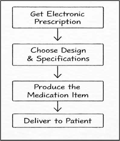
Figure 5: Process of 3D Printing
Elements Influencing Drug Release:
Standards of Quality:
Advantage & Disadvantage of FDM
Table 1: Advantage & Disadvantage of FDM
| Sr.no | Advantages | Disadvantages |
|---|---|---|
| 1 | Affordable and simple to use. | Thermal breakdown of drugs sensitive to heat. |
| 2 | Elevated accuracy and consistency. | Restricted compatibility of drug-excipient pairings. |
| 3 | Facilitates customized healthcare and adjusted medication level. | Problems related to filament (brittleness, changes in diameter). |
| 4 | Lowers the chances of human mistakes compared to conventional compounding. | Demands precise regulation of printing parameters. |
| 5 | Adept at creating intricate and multi-drug delivery mechanisms. | Printers with multiple nozzles for the production of various drugs. |
| 6 | Suitable for various pharmaceutical excipients and polymers. | Environments free from contamination. |
Semisolid Extrusion (SSE) 3D Printing in Drug Delivery
Semisolid extrusion (SSE) is an innovative additive fabrication method originating from fused deposition modelling (FDM) (8,31). Unlike FDM, which relies on thermoplastic filaments heated to high temperatures, SSE operates at or near room temperature by extruding semisolid materials through a controlled nozzle, building three-dimensional structures layer by layer (8,30). This temperature-sensitive method renders SSE especially appropriate for pharmaceutical uses, as it reduces thermal degradation of active pharmaceutical ingredients (APIs) (8,13). Nonetheless, keeping ideal temperature conditions is essential to avoid excessive softening, distortion, or loss of structural stability during printing (8).
SSE systems generally employ syringe-driven extrusion methods and can be classified into three primary categories (8,31):
Application
Facilitates the production of tablets featuring several compartments (16).
Supports:
Customized Pediatric Dosage Forms
Manufacturing of chewable tablets or gummies with adjustable (13,23,32):
Process of SSE 3D Printing (8,30):
Fig 6. shows step by step process of SSE 3D Printing
Material Preparation
Loading into Syringe System
Layer-by-Layer Deposition
Solidification and Shape Retention
Ensure the extruded filament retains its shape immediately.
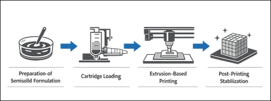
Figure 6: SSE 3D Printing
Advantage & Disadvantage of SSE
Table 2: Advantage & Disadvantage of SSE
| Sr.no | Advantages | Disadvantages |
|---|---|---|
| 1 | Facilitates tailored healthcare through individualized medication formulations. | Demands accurate rheological regulation of material. |
| 2 | Functions at low temperatures, safeguarding heat-sensitive medications. | Confined to semisolid formulations, limiting material variety. |
| 3 | Facilitates intricate shapes and multi-drug solutions. | Risks of shape distortion if viscosity or temperature is not properly adjusted. |
| 4 | Enables regulated drug release patterns (instantaneous + prolonged). | Reduced resolution in comparison to certain alternative 3D printing methods. |
| 5 | Affordable and fairly straightforward in comparison to alternative 3D printing techniques. | Challenges in forecasting filament growth or contraction. |
| 6 | Suitable for various pharmaceutical excipients and polymers. | Increasing industrial production continues to be difficult. |
Stereolithography (SLA) 3D Printing in Drug Delivery
Stereolithography (SLA) is a sophisticated and accurate additive manufacturing method based on photopolymerization, which transforms liquid photopolymer resins into solid forms through a concentrated ultraviolet (UV) light source. It was one of the earliest 3D printing technologies created and still stands as one of the most precise techniques available today for producing intricate shapes with exceptional surface quality (33). In SLA, a digital 3D model is converted into thin cross-sectional layers, and each layer is cured in sequence with a UV laser beam. The laser meticulously scans the surface of a liquid resin, triggering a chemical reaction that converts the resin into a solid polymer (33,34). A key feature of SLA is its remarkable accuracy and detail, with layer thickness generally between 0.01 and 0.02 mm and laser spot sizes ranging from 0.011 to 0.075 mm. This elevated level of precision enables the creation of microscale features, making SLA especially important in biomedical and pharmaceutical fields where accurate dosing, structure, and functionality are essential (33,35).
In contrast to thermal-based 3D printing techniques such as fused deposition modelling (FDM), stereolithography (SLA) operates at or near room temperature. This represents a major benefit in pharmaceutical sciences, as it stops the deterioration of thermolabile (heat-sensitive) medications, such as numerous biologics and peptides (35,36). As a result, SLA has emerged as a promising approach for advancing personalized medicine, enabling the customization of drug dosage forms to match individual patient needs in terms of dose, shape, size, and release profile (8,21,23).
Applications in Drug Delivery
Process of SLA 3D Printing (31,33):
Fig 7. shows step by step process of SLA 3D Printing.
Laser Scanning
Stratum Development
Polymerization Science
Layer beneath.
Post-Processing
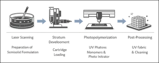
Figure 7: SLA 3D Printing
Advantage & Disadvantage of SLA
Table 3: Advantage & Disadvantage of SLA
| Sr.no | Advantages | Disadvantages |
|---|---|---|
| 1 | Creates components with less roughness in comparison to alternative 3D printing techniques. | Restricted Biocompatible Substances. |
| 2 | Appropriate for Temperature-Sensitive Medication. | Only a limited number of resins are authorized for pharmaceutical or internal medical applications. |
| 3 | Regulated Medication Dispersion. | Post-Processing Specifications. |
| 4 | Individualized Healthcare. | Extra processes such as curing and cleaning add to both time and expense. |
| 5 | Facilitates individualized dosage and personalized medication formulations. | Resins can be influenced by surrounding light and temperature, impacting their stability. |
| 6 | Aids in the production of: Polypills with multiple layers. | In systems with multiple drugs (polypills), diffusion of drugs between layers may take place. |
3D Inkjet Printing in Drug Delivery
Inkjet-based 3D printing is a sophisticated additive manufacturing technique that allows for the accurate and controlled application of materials as tiny droplets to create three-dimensional structures one layer at a time. Drawing from traditional 2D inkjet printing, this method removes the necessity for Molds, masks, or intricate tooling, rendering it especially ideal for quick prototyping and tailored manufacturing (30,37). Fundamentally, inkjet 3D printing functions by spraying minuscule droplets of liquid substances like pharmaceutical solutions, polymer mixtures, or adhesives onto a surface or powder layer with precision and consistency. These droplets harden upon deposition via processes like solvent evaporation, cooling, or chemical reactions, eventually creating a solid structure (37,38). The motion of either the printhead or the build platform guarantees precise spatial arrangement of each droplet, enabling exceptional accuracy in both shape and material makeup.
This technology can generally be divided into two primary approaches:
A key importance of inkjet-based 3D printing is its remarkable dosing precision, which is particularly vital in pharmaceutical uses. It enables precise control over the amount of active pharmaceutical ingredient (API) deposited in each layer, ensuring that the final dosage form closely matches the intended drug content. Research has shown that printed tablets frequently attain nearly optimal drug loading with little variation, emphasizing the consistency and dependability of the method (37,38). A significant benefit is its versatility in materials. Inkjet 3D printing is capable of using various materials, such as polymers (like polyvinylpyrrolidone), resins, ceramics, and inks intended for edible pharmaceuticals. This adaptability enables researchers and producers to customize formulations based on particular therapeutic requirements, like immediate or prolonged drug release (30,37).
Application
Oral Dosage Forms
Personalized Medicine
Systems for Regulated Drug Release
Oral dispersible Tablets (ODTs)
Systems Controlled by Morphology
SEM research indicates:
Procedure of Printing (Operational Mechanism) (37,38):
Fig 8. shows step by step process of Inkjet 3D Printing.
Droplet Deposition
The printhead ejects microdroplets of build material (e.g., photopolymer, wax, or binder) onto a substrate or powder bed with high precision.
Layer Formation
The deposited droplets solidify or cure upon contact, forming a thin, uniform layer that matches the digital design.
Layer-by-Layer Construction
The process repeats sequentially, depositing and solidifying new layers to build the complete 3D structure.
Post-Processing (Optional)
Steps such as curing, cleaning, or sintering may follow to enhance mechanical strength and surface finish.
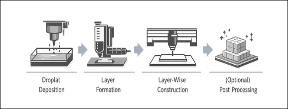
Figure 8: Inkjet 3D Printing
Advantage & Disadvantage of Inkjet Printing
Table 4: Advantage & Disadvantage of Inkjet Printing
| Sr.no | Advantages | Disadvantages |
|---|---|---|
| 1 | Elevated Precision and Exactness The drug content is in strong alignment with the expected values. |
Mechanical Fragility (Binder Jetting) Printed components are initially delicate (“green” condition). |
| 2 | Support Structures Not Required (Binder Jetting) The powder bed inherently supports the structure. |
Requirement for post-processing Heating process or infusion necessary to enhance durability. |
| 3 | Flexibility of Materials Suitable for polymers, ceramics, and pharmaceutical inks. |
Surface Texture Coarser surface in comparison to SLA or SLS. |
| 4 | Tailored Medication Dispersion Release profiles can be influenced by geometry and porosity. |
Inconsistency in Quality Assessment Parameters such as hardness and friability are not consistently assessed. |
| 5 | Rapid Printing Procedure Particularly in binder jetting (no heat necessary during printing). |
Porosity Issues Can influence medication release and structural integrity. |
Binder Jetting 3D Printing in Drug Delivery
Binder Jetting is a powder-based additive manufacturing method that originates from inkjet printing technology. It constructs three-dimensional items by selectively applying a liquid binding substance onto a layer of powder particles, one layer at a time (30,37). In contrast to other 3D printing techniques like laser systems, Binder Jetting avoids high temperatures during the printing process, which allows it to be used with materials sensitive to heat (9,37). During this procedure, a fine coat of powder (like metals, ceramics, polymers, or pharmaceutical excipients) is evenly distributed on a construction platform. A printhead subsequently travels across the surface and applies a liquid binder in designated areas following the digital design. The binder functions as an adhesive, connecting the powder particles (37).
A significant characteristic of Binder Jetting is that the unbound powder around the model inherently supports the structure, removing the requirement for extra support This enables the creation of intricate shapes and internal designs (37). Nevertheless, the printed components are initially delicate (“green parts”) and typically need post-processing like drying, sintering, or infiltration to attain sufficient mechanical strength (30,37).
Applications
Pharmaceutical Applications
Applications in Industry
Biomedical Uses
Process Binder Jetting (Powder-Based Inkjet Method) (9,21,23,37):
Fig 9. shows step by step process of Binder Jetting 3D Printing.
Powder Spreading
Binder Deposition
Layering
Part Formation
Post-Processing
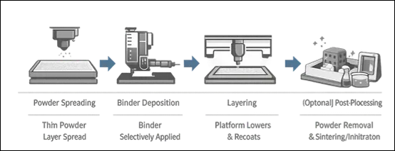
Figure 9: Binder Jetting 3D Printing
Advantage & Disadvantage of Binder Jetting
Table 5: Advantage & Disadvantage of Binder Jetting
| Sr.no | Advantages | Disadvantages |
|---|---|---|
| 1 | High Precision and Accuracy Drug content closely matches theoretical values. | Mechanical Weakness (Binder Jetting) Printed parts are initially fragile (“green” state). |
| 2 | No Need for Support Structures (Binder Jetting) Powder bed supports the structure naturally. | Need for post-processing Heat treatment or infiltration required to improve strength. |
| 3 | Versatility of Materials Compatible with polymers, ceramics, and pharmaceutical inks. | Surface Roughness Grainy texture compared to SLA or SLS. |
| 4 | Customizable Drug Release Geometry and porosity can control release profiles. | Variability in Quality Testing Parameters like hardness and friability not consistently evaluated. |
| 5 | Fast Printing Process Especially in binder jetting (no heat required during printing). | Binder Dependency High-viscosity binders may slow disintegration. |
Comparison of various 3D printing technologies
| Sr. no. |
Various techniques | Advantage over other technique |
Disadvantage over other technique |
Interpretation | Conclusion |
|---|---|---|---|---|---|
| 1 | FDM (Fused Deposition Modelling) | Cost-effective and widely accessible (13,22). Easy operation and scalability (22). Good for polypills and complex geometries (16,26). Strong mechanical properties (16,26). |
High temperature limits use with heat-sensitive drugs (inferior to SSE, SLA, Inkjet) (13,22). Filament preparation challenges (22). Limited drug–polymer compatibility (14). |
Worst for temperature sensitivity (14). Moderate Precision and accuracy (14). Best Mechanical strength (31,33). |
Economical, solid dosage forms (2). |
| 2 | SSE (Semisolid Extrusion) | Operates at low temperature (better than FDM, SLS) (30,31). Suitable for heat-sensitive drugs (30). Enables multi-drug and multi-layer systems (12). Flexible formulation options |
Lower resolution than SLA and Inkjet (8). Limited to semisolid materials (8). Shape deformation risks (8). |
Best for temperature sensitivity. Lower Precision and accuracy. Best Material flexibility (33,35). |
Heat-sensitive and pediatric drugs (13,22). |
| 3 | SLA (Stereolithography) |
Extremely high precision and surface finish (better than FDM, SSE) (33,35). No heat → suitable for thermolabile drugs (35,36). Excellent for microstructures(microneedles) (31,33). |
Limited biocompatible resins (33). Requires post- processing (33). Risk of drug diffusion between layers (35). |
Best for temperature sensitivity (37,38). best Precision and accuracy (9). Modrate Mechanical strength (9). Limited Material flexibility (37,38). |
high precision and microstructures (9). |
| 4 | SLS (Selective Laser Sintering) | No need for support structures Can produce complex internal geometries Good mechanical strength (9). |
High temperature → drug degradation (worse than SSE, SLA) Expensive equipment Limited pharmaceutical polymers (9,30). |
Worst for temperature sensitivity. Moderate Precision and accuracy. Best Mechanical strength. Limited Material flexibility (9,30). |
strong and complex structure (14). |
| 5 | Inkjet 3D Printing | Highest dosing precision (better than all) No heat required (37,38) Suitable for personalized medicine Fast and |
Weak mechanical strength (especially binder jetting) Nozzle clogging issues. Limited viscosity range (37). |
Weak mechanical strength (especially binder jetting) Nozzle clogging issues. Limited viscosity range (37). |
Precise dosing and drug release. |
| 6 | Binder Jetting | No heat → ideal for sensitive drugs. High yield speed. Complex porous structures possible. No support required (37). |
Very fragile initial (green parts). Requires post- processing (37). Surface roughness Binder dependency (37,38). |
1.Best for temperature sensitivity. 3.Lower Mechanical strength. |
porous structures and fast production (30,37). |
Table no. 6 comparison of various 3D printing technologies
MATERIALS:
Polycaprolactone (PCL; molecular weight 50,000 Da, melting point 58 °C) was sourced from Polysciences Inc. This molecular weight was chosen based on previous research demonstrating appropriate melt viscosity for 3D printing uses. Timolol maleate (TML), phosphate-buffered saline (PBS, pH 7.4), dexamethasone (pharmaceutical secondary standard), β-carotene (≥93% UV), sodium alginate, l-α-phosphatidylcholine (derived from soybeans), calcium chloride dihydrate, sodium carbonate, sodium bicarbonate, acetic acid, tartrazine, and penicillin–streptomycin were obtained from Sigma-Aldrich.
Photo None: Initiators Such as Phenyl (2,4,6-Trimethylbenzoyl) Phosphine Oxide (Irgacure 819) and Lithium Phenyl-2,4,6-Trimethylbenzoylphosphinate (LAP) Were Obtained from Sigma-Aldrich. Polyethylene Glycol 400 (PEG 400) and Polyethylene Glycol Diacrylate (PEGDA, Mn 575), Along with Polyvinyl Alcohol (PVA; Molecular Weight 89–98 kDa, >99% Hydrolyzed), Were Used as Received.
Dulbecco’s Modified Eagle’s Medium (DMEM) and fetal calf serum were obtained from Thermo Scientific (Gibco, UK). Dulbecco’s phosphate-buffered saline (PBS) was sourced from both Sigma-Aldrich and Fisher Scientific. CellTiter 96® Aqueous One Solution Reagent was supplied by Promega (Southampton, UK).
Trifluoroacetic acid (TFA, ≥99.0%, HPLC grade) and acetonitrile (ACN, ≥99.9%, HPLC grade) were employed for analytical purposes. Type A gelatin (gel strength 300) and methacrylic anhydride were procured from Sigma (St. Louis, MO, USA), while sodium hyaluronate (95%) was obtained from Acros Organics.
Moxifloxacin hydrochloride was supplied by Sa novel İlaç, and sterile distilled water was provided by Polavaram İlaç San. ve Tic. A.Ş. All reactions, solution preparations, and polymer purification processes utilized ultrapure water.
General Selection Criteria for Materials (All Technologies)
Safety & Biocompatibility:
Mechanical Properties
Capacity to facilitate:
Criteria Particular to Technology
Table 7: Material Selection Criteria for Various Technology
| 3D Printing Technique | Type of Material | Selection Standards | Obstacles / Benefits |
|---|---|---|---|
| Fused Deposition Modelling (FDM) | Thermoplastic polymers such as PVA, PLA, HPMC | • High thermal stability (extrusion temperature ~150–250°C) • Good melt flowability and viscosity • Ability to form filaments • Drug should be heat-stable |
Obstacles: • Not suitable for heat-sensitive drugs • Risk of drug degradation during extrusion |
| Stereolithography (SLA) | Photopolymerizable resins | • Photo-reactivity under UV light • Low photoinitiator toxicity • Biocompatibility of resin • Transparency for light penetration | Obstacles: • Limited pharmaceutically approved resins • Possible toxicity from residual monomers |
| Selective Laser Sintering (SLS) | Powdered polymers such as PVP blends, nylon | • Good powder flowability • Thermal stability against laser heat • Proper particle size distribution • Sintering without API degradation | Obstacles: • High-temperature exposure • Limited excipient options |
| Binder Jet / Drop-on-Demand Inkjet Printing | Powder beds with liquid inks | • Low viscosity for droplet formation • Proper surface tension • Drug–solvent compatibility • Fast drying capability | Obstacles: • Nozzle clogging • Drug instability in solution |
| Pressure-Assisted Printing / Semi-Solid Extrusion (SSE) | Hydrogels, gels, pastes | • Controlled viscosity • Shear-thinning behavior • Low-temperature processing compatibility | Benefits: • Suitable for heat-sensitive drugs • Wide range of excipients available |
Selection of excipients:
Table 8: Selection of Excipient
| Technology | Typical Excipients | Key Selection Criteria | Limitations |
|---|---|---|---|
| FDM | HPMC, PVA, HPC, PEG | Thermal stability, plasticity | Heat-sensitive drugs |
| SLA | Photocurable resins, photo initiators | Cure under UV, low toxicity | Limited approved excipients |
| SLS | PVP, lactose, mannitol | Powder flow, thermal stability | High temp, limited excipients |
| Inkjet | PVP, HPMC, water/ethanol | Low viscosity, fast drying | Nozzle clogging, API stability |
| SSE | Alginate, HPMC gels, thickeners | Shear-thinning, shape retention | Mechanical strength |
| DPE | Plasticizers, binders, fillers | Flow, compressibility | Extrudate consistency |
Suitable method:
A conclusion that sums it all up-3D printing, especially techniques like FDM, SLA, SLS, and inkjet printing, Binder jetting is truly a game-changer for ophthalmic drug delivery. Inkjet-based 3D printing is often considered the most pharmaceutically optimal because it combines several critical advantages. It contains a higher dose accuracy. Drug content closely matches theoretical values essential for safety and efficacy. It Operates at room temperature and Ideal for heat-sensitive drugs (proteins, peptides, vaccines). Inject printing having a better than the FDM& SLS. Mainly Excellent for personalized medicine & easily adjustable dose for each patient. Inkjet printing Suitable for on-demand manufacturing in hospitals/pharmacies. It mainly useful to design immediate and sustained release products. Inject printing was Rapid production.
FDM is simply and widely available technique at low cost 7 easy to use. SSE, SLA, Inkjet these techniques are very suitable for the heat sensitive drugs. More accurate and precise result can be obtained in the SLA. Pediatric and flexible dosage form are prepared by using SSE technique
CONCLUSION
Ocular drug delivery is widely recognized as one of the most complex areas in pharmaceutical research, largely due to the eye’s intricate anatomical and physiological barriers. Conventional ophthalmic formulations often suffer from low bioavailability and require repeated dosing, which can negatively impact patient compliance. Ocular inserts offer a promising solution by enabling sustained and controlled drug release directly at the ocular surface. The incorporation of advanced fabrication methods, particularly 3D printing, has further strengthened the potential of these systems.
3D printing provides precise control over device geometry, drug distribution, and internal structure, making it possible to design highly tailored ocular inserts with enhanced therapeutic outcomes. Recent studies have demonstrated the feasibility of employing different additive manufacturing techniques to produce ocular drug delivery devices capable of improving bioavailability and maintaining prolonged drug release. However, challenges remain, including the identification of suitable printable materials, navigating regulatory requirements, and scaling up production for widespread use.
Despite these hurdles, continuous progress in additive manufacturing and biomaterials is expected to broaden the scope of 3D printing in ophthalmic drug delivery. In summary, the integration of 3D printing with ocular drug delivery systems represents a forward-looking approach to developing next-generation ophthalmic therapies. Ongoing innovation in this field may ultimately pave the way for personalized and highly effective treatments for a variety of ocular diseases.
Acknowledgement
We would like to thanks All the teaching and non-teaching staff of the Pharmaceutics Department of Adarsh College of Pharmacy, Vita. We also thanks to Adarsh College of Pharmacy, Vita for providing instrumental facilities.
REFERENCES







Anurag Mangale, Rushikesh Jangam, Aniket Katkar, Vaibhav Patil, Pruthviraj Jagtap, Adesh Magar, Prajwal Gaikwad, 3D Printing Technology in the Design and Development of Ocular Inserts for Advanced Ophthalmic Drug Delivery Systems, Int. J. of Pharm. Sci., 2026, Vol 4, Issue 11, 2403-2422. https://doi.org/10.5281/zenodo.24276286
 10.5281/zenodo.24276286