

We use cookies to ensure our website works properly and to personalise your experience. Cookies policy
Department of pharmacology, Indira Gandhi Institute of Pharmaceutical Sciences, perumbavoor.
Inflammation is a protective physiological response to harmful stimuli, including infection and tissue injury. However, excessive or persistent inflammation may contribute to the pathogenesis of various diseases. Although several anti-inflammatory agents are available, their prolonged use may be associated with adverse effects. Consequently, natural compounds with potential anti-inflammatory properties have attracted considerable research interest. Icaritin is a naturally occurring prenylated flavonoid derived from Epimedium species and has been reported to exhibit several pharmacological activities, including antioxidant, anticancer, and anti-inflammatory effects. The present study aimed to evaluate the cytotoxic and anti-inflammatory activities of serving as the standard drug. Anti-inflammatory activity was evaluated using the protein denaturation inhibition assay, with aspirin as the reference standard. Icaritin demonstrated an IC50 value of 107.47 µg/mL in the MTT assay. In the protein denaturation assay, icaritin exhibited 69.31 ± 2.45% inhibition at a concentration of 100 µg/mL, compared with 76.21 ± 3.14% inhibition observed with aspirin.These findings indicate that icaritin possesses promising preliminary anti-inflammatory activity. Further studies are warranted to elucidate its mechanism of action and assess its therapeutic potential.
INTRODUCTION TO INFLAMMATION
Inflammation is the local response to the mammalian tissues to injury due to any agents.it is a body defense reaction in order to eliminate or limit the speed of injurious agents, followed by removal of necrotized cells and tissues. Inflammation will elicit vascular and cellular changes that serve to eradicate the underlying cause of damage, remove unhealthy tissue caused by the injury/disease causing inflammation, and promote healing. In addition to being the body’s natural response to substances it considers potentially harmful,inflammation also has distinct signs/symptoms. Inflammation may cause by some agents such as:[1]
• Infective agents
• Immunological agents
• Physical agents
• Chemical agents
• Inert materials
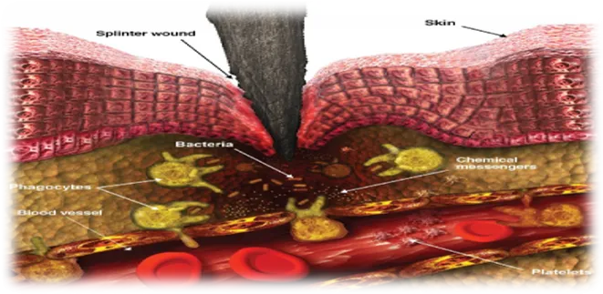
Figure None: 01 Inflammation
SIGNS OF INFLAMMATION:
There are four cardinal signs of inflammation as;
Rumor (redness)
Tumor (swelling)
Calor (heat)
Dolor (pain)
CAUSES AND TRIGGERS OF INFLAMMATION
Inflammation can be initiated by a wide range of harmful or potentially harmful agents. Infective causes include bacteria, viruses, fungi and parasites. Components of microorganisms may be recognised by immune cells as signals of infection and can activate inflammatory pathways. The response helps recruit immune cells and antimicrobial substances to the affected region. The type and severity of inflammation may vary according to the organism, the site of infection and the host's immune condition.[2]
Physical causes include mechanical injury, cuts, burns, excessive heat or cold, radiation and other forms of tissue damage. Chemical irritants, toxic substances and certain drugs may also injure cells and initiate inflammation. In these situations, damaged cells release endogenous signals that alert nearby immune and vascular cells. Foreign or inert materials, such as particles that cannot be readily removed, may produce a persistent local reaction.[3]
A single inflammatory episode may involve more than one trigger. For example, tissue injury can be followed by microbial infection, while a foreign material may cause both mechanical irritation and an immune response. Identifying the initiating cause is important because treatment should address the source of inflammation whenever possible, in addition to controlling symptoms. The categories of triggers described here follow the broad classification used in the supplied project document.[4]
TYPES OF INFLAMMATION
There are several different stages involved in the inflammatory response that includes the different time frames that each of these responses may take along with several factors related immunological status. This overall inflammation process may be classified into either an acute or chronic inflammation.
Acute Inflammation
It is a rapid and usually short-duration of reaction to tissue injury or infection. It commonly begins within a short period or hours after the harmful stimulus. Its main features are changes in small blood vessels, movement of fluid and plasma proteins into tissues, and recruitment of leukocytes, particularly neutrophils. The purpose is to deliver protective components to the site and remove the initiating cause as quickly as possible.[5]
The early vascular response includes brief changes in vessel tone followed by vasodilation. Blood flow increases, which contributes to redness and heat. Endothelial cells lining the blood vessels become more permeable, allowing protein-rich fluid to enter the tissue. This fluid accumulation produces swelling and can dilute harmful substances. The movement of leukocytes from blood vessels into tissues is called leukocyte recruitment or extravasation.[6]
Neutrophils are commonly among the first circulating cells to reach an acute inflammatory site. They move towards chemical signals, adhere to the vascular endothelium, pass through the vessel wall and migrate towards the source of injury. They can engulf microorganisms and cellular debris through phagocytosis and release substances that help destroy harmful agents. Monocytes may subsequently enter tissues and develop into macrophages, which contribute to clearance and repair.[6]
Acute inflammation may end in complete resolution when the stimulus is removed and tissue damage is limited. In other situations, it may lead to abscess formation, healing with scar tissue or progression to chronic inflammation. The final outcome depends on the nature of the injury, the extent of tissue destruction and the ability of the tissue to regenerate. Although acute inflammation is protective, an uncontrolled systemic response can become harmful.[7]
Chronic Inflammation
Chronic inflammation is a prolonged inflammatory response that may continue for weeks, months or years. It can develop when the initiating agent is not eliminated, when repeated injury occurs or when immune regulation remains abnormal. Persistent infections, long-term exposure to irritants, autoimmune reactions and some metabolic disturbances can contribute to chronic inflammation. Unlike acute inflammation, which is often dominated by neutrophils, chronic inflammation commonly involves lymphocytes, plasma cells and macrophages.[8]
Macrophages have several important functions in chronic inflammation. They engulf foreign material and damaged cells, release cytokines and other mediators, and influence tissue repair. Lymphocytes regulate immune responses through cell-to-cell interactions and soluble signals. When these cells remain active for a long period, the mediators they release may cause continuing tissue injury. At the same time, repair processes may be activated, resulting in the formation of new connective tissue and, in some cases, fibrosis.[9]
Treatment of chronic inflammatory disorders often requires control of the underlying cause and careful regulation of immune activity. Long-term therapy must balance disease control against adverse effects, particularly when drugs suppress protective immune functions. This need for effective and safer treatment options provides a rationale for investigating compounds with potential anti-inflammatory activity.[10]
INFLAMMATORY MEDIATORS
Inflammatory mediators are chemical substances that initiate, amplify, regulate or resolve the inflammatory response. They may be released by immune cells, damaged tissues, platelets or vascular cells, or generated from inactive precursors present in plasma. Their actions include changing blood vessel diameter and permeability, attracting leukocytes, producing pain and fever, and regulating tissue repair. The response depends on the type, amount and timing of mediator release.[11]
Histamine is stored mainly in mast cells and is released during several forms of tissue irritation and immune activation. It can produce vasodilation and increased vascular permeability. Bradykinin contributes to pain and vascular changes. Prostaglandins are lipid mediators formed from arachidonic acid through cyclooxygenase enzymes. They participate in pain, fever and vascular responses.[12]
Cytokines are small signalling proteins that coordinate communication between immune and other cells. Tumour necrosis factor-alpha (TNF-α) and interleukin-6 (IL-6) are important pro-inflammatory cytokines. They can influence immune-cell activation, acute-phase responses and the expression of other inflammatory mediators. Interleukin-1 beta (IL-1β) is another mediator involved in inflammatory signalling and fever. Chemokines form a group of signalling proteins that guide leukocytes towards sites of inflammation.[13]
Nitric oxide is a signalling molecule that can have different effects depending on its source and concentration. During inflammation, inducible nitric oxide synthase may produce increased amounts of nitric oxide. Excessive production can contribute to tissue injury under certain conditions. Measurement of mediators such as TNF-α, IL-6, IL-1β and nitric oxide can help researchers understand whether a test compound modifies inflammatory activity.[14]
MOLECULAR PATHWAYS INVOLVED IN INFLAMMATION
Inflammation is regulated by interconnected molecular pathways rather than by a single mechanism. When cells detect microbial components or signals released from injured tissues, intracellular signalling proteins are activated. These signals may reach the nucleus and alter the expression of genes involved in cytokine production, enzyme activity, cell survival and immune-cell recruitment. Understanding these pathways helps explain how anti-inflammatory medicines produce their effects.[15]
The nuclear factor-kappa B (NF-κB) pathway is an important regulator of inflammatory gene expression. Inactive cells, NF-κB is retained in a resting state by inhibitory proteins. Appropriate inflammatory stimulation activates signalling events that allow NF-κB to enter the nucleus, where it can promote the transcription of genes encoding cytokines, adhesion molecules and inflammatory enzymes. Excessive or prolonged NF-κB activity is associated with several inflammatory disorders. Compounds that reduce this pathway may therefore lower the expression of selected inflammatory mediators.[15]
Mitogen-activated protein kinase (MAPK) pathways, including p38, extracellular signal-regulated kinase (ERK) and c-Jun N-terminal kinase (JNK), also regulate cellular responses to stress and inflammatory stimuli. Their activation can influence cytokine production, cell differentiation and survival. The pathways interact with NF-κB and other signalling systems, so the final response depends on the combination and duration of signals received by the cell.
Arachidonic acid metabolism is another major pathway. Phospholipase A2 releases arachidonic acid from membrane phospholipids. Cyclooxygenase enzymes convert arachidonic acid into prostanoid precursors, which are further converted into prostaglandins and thromboxanes. Lipoxygenase enzymes produce leukotrienes and related compounds. Drugs that inhibit cyclooxygenase reduce prostaglandin formation, while corticosteroids act at an earlier level and influence several inflammatory processes. These mechanisms provide important targets for anti-inflammatory drug development.[16]
Currently Available Anti-Inflammatory Agents
A number of drugs can be purchased without a prescription for inflammation. They include over-the-counter non-steroidal anti-inflammatory medications (NSAIDs) such as ibuprofen, naproxen, and aspirin, and prescription NSAIDs include celecoxib, diclofenac and indomethacin. While NSAIDs are effective for inflammation, severe acute inflammatory conditions may be treated with corticosteroid medications and/or topical therapies (e.g., diclofenac gel) for localized pain.
More recently, specific drugs targeting inflammation, such as monoclonal antibodies and small molecule inhibitors, have been developed. Drugs targeting tumor necrosis factor-α (TNF-α), interleukins, Janus Kinase (JAK) signaling pathway, and others have shown great success in treating several immune-mediated conditions. However, there are drawbacks to these drugs such as their high price, poor effectiveness in some cases, immunosuppressive nature, side effects, and the development of resistance against these drugs.[17]
Non-Steroidal Anti-Inflammatory Drugs (NSAIDs)
They reduce inflammation and pain by blocking cyclooxygenase enzymes; they may also reduce fever.
Corticosteroids (Steroids)
Corticosteroids can be used to treat severe chronic inflammatory conditions and acute inflammation (especially due to autoimmune disease), For example, steroids such as prednisone, cortisone and methylprednisolone are often prescribed for severe inflammation.
Additional Anti-Inflammatory Medications
Topical therapy options include the use of diclofenac patches (Flector patches) or diclofenac gel for localized sprains/strains/arthritis.
Supplemental Enzymes - bromelain, trypsin and rutoside in combination (available as enzyme tablets).
DMARDs (disease-modifying anti-rheumatic drugs) and biologics - used for chronic inflammatory arthritis (e.g., Rituximab and Golimumab).[17]
MOLECULAR MECHANISM OF ANTI-INFLAMMATORY AGENTS
Anti-inflammatory agents work by interfering with the molecular pathways involved in the inflammatory response to reduce inflammation and its associated symptoms. These pathways include: inhibition of the production of molecules and signaling proteins involved in inflammation such as prostaglandins and cytokines; inhibition of the transcription factor NFκB that controls the expression of many inflammatory genes; and blockade of the cyclooxygenase enzymes, which reduce the production of prostaglandins through the inflammatory response and effects of pain, growth and sell repair. A better understanding of the role of these pathways and their specific mechanisms has led to safer and more effective treatment options for inflammatory diseases.[18]
The release of chemicals from tissue and cell migration is the cause of inflammation. Prostaglandins (PGs) and leukotriene’s (LTs) as well as histamine, bradykinin, and now platelet activating factor (PAF) and interleukin-1 are primarily responsible for causing inflammation. Evidence supporting this includes both competitive receptor blockers and inhibitory agents of their synthesis.[19]
NSAIDs And Their Pharmacological Action
Non-steroidal anti-inflammatory drugs are extensively used medicines for pain and inflammatory symptoms. Their principal mechanism is inhibition of COX-1and COX-2 enzymes, which are required for the metabolism of arachidonic acid into prostaglandin precursors. Prostaglandins contribute to sensitisation of pain pathways, fever and local inflammatory changes. Reducing their synthesis can therefore decrease pain, fever and swelling.[20]
Cyclooxygenase-1 (COX-1) is expressed in many tissues and contributes to physiological functions such as protection of the gastric mucosa, regulation of platelet activity and maintenance of renal blood flow. Cyclooxygenase-2 (COX-2) is often increased in response to inflammatory stimuli, although it also performs normal physiological roles in certain tissues. Inhibition of COX-2 can reduce inflammatory prostaglandin production, while inhibition of COX-1 may contribute to some unwanted effects, including gastrointestinal irritation and altered platelet function.[21]
Aspirin is an important NSAID that irreversibly acetylates cyclooxygenase enzymes. Other commonly used NSAIDs generally inhibit the enzymes reversibly. The pharmacological response varies with dose, duration of treatment, drug distribution and individual patient characteristics. In laboratory studies, aspirin is frequently used as a reference standard to compare the activity of a test substance in selected anti-inflammatory assays.[22]
Although NSAIDs provide useful symptomatic relief, they do not necessarily remove the underlying cause of inflammation. Their use should be based on an assessment of expected benefit and potential risk. This is particularly important in individuals with a history of peptic ulcer disease, kidney impairment, cardiovascular disease or other factors that may increase the chance of adverse effects.[23]
Corticosteroids And Targeted Anti-Inflammatory Therapy
Corticosteroids are potent anti-inflammatory medicines that influence several stages of the inflammatory response. After entering cells, they bind to glucocorticoid receptors. The activated receptor complex can alter gene transcription and regulate the release of inflammatory proteins.
These actions may reduce cytokine release, inflammatory enzyme expression, leukocyte migration and other immune responses. Their broad activity makes them beneficial for selected conditions where inflammation is severe or driven by abnormal immune activity.[24]
The benefits of corticosteroids must be considered alongside their potential adverse effects. Prolonged systemic use may be associated with metabolic changes, increased infection risk, bone loss, changes in blood pressure and suppression of the body's own corticosteroid production. Local preparations can reduce systemic exposure in some situations, but prolonged or inappropriate use may still produce local and systemic complications. The dose and duration should therefore be carefully selected.[24]
Targeted biological medicines and small-molecule inhibitors have been developed to interfere with particular inflammatory signals. Examples include medicines that block TNF-α, selected interleukins or intracellular enzymes involved in cytokine signalling. Targeted approaches may provide disease control when conventional medicines are insufficient. However, they may be expensive, may not work equally well in all patients and can alter protective immune responses.[24]
The availability of several drug classes demonstrates that inflammation can be modified at different points in its biological pathway. At the same moment, no single medicine is appropriate for every inflammatory condition. Research into additional agents is therefore important, particularly when the aim is to identify compounds with useful activity, acceptable safety and mechanisms that may complement existing treatment approaches.[25]
LIMITATIONS / DRAWBACKS OF CURRENT THERAPY
NSAIDs are a popular option to help manage arthritis pain and pain from cancer or other medical issues, both acute and chronic. They can be used alone for mild or moderate pain, or in combination with other medications (opioids or adjuvant) for treating severe pain. Unfortunately, the non-selective inhibition of the cyclooxygenase enzymes by NSAIDs creates a significant risk of gastrointestinal, hematologic, and nephrotoxic injury from these drugs after only short or long use, respectively. High-risk patients for NSAID adverse effects are those patients with acute and persistent pain related to serious or life-threatening medical illness (i.e., cancer, HIV/AIDS) because there is an increased risk for having an adverse effect from NSAIDs due to the occurrence of gastrointestinal bleeding, therefore masking the opportunistic infections related to the antipyretic effects of NSAIDs, and possibly leading to life-threatening complications for neutropenia, thrombocytopenic, or otherwise at risk individuals.[26]
Depending on disease severity, patients may receive various doses of oral and/or topical corticosteroids to control the underlying inflammation due to their pharmacological properties. Most patients can be treated with sufficient topical corticosteroid treatment to allow for either a decrease or complete withdrawal of systemic corticosteroids. Where long-term treatment has occurred using high-potency topical corticosteroids, the negative effects of treatment frequently outweigh any possible treatment benefit. Negative effects associated with topical corticosteroid usage may be experienced at varying degrees of severity; only those patients with sufficient knowledge of past and present topical steroid usage are able to provide valid reporting on the occurrence of specific negative effects.
Non-Steroidal Anti-Inflammatory Drugs (NSAIDs) decrease inflammation and pain by blocking cyclooxygenase (COX) formation of prostaglandins, which act to produce fever and pain as well as preserving blood flow to the kidney, creating a delicate physiological balance. This blockade can result in excessive inhibition of prostaglandin formation, resulting in decreased protection of the gastrointestinal tract (gastric irritation, ulcers, and bleeding), decreased renal blood flow (resulting in kidney damage and possible kidney failure), promotion of hypertension and heart attack from promotion of thromboxane production resulting in clot development, bronchospasm, dizziness, headache, and confusion, in high enough concentrations, such as in overdose or prolonged use, multi organ toxicity can occur,exact cause and effects are unknown.[27]
INTRODUCTION TO ICARITIN
Icaritin is a flavonoid type compound that belongs to the prenylated flavonoids which have been gaining popularity among researchers in recent years for a number of medicinal uses. Icaritin derived from epimedium is primarily obtained from harvesting leaves of the plant.Icaritin is yellowish in color with a crystalline powder consistency and is highly soluble in organic solvents such as methanol and ethanol.[28]
Icaritin has various effects on living organisms such as anticancer effects, immunomodulatory properties, osteogenic activity, antioxidant effects, and anti-inflammatory effects. Various clinical drug possibilities also exist, even though they have some disadvantages such as low bioavailability and poor solubility, and will continue to be viable opportunities for development of naturally sourced drugs to treat malignancies and nflammatory disorders. The goal of this review is to update and expand the current knowledge of Icaritin and provide a foundation for future research into the use of Icaritin.[28]
Icaritin, chemically known as 3, 5, 7-trihydroxyflavone, is a flavonoid compound derived from Herba Epimedii, which is frequently used in East Asian traditional medicine and has been characterized as a major active ingredient of Icariin. The pharmacological effect of Icaritin is documented in several reports to have a powerful inhibition effect on various cancer cells including human cervical, liver, prostate, and breast carcinoma cells. Besides the above mentioned anticancer effect, it has also showed a reduction in steroid-associated osteonecrosis. In another research, it has been found to have an immunosuppressive effect on T cells.
The pharmacokinetics of oral administration of Icaritin indicates a half-life of 8.3 ± 1.0 h, which is much longer than previous reports of other flavones. The long half-life indicates a potential pharmacokinetics within a therapeutically range which means a big potential for pharmaceutical products based on Icaritin in the medical field.
In addition, Icaritin has been well found to be distributed in liver and excreted via urine. In case of intraperitoneal administration, it shows a half-life of 3.14 ± 0.34 h and is well distributed in kidneys and liver indicating low systemic clearance and long half-life, making it a suitable candidate drug to treat for instance osteonecrosis.
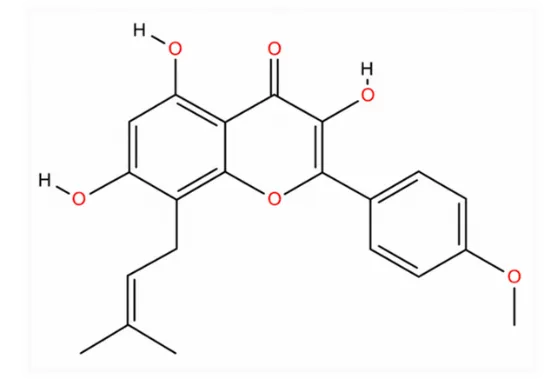
Figure None: 02 Chemical Structure
Overall, based on the long half-life and good pharmacokinetics of Icaritin, it can be concluded that this compound has great potential of treatment effect of diseases.[29]
Pharmacological Action Icaritin:
Lungs:
Icaritin, an active bioflavonoid compound derived from Epimedium, may exhibit both prophylactic and therapeutic efficacy against pulmonary fibrosis in murine models. The drug acts by up regulating PPARγ to activate antifibrotic signaling, in turn leading to reduced collagen deposition and ultimately decelerated progression of fibrosis in the lung parenchyma. This study seeks to further assess the role of Icaritin in regulating pulmonary fibrosis and delineate its potential as a remedial candidate for pulmonary fibrosis to serve as a basis for future applications of Icaritin in treating pulmonary fibrotic disorders and other fibrotic diseases.
Icaritin can ameliorate pulmonary fibrosis through up regulation of PPAR-γ and blockade of the TGFβ signaling pathway. The up regulation of PPAR-γ and blockade of the TGFβ pathway correlate to enhanced autophagy in lung tissues, leading to reduced pulmonary fibrosis and decreased collagen deposition. These findings provide insight into the underlying mechanism of action of Icaritin.[30]
Liver:
Icaritin has a significant hepatoprotective effect. Efficacy tests showed that Icaritin can significantly reduce the level of serum aspartate aminotransferase and alanine aminotransferase, suppress the activation of HSC and limit the deposition of collagen in the fibrotic liver. The apoptosis of activated HSCs can be promoted by Icaritin, which makes the profibrotic HSC disappear and leads to the resolution of fibro genesis. Western blot results show that Icaritin down-regulates the expression of Bcl-2, and up-regulates Bak-1, Bmf and Bax to promote the mitochondrial apoptosis of activated HSCs. This research provides a solid theoretical basis for the study of the molecular mechanism of the liver protective effect of Icaritin and lays a foundation for the further development of Icaritin as the specific auxiliary medicine for the treatment of liver lesions.[31]
Skin:
Icaritin activates AMPK pathway; inhibits Wnt/β-catenin pathway. The AMPK pathway promotes skin cell apoptosis; the Wnt/β-catenin pathway promotes proliferation and inhibits apoptosis. The inhibition of β-catenin leads to the inhibition of Wnt/β-catenin pathway and promotes the apoptosis of skin keratinocytes, thereby alleviating skin lesions. The activation of AMPK leads to an increase in Th cells, thereby promoting the expression of IL-12 and inhibiting the expression of IL-4.
Various studies conducted (in-vitro & in-vivo):
Icaritin (ICT), as an important bioactive constituent of Herba Epimedii, has been shown to both inhibit neuroinflammation and regulate microglial polarization in the brain. However, it has yet to be determined whether it can regulate microglial polarization in the treatment of cerebral ischemia. Additionally, the specific mechanism of ICT involved in microglia polarization has not been fully disclosed.[32]
This study aimed to explore the role of ICT in ameliorating microglial activation and the underlying mechanisms involved, so as to provide possible therapeutic strategies for treatment of acute ischemic stroke. The in vitro and in vivo methods were employed to establish the transient middle cerebral artery occlusion (tMCAO) rat model. The behavior recovery was evaluated with Garcia scale and rotarod test. The brain infarct was assessed with Nissel staining and the infarct volume was measured.[33]
The inflammatory mediators were performed with enzyme-linked immunosorbent assays (ELISAs). The expressions of GPER, ERK and p-EPK, NF-κB and pro-inflammatory cytokines as well as anti-inflammatory cytokines were performed with western blotting. In addition, immunofluorescence staining and quantitative PCR (qPCR) were performed to detect the expression of GPER, TNF-α and IL-10 in microglial cells. This study demonstrated that ICT ameliorated the activity of microglial cells and microglial-mediated neuroinflammation following cerebral ischemia through the regulation of microglial cell polarization by targeting the GPER-ERK-NF-κB signaling pathway and it also revealed a novel pharmacological mechanism of ICT in cerebral ischemia. Our data suggested that ICT may be a promising therapeutic strategy for the treatment of cerebral ischemia through modulation of microglial polarization.[34]
Background Cerebral ischemic stroke is a serious disease associated with neurodegeneration, but no effective treatment is available. Therefore, an effective treatment would be beneficial to lessen complications and improve the quality of life after acute ischemic strokes.[35]
Methods In the present study, the effect of ICT treatment on cerebral infarct volume and inflammatory responses was first evaluated by specific assays in vivo and in vitro. The mechanisms involved in these effects were examined.[36]
Results ICT treatment decreased cerebral infarct volume, reduced brain water content, and lessened the fluorescence intensity of FJC. In addition, ICT treatment improved Garcia score, increased latency to fall and rotation speed in rotarod test, and decreased IL-1β, TNF-α, Iba1, CD40, CD68, and p-P65-NF-κB, and increased mRNA expression levels of IL-10 and IL-4, and protein levels of CD206 and p-ERK. However, these changes were antagonized with inhibition of GPER and ERK signaling pathway.
Conclusion it is concluded that ICT treatment not only inhibited microglial activation but also promoted M2 polarization through GPER-ERK-NF-κB signaling pathway, contributing to the amelioration of cerebral ischemic stroke complications.[37]
PLAN OF WORK
AIM AND OBJECTIVES
AIM:
Pharmacological evaluation of the anti-inflammatory activity of Icaritin-a natural Isoflavone.
Objective:
1.To study the cytotoxic effect of Icaritin by MTT assay using raw human tumor (HEK293) cell lines.
2.To study the anti-inflammatory activity of Icaritin by protein inhibition assay.
REVIEW OF LITERATURE
1. Song et al. (2026) reviewed the therapeutic potential of Epimedium and its bioactive compounds, including Icaritin, in respiratory diseases. The review highlighted their anti-inflammatory, antioxidant, immunomodulatory, and anti-fibrotic properties, emphasizing their ability to regulate multiple signaling pathways. The authors concluded that these compounds show promise as potential therapeutic agents for respiratory disorders.
2. Che et al. (2026) developed a nanoparticle system combining tanshinone IIA and Icaritin to enhance anti-PD-1 immunotherapy in melanoma lung metastasis. The study demonstrated that the formulation promoted vascular normalization, restored tumor-infiltrating T-lymphocyte function, and improved the therapeutic efficacy of anti-PD-1 treatment. These findings suggest that Icaritin-based combination therapy has promising potential in cancer immunotherapy.
3. Su et al. (2025) investigated the therapeutic effects of Icaritin on inflammation and oxidative stress. The study demonstrated that Icaritin reduced inflammatory responses and oxidative damage by regulating key cellular signaling pathways, highlighting its potential as a promising therapeutic agent for inflammation-related diseases.
4. Zhao et al. (2025) reviewed the pharmacological properties and molecular mechanisms of Icariin and its related metabolites, including Icaritin, in fibrosis management. The review highlighted their anti-inflammatory, antioxidant, and anti-fibrotic activities, emphasizing that these compounds regulate multiple signaling pathways to reduce tissue fibrosis and inflammation.
5. Huang et al. (2024) investigated the therapeutic effects of Icaritin in recurrent spontaneous abortion in mice. The study found that Icaritin restored the balance between Treg and Th17 cells by regulating the TGF-β/SMAD signaling pathway, thereby reducing inflammation and improving pregnancy outcomes. These findings highlight the immunomodulatory and anti-inflammatory potential of Icaritin.
6. Alharbi et al. (2023) conducted a cross-sectional study to assess patients' knowledge and use of oral non-steroidal anti-inflammatory drugs (NSAIDs) among individuals with rheumatic disorders in Saudi Arabia. The study found that although NSAIDs were widely used for pain management, many patients had limited knowledge regarding their proper use and potential adverse effects, highlighting the need for improved patient education and safe NSAID use.
7. Bi et al. (2022) reviewed the anti-inflammatory and immunoregulatory effects of Icariin and Icaritin. The review highlighted that these compounds suppress inflammatory cytokines, regulate immune cell function, and modulate key signaling pathways such as NF-κB, MAPK, and PI3K/Akt. The authors concluded that Icariin and Icaritin have significant therapeutic potential for the treatment of inflammatory and immune-related diseases.
8. Yu et al. (2022) evaluated the anti-inflammatory effects of Icaritin in a rat model of cerebral ischemia. The study showed that Icaritin reduced neuroinflammation by promoting microglial polarization through the GPER-ERK-NF-κB signaling pathway. These findings indicate that Icaritin has potential as a therapeutic agent for neuroinflammatory and ischemic brain diseases.
9. Mohan et al. (2019) described inflammation as a protective response of vascularized tissues to injury or infection. The textbook explains the mechanisms of acute and chronic inflammation, the role of inflammatory mediators, and the processes involved in tissue repair, providing a fundamental understanding of inflammatory diseases.
MATERIALS AND METHODS
LIST OF MATERIALS AND EQUIPMENT’S USED IN MTT ASSAY
| SL.NO | CHEMICALS | BIOLOGICAL MATERIALS |
CONSUMABLES | EQUIPMENTS |
|---|---|---|---|---|
| 1 | MEM (Minimum Essential Medium) | Fetal Bovine Serum (FBS) | 96-well microtiter plate | CO₂ incubator |
| 2 | L-glutamine | HEK293 human cell line | Cell culture flasks | laminar airflow cabinet |
| 3 | Balanced Salt Solution (BSS) | Sterile pipette tips | Inverted tissue-culture microscope | |
| 4 | Sodium carbonate (Na₂CO₃) | Serological pipettes | Micropipettes | |
| 5 | Non-essential amino acids | Microcentrifuge/collection tubes | Multichannel pipette (if available) | |
| 6 | Sodium pyruvate | Sterile culture medium containers | Microplate reader | |
| 7 | Glucose | Water bath | ||
| 8 | Penicillin | Freezer | ||
| 9 | Streptomycin | |||
| 10 | MTT (3-(4,5-dimethylthiazol-2-yl)-2,5-diphenyltetrazolium bromide) | |||
| 11 | DMSO (Dimethyl sulfoxide) |
LIST OF MATERIALS AND EQUIPMENTS USED IN PROTEIN DENATURATION PREVENTION ASSAY
| SL; NO | CHEMICALS | SOLUTIONS | CONSUMABLES | EQUIPMENTS |
|---|---|---|---|---|
| 1 | Bovine Serum Albumin (BSA) | BSA solution0.4% (w/v) | Test tubes / assay tubes | Water bath – for heating at 72°C |
| 2 | Sodium chloride (NaCl) | Tris Buffered Saline (TBS) – 0.05 M Tris + 0.15 M NaCl, pH 7.6 | Micropipette tips | UV-Visible spectrophotometer – absorbance at 660 nm |
| 3 | Glacial acetic acid | Different concentrations of test samples | Pipettes | pH meter |
| 4 | Dimethyl sulfoxide (DMSO) – negative control | DMSO control | Cuvettes | Micropipettes |
| 5 | Aspirin – positive control | Aspirin control | Measuring cylinder | Thermometer |
| 6 | Test extracts / isolated compounds | Beakers | Test tube rack | |
| 7 | Distilled water |
METHODOLOGY
1.Cytotoxicity property of extract samples:
Cell culture technique
The Human tumor cell lines HEK293 cells were purchased from the National Centre for Cell Sciences (NCCS), Pune, India. Cells were maintained in MEM supplemented with 2 mM l-glutamine and balanced salt solution (BSS) adjusted to contain 1.5 g/L Na2CO3, 0.1 mM nonessential amino acids, 1 mM sodium pyruvate, 2 mM l-glutamine, 1.5 g/L glucose, 10 mM HEPES (4-(2-hydroxyethyl)-1-piperazine ethane sulfonic acid) and 10% fetal bovine serum (GIBCO, USA). Penicillin and streptomycin (100 IU/100µg) were adjusted to 1 mL/L. The cells were maintained at 37oC with 5% CO2 in a humidified CO2 incubator.
Evaluation of cytotoxicity
The inhibitory concentration (IC50) value was evaluated using an MTT assay. Cultured cells (1×10⁵) were seeded in a 96-well plate and incubated for 48 h at 37 °C at 5% CO2 incubator. After 48 h, monolayer was washed with medium and 100 µL of different test concentrations of samples were added on to the monolayer and the cells were further incubated at the same conditions. The cultured medium was removed, and 100 µL of the MTT solution was added to each well and incubated at 37 °C for 4 h. After removal of the supernatant, 100 µL of DMSO was added to each of the wells and incubated for 10 min to solubilize the formazan crystals. The optical density was measured at 590 nm. The percentage growth inhibition was calculated and results were expressed as IC50 values using dose-response curve.[40]
2.Protein denaturation prevention assay:
The anti-inflammatory activities of the crude extracts and isolated compounds were determined based on (Williams et al., 2008). BSA (Bovine serum albumin) solution (0.4%, w/v) was prepared in Tris Buffered Saline (0.05M Tris and 0.15M sodium chloride, pH 7.6 at 25 o C). The pH was adjusted by glacial acetic acid. Different concentrations of samples were taken and added 1 mL of 0.4%, w/v BSA in that negative (DMSO) and positive (aspirin) controls were taken in a similar manner. The solutions were then heated in a water bath at 72 °C for 10 minutes, and cooled for 20 minutes under laboratory conditions. The absorbance of the solutions was measured at 660 nm with buffer as blank. The percentage inhibition of precipitation (protein denaturation) was determined and expressed the results as IC50 value.[41]
RESULT AND DISCUSSION
1. Cytotoxic effect of Icaritin by MTT Assay
Table 1: Cytotoxicity Properties of Icaritin by MTT Assay IC50-107.47Μg/ml HEK293 Cell Lines.
| SL.No | Samples | Raw 264.7 |
|---|---|---|
| 1 | Doxorubicin | 31.57 |
| 2 | Icaritin | 107.47 |
2. Anti-inflammatory activity of Icaritin by protein inhibition assay
Table 2: Protection of Protein Denaturation Assay in Protein Inhibition
| SL.No | Sample name | Inhibition of Protein Denaturation (%) |
|---|---|---|
| 1 | Icaritin | 69.31 ± 2.45 |
| 2 | Std.Aspirin | 76.21 ± 3.14 |
Anti-inflammatory MTT assay calculations
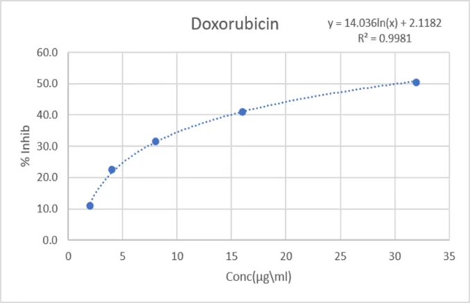
Doxorubicin was taken as Std for comparison. The results showed IC 50 value of Icaritin: IC₅₀ = 107.47µg/ml.
Protein denaturation assay calculations
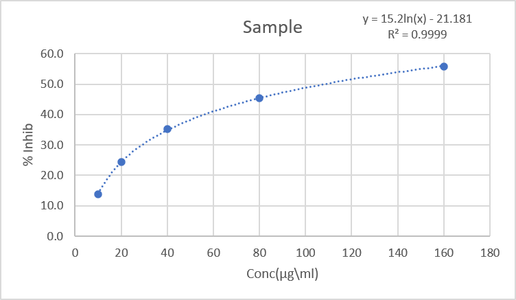
Icaritin protected cell from protein precipitation at a dose of 100 µg/ml. Protein precipitation was only 69.31 ± 2.45 when compared with Std Aspirin 76.21 ± 3.14.
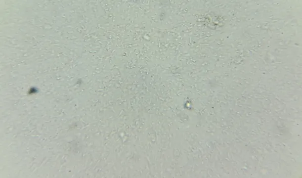
MTT Assay for cytotoxicity test images
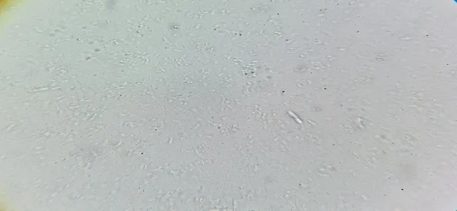
Figure 4: Blank (Its Control without Any Sample or Standard)
Figure None: 5 (Test Sample Treated)
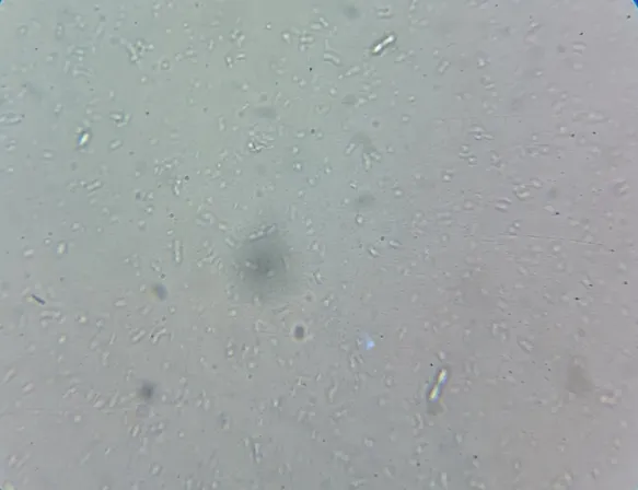
Figure None: 6 (Standard Drug Doxorubicin Treated)
SUMMARY AND CONCLUSION
SUMMARY
REFERENCES





Dr. J. Nandhakumar, Dr. Tharani M., Nehla Rafeeq P. R., Shanima Shamsudeen, Sajeena V., Pharmacological Evaluation of the Anti-Inflammatory Activity of Icaritin-A Natural Isoflavone, Int. J. of Pharm. Sci., 2026, Vol 4, Issue 11, 2388-2402. https://doi.org/10.5281/zenodo.24272366
 10.5281/zenodo.24272366