
We use cookies to ensure our website works properly and to personalise your experience. Cookies policy
Department of Pharmacology, AISSMS College of Pharmacy, RB Motilal Kennedy Rd, near RTO Pune, Sangamvadi, Pune, Maharashtra 411001.
Inflammation is a biologically heterogeneous reaction caused by tissue-damaging stimuli like pathogens, toxic chemicals, or trauma. Although the use of non-steroidal anti-inflammatory drugs (NSAIDs) for anti-inflammatory purposes is widespread, they typically possess undesirable side effects. Plant-based compounds have proved to be great candidates as more acceptable alternatives with less toxicity in their anti-inflammatory action. The present study assesses the in vitro anti-inflammatory activity of plant extracts by protein denaturation test and heat-induced hemolysis test. The egg albumin denaturation test is used to quantify the inhibition of protein denaturation by plant extracts, which is a part of inflammatory reactions. Likewise, the heat-induced hemolysis test is used to assess the resistance of red blood cell membranes of plant extracts to heat stress. The results reveal a substantial reduction in protein denaturation as well as membrane stabilization that signifies the future of plant extracts as potential anti-inflammatory drugs. These results highlight the importance of natural compounds in designing new safe anti-inflammatory drugs.
Protein Denaturation Assay
Inflammation is a somatic reaction to damage, infection, or injury, and it is marked by heat, redness, pain, swelling, and compromised physiological processes1. It is a normal defense process when the tissues are injured by physical injury, toxic compounds, or microbe infections. The aim of the body is to remove the irritants, kill invading pathogens, and facilitate tissue repair1. This is a result of the liberation of chemical mediators from injured tissue and migrating cells. Non-steroidal anti-inflammatory drugs (NSAIDs) are the most widely prescribed drugs used in the treatment of inflammatory diseases, but they have side effects, most notably gastric irritation leading to the formation of gastric ulcers2. Inflammation is a multifaceted biological response of vascular tissues to possibly injurious stimuli, acting as the body's defense mechanism to eliminate noxious agents and stimulate the process of healing3. Initiation of inflammation induces cells to secrete diverse inflammatory mediators like histamine, serotonin, SRS-A, prostaglandins, and several plasma enzyme systems like the complement system, clotting system, fibrinolytic system, and kin in system4. These mediators work together to induce vasodilation and raise blood vessel permeability, which enables blood to circulate more freely, plasma proteins, and fluids to ooze from the damaged tissues, and leukocytes mainly neutrophils to leave the blood vessels. Inflammation may be divided into two categories: acute and chronic. Acute inflammation is the immediate reaction of the body towards harmful stimuli, leading to an elevated plasma and leukocyte entry into the injured tissues. This is a process that is triggered by existing cells in the tissues, which are marked by vasodilation and increased capillary permeability caused by inflammatory mediators5. Chronic inflammation, on the other hand, entails concurrent tissue destruction and repair, which is a chronic inflammatory. Process that causes progressive cellular changes at the site of inflammation.6 The integration of natural products has been a key factor in shaping contemporary medicine. The international world is now rediscovering traditional medicine as a consequence of current extensive studies of various plant species and their active medicinal components. The study of anti-inflammatory natural plants offers the possibility of uncovering and exploring natural compounds to replace synthetic medicines.7 The search for the anti- inflammatory properties of natural plants opens the door to utilizing this large body of information and to considering long-term healing alternatives. Natural plants have been used in traditional medicine for centuries for their therapeutic qualities.8 Natural plant-based anti- inflammatory chemicals have been used by humans for centuries and are usually harmless, possibly unveiling substances that will prove to be less toxic and with fewer side effects than synthetic medication. This is particularly significant in chronic disease, where long-term use of anti-inflammatory drugs could have detrimental effects on health.9 A wide range of bioactive compounds, including polyphenols, flavonoids, terpenoids , and alkaloids, have been found to be present in plants. These bioactive compounds may act on different pathways of inflammation and show promising anti-inflammatory activity. Studying and investigating natural plants can provide new bioactive compounds with potential anti-inflammatory effects.10 Natural flora has been a source of great inspiration for the discovery of new drugs. Plant-derived natural compounds have been used to develop drugs, such as aspirin and other nonsteroidal anti-inflammatory drugs (NSAIDs). Studying naturally occurring anti-inflammatory plants may allow one to discover novel compounds or novel structures as potential leads for drug development and discovery, resulting in improved and better-targeted anti-inflammatory drugs.8 This approach centers on in vitro anti-inflammatory methods that can analyze natural anti-inflammatory compounds under different conditions. They are easy to maintain and are cost-effective, dependable, and easy to use. In this regard, the egg albumin denaturation method can be useful for researchers in this field. Thus, the improved egg albumin denaturation technique is as described below.
Principle of In-vitro Egg Albumin Assay
The major objective of the egg albumin denaturation assay is to identify if agents or chemicals can inhibit or delay egg albumin from undergoing denaturation under certain conditions. Denaturation is defined as the alteration of the structure of a protein leading to a loss of biological activity.11 Egg albumin serves as a model protein in this experiment, and denaturation is caused by exposure to excessive heat, pH levels, or other denaturing substances. Under denaturation, the native conformation of egg albumin is destroyed, changing its physical properties and resulting in loss of functional activity. Egg albumin denaturation assay determines a compound or drug's capacity to inhibit or mitigate egg albumin denaturation in order to gauge its anti-inflammatory activity. The assay is based on the principle that compounds possessing anti-inflammatory characteristics can inhibit such denaturation effectively.12 Stabilize protein structures and avoid denaturation, which are usually implicated in inflammation and tissue injury. Thus, such chemicals or agents with significant ability to inhibit denaturation of egg albumin in this assay would have anti-inflammatory potential13. Protein denaturation has been speculated as one of the causes of inflammation. Inhibitors of protein denaturation are the NSAIDs and these also block COX enzyme14. Different concentrations of the test sample can be incubated with egg albumin solution under experimental conditions, and reactions allowed to take place, followed by determination of absorbance for calculation of the percentage inhibition. IC50 values can be calculated using GraphPad Prism software, with diclofenac sodium as a reference drug.15
MATERIALS
Chemical and Reagents:
Egg albumin solution
Phosphate buffered saline
Distilled water/DMSO
Equipment:
Clean pipettes and pipette tips
Test tubes or Khan tubes
Incubator
Spectrophotometer
Water bath
Egg Albumin
METHOD
Denaturation Assay:
The maceration method was used to make the plant extract, a common homemade tonic preparation method that has become popular due to its affordability in extracting essential oils and bioactive compounds. The maceration process is usually done in a number of steps for small-scale extraction. The plant material leaves, stem bark, or root bark is first ground into fine particles to increase the surface area for maximum solvent mixing. Next, a suitable solvent, referred to as menstruum, is added to a sealed container, with water being the most polar solvent. The liquid is then strained, and the solid residue, or marc, is pressed to obtain any remaining solutes. The pressed and strained liquid is blended and filtered to eliminate impurities. Shaking at regular intervals during maceration facilitates extraction by (a) enhancing diffusion and (b) renewing the solvent in contact with the sample for better yield.16 Fresh plant material is harvested and washed under running water to remove visible impurities. The purified samples are dried for one to two days in a shaded place, not exposed to direct sunlight. The dried plant material is ground in a mechanical blender. The powdered substance that is left is kept in an airtight container. Distilled water is added to the powdered plant substance in the ratio of 3:1 (volume and weight) and shaken, followed by keeping in a tightly covered container for 48 hours. Filtering is done through three layers of muslin cloth, and the samples are kept in the fume cupboard for 48 hours, upon which the solution will evaporate and remove all solvents. A spatula is employed to scratch the resulting extract. dried powder after water had evaporated. Amber-colored vials were then employed to store aqueous plant powder dry for later use. One gram of the aqueous plant extract powder was dissolved into 1 mL of distilled water. This concentration is an undiluted extract, which was the highest point of the dilution series, and 1 mL was used to produce a two-fold dilution series. The series of dilutions was conducted using a two-fold process in which each concentration was half the previous one. This process was repeated for a total of 10 points, thus creating different concentrations of the aqueous plant extract from 1 g/mL to 2 x 10^3 g/ml. Then, 2 mL of the prepared solution was pipetted into the corresponding test tubes to perform the assay.16 Preparation of 1% egg albumin solution: A 1% egg albumin solution is prepared by using fresh hen eggs or by using egg albumin powder that can be bought commercially. To prepare this solution using a fresh hen's egg, gently crack the egg, take 1 mL of the clear portion, and add it to 100 mL of w/v distilled water, and mix well. The clear portion of the egg is egg albumin. Cold water should be used in this preparation since boiling water will lead to coagulation.16
Procedure
The anti-inflammatory effect of unidentified crude extracts can be determined in vitro for their capacity to inhibit egg albumin denaturation (protein). To prepare the reaction mixture, mix 0. 2 mL of the 1-2% egg albumin solution (fresh hen's egg or commercial egg albumin powder) with 2 mL of the sample extract or standard (Diclofenac sodium) at different concentrations, and 2. 8 mL of phosphate-buffered saline (pH 7. 4) to achieve a total volume of 5 mL. The control was made by combining 2 mL of triple-distilled water, 0. 2 mL of 1-2% egg albumin solution, and 2. 8 mL of phosphate-buffered saline to a total volume of 5 mL. The reaction mixtures were incubated at 37 ± 2°C for 30 minutes and subsequently heat-treated in a water bath at 70 ± 2°C for 15 minutes. After cooling, absorbance at 280 nm was recorded on a proper UV/Vis spectrophotometer with triple-distilled water as a blank.1,15,17
Percentage inhibition of denaturation of egg albumin was determined using the formula:
Percentage inhibition = (Absorbance of control – Absorbance of test sample) / Absorbance of control x 100.
Observation
Table No. 1 - Absorbance values
|
Sr. No. |
Concentration(μg/ml) |
Absorbance(nm) |
|
|
Test Sample |
Standard |
||
|
1 |
100 |
0.923 |
0.312 |
|
2 |
200 |
0.865 |
0.156 |
|
3 |
300 |
0.746 |
0.092 |
|
4 |
400 |
0.635 |
0.044 |
|
5 |
500 |
0.468 |
0.026 |
Table No.2 - % Inhibition of Protein Denaturation
|
Sr No. |
Concentration(μg/ml) |
%inhibition of Protein Denaturation |
|
|
Test Sample |
Standard |
||
|
1 |
100 |
40.9 |
80.02 |
|
2 |
200 |
44.6 |
90.01 |
|
3 |
300 |
52.2 |
94.11 |
|
4 |
400 |
59.3 |
97.18 |
|
5 |
500 |
70.03 |
98.33 |
Absorbance of Heated Control – 0.99
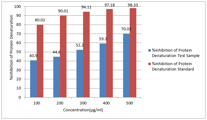
Figure No.1 - Effect of Sample and Standard in Protein Denaturation Assay
Heat Induced Hemolysis Assay
Introduction to Heat Induced Hemolysis Assay
Heat induced hemolysis assay is a vital experimental method utilized to identify the stability of the red blood cell membrane following thermal stress exposure. In biomedical research, this is significantly important in identifying the impacts of numerous chemicals on the red blood cell's integrity and in disease mechanisms against hemolysis. Hemolysis, or red blood cell breakdown and spilling of their contents in the medium, may give insight into the physiological and pathologic processes of erythrocyte loss and malfunction. Heating erythrocytes is a try by researchers to investigate conditions leading to membrane instability and decay.22 This test has special use in measurement of the protective effect of antioxidants, drugs, or other therapeutic agents capable of strengthening cell membranes. In this assay, blood samples are typically subjected to progressively higher temperatures, and hemolysis is quantitated by the measurement of hemoglobin released into the supernatant, an index of membrane damage. The heat-induced hemolysis assay not only provides a convenient and effective tool to study red blood cell stability but also provides valuable information regarding the potential hemoprotective activity of most compounds. It has thus become a leading assay in clinical and experimental hematology, enabling the development of strategies to preserve erythrocyte integrity under different conditions of stress.23,24
Principles And Mechanism of Heat Induced Hemolysis
The principles and mechanisms of heat-induced hemolysis are involved with the manner in which red blood cell membrane structure is affected by rise in temperature. Hemolysis means bursting or lysis of red blood cells, resulting in release of hemoglobin into the medium. Red blood cell membranes, under normal physiological conditions, are compact structures made up of a lipid bilayer that is mixed with proteins, and this accounts for strength as well as flexibility. But excess heat will disrupt the balance and cause structural changes which weaken membrane stability.25 High temperatures enhance lipid bilayer fluidity, and this may distort protein conformation and interfere with membrane organization. Denaturation of membrane proteins may also lead to disruption of interactions for cell integrity. In this case, the stress is heat that overloads the body's compensatory mechanisms for cell homeostasis, leading to membrane lysis. Heat exposure will also cause oxidative stress, making red blood cells hemolytic susceptible by initiating reactive oxygen species production that further destabilizes the membrane. The mechanisms involved are mechanical disruption and biochemical alterations, leading to loss of cell membrane integrity and subsequent leakage of cellular contents. Knowledge of these principles is necessary for the optimization of assay conditions to measure red blood cells' stability and resistance to thermal stress accurately.26,27
Method
1. Red Blood Cell (RBC) Preparation: RBCs are separated from fresh blood by centrifugation and washed in saline or phosphate-buffered saline (PBS) to eliminate plasma and other material.
2. Preparation of Suspension: A 10% v/v suspension of RBC is prepared using an isotonic buffer.
3. Assay Setup: Drug tests are mixed with suspension of RBC in various concentrations and incubated at high temperature (typically 56°C) for a particular duration of time (typically 30 minutes).
4. Measurement of Hemolysis: Heated mixture is cooled and centrifuged. Supernatant is then assayed for absorbance at a particular wavelength (typically 560 nm) to calculate the degree of hemolysis.
5. Inhibition Calculation: Percentage inhibition of hemolysis is calculated by dividing the absorbance value of the test samples by the value of a control sample in which complete hemolysis is achieved.28,29
Procedure
Assay mixture was 2 mL of test sample or standard (Diclofenac) in a range of 100, 200, 300, 400, and 500 μg/mL in isotonic PBS and 2 mL of 10% RBC suspension. Reaction mixtures were incubated at 54 °C in a water bath for 20 minutes, centrifuged at 1500 rpm for 5 minutes, and supernatant was measured on a spectrophotometer at 540 nm.
Percentage hemolysis inhibition was calculated with the formula:
Inhibition of hemolysis (%) = 100 × (1 - OD2 - OD1 / OD3 - OD1)
Where, OD1 = unheated test sample, OD2 = heated test sample, and OD3 = heated control sample.
Observation
Table No.1 - Absorbance values of Unheated Test tubes
|
Sr. No |
Concentration(μg/ml) |
Absorbance(nm) |
|
|
Test Sample |
Standard |
||
|
1 |
100 |
0.515 |
0.378 |
|
2 |
200 |
0.447 |
0.332 |
|
3 |
300 |
0.398 |
0.283 |
|
4 |
400 |
0.355 |
0.248 |
|
5 |
500 |
0.27 |
0.202 |
Table No.2 - Absorbance values of Heated Test tubes
|
Sr. No |
Concentration(μg/ml) |
Absorbance(nm) |
|
|
Test Sample |
Standard |
||
|
1 |
100 |
0.914 |
0.766 |
|
2 |
200 |
0.861 |
0.685 |
|
3 |
300 |
0.799 |
0.59 |
|
4 |
400 |
0.712 |
0.471 |
|
5 |
500 |
0.598 |
0.38 |
Absorbance of Heated Control – 0.99
Table No.3 - %Inhibiton of Hemolysis
|
Sr. No |
Concentration(μg/ml) |
Absorbance(nm) |
|
|
Test Sample |
Standard |
||
|
1 |
100 |
16.4 |
36.9 |
|
2 |
200 |
24.1 |
46.6 |
|
3 |
300 |
32.5 |
56.7 |
|
4 |
400 |
44.1 |
70.1 |
|
5 |
500 |
54.6 |
77.5 |
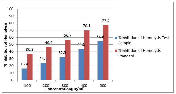
Figure No.1 – Effect of Sample and Standard in Heat Induced Hemolysis Assay
REFERENCES


Shubhangi Deshpande*, Aditya Rathi, Sanskruti Dabade, Vishwajeet Mathane, Aryan Agarwal, Atharva Taye, Investigating the Anti-Inflammatory Effects of Natural Extracts Using Protein Denaturation Assay and Heat Induced Hemolysis Assay, Int. J. of Pharm. Sci., 2025, Vol 3, Issue 7, 2918-2926. https://doi.org/10.5281/zenodo.16277443
 10.5281/zenodo.16277443