

We use cookies to ensure our website works properly and to personalise your experience. Cookies policy
Bhujbal Knowledge City MET’s Institute of Pharmacy, Adgaon , Nashik, Maharashtra 422003.
Background: The global outbreak of SARS-CoV-2 in 2019 profoundly disrupted healthcare systems and resulted in significant morbidity and mortality. While initially categorized as a respiratory infection, emerging evidence has revealed that COVID-19 affects multiple organ systems, particularly the cardiovascular system. Long COVID, or post-acute COVID-19 syndrome (PACS), has been associated with spectrum of persistent syndrome, with cardiac complications identifies as a major concern. Previous research has established the occurrence of acute cardiovascular events such as myocarditis, myocardial injury, arrhythmias, pericarditis, thromboembolism and acute coronary syndrome during COVID-19 infection. These effects have been linked too direct viral invasion, systematic inflammatory responses, endothelial dysfunction, and coagulopathies. However, data specific to regional populations, including Maharashtra, remain limited.Objective: This study aimed to assess and compare the incidence and type of cardiovascular complications in hospitalized COVID-19 patients Vs Hospitalized non COVID-19 patients at a tertiary care hospital.Materials and Method: The retrospective study conducted in tertiary care hospital over an 8-month period. The study included 320 adult patients (160 per group), matched for age, sex, and comorbidities. Patient with end stage renal disease or incomplete cardiac data were excluded. Data collection encompassed demographics, clinical history, inflammatory and cardiac biomarkers (e.g. troponin, BNP), ECG, echocardiography and clinical outcomes. Statistical analysis was performed using the SPSS version 29.0.Results: The study results indicated significantly higher rate of cardiac complications in the COVID-19 cohort (27.5%) compared to non-COVID-19 group (21.3%). Covid patients showed elevated inflammatory and myocardial injury markers, higher heart and respiratory rates, and lower oxygen saturation. Prolonged QT intervals and abnormal ECGs were more frequent in the COVID group. ICU admissions and overall mortality were also significant higher among COVID-19 patients. Logistic regression revealed that COVID-19 infection was an independent predictor of cardiac complications (OR = 2.469, 95% CI: 1.093-5.578, p = 0.030). Conclusion: In conclusion, COVID-19 is associated with a greater risk of cardiac injury and complications in hospitalized patients. These findings highlight the need to vigilant cardiovascular monitoring and intervention strategies in patients with COVID-19 to mitigate morbidity and mortality risks.
The SARS-CoV-2-caused worldwide pandemic of 2019 (COVID-19) began in late 2019 and has swiftly progressed. This pandemic has caused unprecedented healthcare system strain and global morbidity and mortality.[1] SARS-CoV-2, initially thought to be a respiratory illness, can affect multiple organ systems, including the cardiovascular system (Nandini Vishwakarma). Long COVID, also known as post-acute COVID-19 syndrome (PACS), causes many people to have long-term symptoms and effects.[2] Public health is threatened by this prolonged sickness, especially if cardiovascular indications become a major worry.[1] Long COVID can cause a wide range of symptoms that last weeks or months, impacting quality of life and function.[3]
Myocardial damage, myocarditis, pericarditis, arrhythmias, acute coronary syndromes, and thromboembolic events have been extensively studied in the acute phase of COVID-19.[4] The present epidemic requires consideration of cardiovascular consequences in post-COVID-19 syndrome.[2] Direct viral invasion, systemic inflammation, endothelial dysfunction, and thromboembolic events have been shown to damage the heart and vascular system by SARS-CoV-2.[3] These acute cardiovascular problems have raised concerns about long-term cardiovascular difficulties.[5]
The immediate cardiovascular consequences of COVID-19 are serious, but the post-acute cardiac difficulties are crucial for patient management and healthcare resource planning.[1] Long COVID syndrome can cause a variety of symptoms for weeks or months, including cardiovascular difficulties.[2] Chest discomfort, palpitations, shortness of breath, weariness, and exercise intolerance can lower quality of life.[3] Understanding the occurrence and characteristics of post-COVID cardiac problems is essential for clinical practice, early detection, and appropriate therapy.[2]
Previous meta-analyses have illuminated cardiovascular problems with lengthy COVID therapy. Shrestha et al. (2023) found that post-COVID patients had higher probabilities of cardiovascular disorders than healthy controls. Electrophysiological anomalies, coronary vascular illnesses, thromboembolic disorders, and pericarditis and myocarditis are examples.[2] A 2025 meta-analysis by Huang et al. indicated that 15% of COVID-19 survivors have post-acute cardiovascular effects. It was also found that COVID-19 infection increases the likelihood of several illnesses. The pooled analysis showed that COVID-19 patients had a higher odds ratio of chest discomfort (OR 4.0) than controls.[3] A comprehensive meta-analysis by Zhang et al. (2025) found that COVID-19 patients at least four weeks after diagnosis had a significantly higher risk of various cardiovascular events than non-COVID-19 controls. Thromboembolic disorders (HR 3.12), coronary heart disease (HR 1.61), stroke (HR 1.71), arrhythmia (HR 1.60), cardiomyopathy (HR 1.71), myocarditis (HR 6.11), hypertension (HR 1.70), heart failure (HR 1.72), and cardiogenic shock (HR 2.09) were found. Rajotiya et al. examined cardio-pulmonary symptoms one year after severe COVID-19 in Northern India in a case-control study. In COVID, pulmonary problems and cardiac biomarker alterations were found.[6] Long-term COVID has a major cardiovascular consequence. Despite the growing volume of research on post-COVID cardiovascular issues, regional data is needed to better understand the epidemiology and consequences of this emerging health challenge. Many meta-analyses have provided global or multi-regional estimates, but few have focused on the Indian subcontinent or its states or regions.[7]
The proposed meta-analysis will fill this gap in the literature by reviewing and synthesizing Maharashtra research on cardiac problems after SARS-CoV-2 infection. In this retrospective analysis of hospitalized patients, acute cardiac injury, arrhythmias, heart failure, and elevated cardiac biomarkers are compared between COVID-19-positive patients and a control group without COVID-19. This comparative study seeks to determine SARS-CoV-2 infection's burden and cardiovascular involvement in acute hospital care, excluding general sickness.
MATERIALS AND METHODS
Study design and population:
The study employs a Retrospective cohort design with two distinct groups: patients who have been hospitalized with confirmed COVID-19 infection and a control group comprising patients without any history of COVID-19. Both cohorts will be matched for demographics such as age, sex, and key comorbid conditions (e.g., hypertension, diabetes mellitus, obesity) to minimize confounding effects. The control group will be derived from the same hospital settings and will include individuals admitted for non-COVID-19-related illnesses but who share similar baseline characteristics.
Inclusion Criteria:
Adult patients (≥18 years) hospitalized with a confirmed positive RT-PCR test for SARS-CoV-2, Availability of complete cardiac biomarker data (e.g., troponin, BNP) and echocardiography reports, Adult patients (≥18 years) hospitalized during the same period without a history of COVID-19, confirmed through negative screening tests.
Exclusion Criteria:
Patients with incomplete clinical records or missing cardiac biomarker data, Patients with end-stage renal disease requiring dialysis prior to admission (given the impact on biomarker levels).
Data Collection:
Data regarding comparison and control group were recorded from patients and their medical records using structured data collection form.
Statistical Analysis:
Frequency and percentage were used as a summary measure and the Chi-square test was used for the association between independent variables and degree of suspected adverse drug reactions which was considered as a dependent variable. T-test was used for the comparison between the mean differences between the two variables.
Binary logistic regression analysis was performed to identify potential predictors associated with cardiac complications in hospitalized patients. P-value < 0.05 was considered indicate statistically significant difference, equivalent to confidence level of 95%. SPSS 29.0 statistical was used for the analysis.
RESULTS:
The descriptive statistics of this study highlight the demographic and anthropometric profiles of participants in the Covid and Non-Covid groups. Both cohorts consisted of 160 individuals. The mean age of the Covid group was 56.18±15.96 years, while the Non-Covid group had a slightly higher mean age of 57.81±16.55 years, indicating a comparable age distribution. Mean height was marginally lower in the Covid group 167.91±7.25 compared to the Non-Covid group 169.12±6.55 cm. Similarly, the average BMI values were closely aligned between the two groups, with the Covid group showing a mean BMI of 20.27±6.79 and the Non-Covid group recording 20.31±6.07. Patients in each group are broadly gender-balanced. In the Covid group, 86 (53.8%) were female and 74 (46.3%) were male. In the non-Covid group, 90 (56.3%) were female and 70 (43.8%) were male.
Table 1. Comparative analysis of the clinical parameters and biochemical parameters.
|
Parameters |
Covid (N=160) |
Non Covid (N=160) |
||||||||
|
Mean |
Median |
Std. Deviation |
Quartile |
Mean |
Median |
Std. Deviation |
Quartile |
|||
|
Q1 |
Q3 |
Q1 |
Q3 |
|||||||
|
Systolic BP |
135.8 |
133.5 |
18.7 |
119.3 |
152.0 |
131.6 |
128.5 |
18.1 |
119.0 |
145.0 |
|
Diastolic BP |
85.6 |
85.5 |
11.0 |
77.0 |
93.0 |
83.4 |
82.0 |
10.6 |
76.0 |
91.8 |
|
Heart Rate |
89.9 |
90.0 |
14.7 |
79.3 |
100.0 |
75.8 |
75.0 |
10.4 |
69.0 |
83.0 |
|
Respiratory Rate |
21.9 |
22.0 |
4.0 |
19.0 |
24.0 |
15.5 |
15.0 |
3.1 |
13.0 |
18.0 |
|
SPO2 (%) |
91.7 |
91.0 |
3.9 |
89.0 |
94.0 |
96.8 |
97.0 |
2.0 |
96.0 |
98.0 |
|
eGFR |
62.9 |
56.0 |
25.7 |
44.0 |
86.0 |
66.5 |
63.5 |
27.0 |
46.3 |
86.8 |
|
Triglycerides |
154.1 |
149.5 |
50.5 |
115.5 |
193.0 |
143.4 |
138.0 |
51.8 |
102.0 |
182.8 |
|
LDL |
123.8 |
123.0 |
34.3 |
102.3 |
146.8 |
117.9 |
114.0 |
32.6 |
94.3 |
142.0 |
|
HDL |
47.3 |
47.0 |
13.0 |
37.3 |
55.0 |
47.9 |
47.0 |
13.2 |
40.0 |
57.0 |
|
Haemoglobin |
13.4 |
13.6 |
1.7 |
12.4 |
14.8 |
13.4 |
13.3 |
1.7 |
12.1 |
14.5 |
|
Troponin Peak (pg/mL) |
14.9 |
11.8 |
10.8 |
7.2 |
20.2 |
5.0 |
4.0 |
3.5 |
2.6 |
6.5 |
|
BNP (pg/mL) |
62.4 |
54.5 |
31.0 |
40.7 |
78.6 |
22.3 |
19.4 |
11.6 |
14.1 |
26.9 |
|
CRP (mg/L) |
21.5 |
20.7 |
8.3 |
14.9 |
26.6 |
5.6 |
5.1 |
3.1 |
3.6 |
6.8 |
|
IL-6 (pg/mL) |
21.3 |
21.4 |
4.4 |
17.8 |
24.4 |
5.1 |
4.6 |
2.6 |
3.5 |
6.3 |
|
Myoglobin (ng/mL) |
62.6 |
57.2 |
29.5 |
40.7 |
79.4 |
21.0 |
19.1 |
10.3 |
13.3 |
26.4 |
|
Creatine Kinase (U/L) |
178.4 |
153.5 |
95.4 |
112.0 |
223.7 |
58.9 |
52.2 |
33.0 |
35.2 |
72.0 |
|
Ferritin |
172.6 |
148.8 |
102.3 |
98.0 |
227.2 |
23.6 |
20.8 |
13.4 |
14.5 |
29.5 |
|
LVEF (%) |
49.1 |
50.6 |
10.0 |
41.8 |
56.9 |
47.7 |
51.8 |
12.1 |
41.2 |
56.6 |
|
QRS Duration (ms) |
98.3 |
94.9 |
15.6 |
87.7 |
108.8 |
97.5 |
94.6 |
16.4 |
85.1 |
108.8 |
|
QT Interval (ms) |
448.6 |
447.6 |
31.2 |
422.9 |
471.2 |
423.3 |
423.9 |
31.4 |
402.4 |
445.6 |
The comparative analysis of clinical and biochemical parameters between Covid and Non-Covid groups in this study reveals significant physiological differences (Table 1.). The Covid group exhibited elevated mean values for systolic (135.8 mmHg) and diastolic (85.6 mmHg) blood pressure, heart rate (89.9 bpm), and respiratory rate (21.9 breaths/min) compared to the Non-Covid group. Markedly lower oxygen saturation (SpO?: 91.7% vs. 96.8%) and reduced renal function (eGFR: 62.9 vs. 66.5 mL/min/1.73m²) were observed in Covid patients. Inflammatory markers such as CRP, IL-6, ferritin, and BNP were substantially elevated in the Covid group, indicating heightened systemic inflammation and cardiac strain. Moreover, myocardial injury markers including troponin, myoglobin, and creatine kinase were significantly higher in Covid cases, suggesting greater cardiovascular involvement. Although lipid profiles and haemoglobin levels remained comparable, the prolonged QT interval in the Covid group (448.6 ms vs. 423.3 ms) indicates potential cardiac conduction abnormalities.
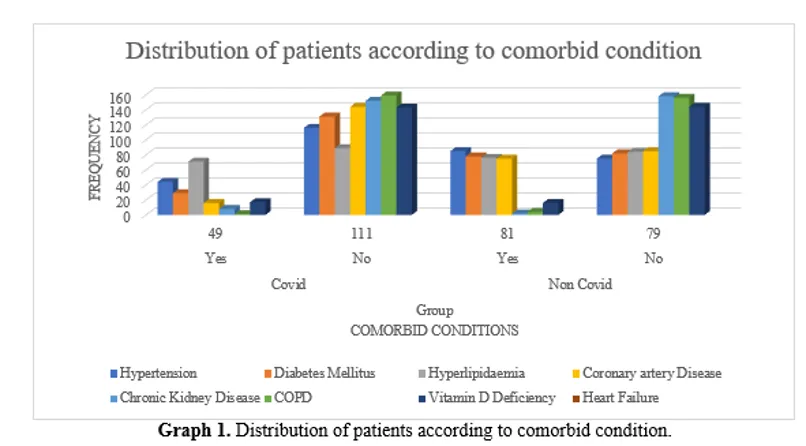
Graph 1. Distribution of patients according to comorbid condition.
Comorbidities differed greatly between Covid and Non-Covid groups (Graph 1.). Covid patients had 49 (30.6%) hypertension, compared to 81 (50.6%) in the Non-Covid cohort. Diabetes mellitus was found in 44 (27.5%) Covid patients and 85 (53.1%) Non-Covid patients. Hyperlipidemia was identified in 29 (18.1%) Covid cases and 78 (48.8%) Non-Covid cases. Coronary artery disease affected 71 (44.4%) Covid and 76 (47.5%) Non-Covid patients nearly equally. Chronic renal disease was more common in Non-Covid patients, at 75 (46.9%) against 16 (10.0%) in Covid. COPD was rare in both groups but more common in Covid patients 8 (5.0%) than Non-Covid patients 2 (1.3%). Only 1 (0.6%) Covid and 4 (2.5%) non-Covid had vitamin D insufficiency. The Covid and Non-Covid cohorts had 17 (10.6%) and 16 (10.0%) heart failure patients, respectively.
Table 2. Distribution of the patients according to the complications.
|
Cardiac Complications |
|||
|
Group |
N |
% |
|
|
Covid |
Yes |
44 |
27.5% |
|
No |
116 |
72.5% |
|
|
Non Covid |
Yes |
34 |
21.3% |
|
No |
126 |
78.8% |
|
The Covid group experienced a greater incidence of cardiac problems than the non-Covid group. 44 (27.5%) of Covid-19 patients had cardiac problems, whereas 116 (72.5%) did not. In contrast, 34 (21.3%) non-Covid individuals reported cardiac problems, whereas 126 (78.8%) did not (Table 2.).
Table 3. Distribution of the patients according to cardiac complications across groups.
|
Cardiac Complications |
Group |
|||||
|
Covid |
Non Covid |
|||||
|
Yes |
Yes |
|||||
|
N |
% |
N |
% |
|||
|
Cerebrovascular |
Stroke |
12 |
7.5% |
12 |
7.5% |
|
|
TIA |
7 |
4.4% |
7 |
4.4% |
||
|
None |
141 |
88.1% |
141 |
88.1% |
||
|
Inflammatory Heart Disease |
Pericarditis |
3 |
1.9% |
3 |
1.9% |
|
|
Myocarditis |
2 |
1.3% |
2 |
1.3% |
||
|
None |
155 |
96.9% |
155 |
96.9% |
||
|
Arrhythmia |
Tachycardia |
3 |
1.9% |
1 |
0.6% |
|
|
Bradycardia |
2 |
1.3% |
2 |
1.3% |
||
|
Ventricular Arrthmias |
2 |
1.3% |
1 |
0.6% |
||
|
None |
153 |
95.6% |
156 |
97.5% |
||
|
Thrombotic Disorder |
Pulmonary Embolism |
20 |
12.5% |
14 |
8.8% |
|
|
DVT |
5 |
3.1% |
2 |
1.3% |
||
|
None |
135 |
84.4% |
144 |
90.0% |
||
|
Ischemic Heart Disease |
ACD |
3 |
1.9% |
1 |
0.6% |
|
|
MI |
13 |
8.1% |
11 |
6.9% |
||
|
Ischemic Cardiomyopathy |
7 |
4.4% |
3 |
1.9% |
||
|
Angina |
15 |
9.4% |
10 |
6.3% |
||
|
None |
122 |
76.3% |
135 |
84.4% |
||
|
Other Cardiac Complications |
Heart Failure |
18 |
11.3% |
12 |
7.5% |
|
|
Cardiac Arrest |
15 |
9.4% |
7 |
4.4% |
||
|
Cardiogenic Shock |
5 |
3.1% |
1 |
0.6% |
||
|
Cardiomyopathy |
5 |
3.1% |
3 |
1.9% |
||
|
None |
117 |
73.1% |
137 |
85.6% |
||
Covid-19 patients had a higher burden of particular cardiac problems. Stroke 12 (7.5%) and transient ischemic attacks 7 (4.4%) were similar in both groups. Pericarditis was detected in 3 (1.9%) and myocarditis in 2 (1.3%) patients in both cohorts. Arrhythmias were significantly more prevalent in Covid. 3 (1.9%) Covid patients had tachycardia and 2 (1.3%) had ventricular arrhythmias, compared to 1 (0.6%) non-Covid patients. Bradycardia was equal in both groups 2 (1.3). Covid patients had higher thrombotic events, including pulmonary embolism (12.5%) and deep vein thrombosis (3.1%) compared to non-Covid patients (8.8%).
Covid patients had greater ischemic heart disease. Acute coronary disease (ACD) was found in 3 (1.9%) Covid patients compared to 1 (0.6%) non-Covid patients, myocardial infarction in 13 (8.1%) versus 11 (6.9%), ischemic cardiomyopathy in 7 (4.4%) versus 3 (1.9%), and angina in 15 (9.4%) versus Covid patients had more other cardiac problems. Compared to 12 (7.5%), 18 (11.3%) had heart failure, 15 (9.4%) had cardiac arrest, 5 (3.1%) had cardiogenic shock, and 5 (3.1%) had cardiomyopathy (Table 3.).
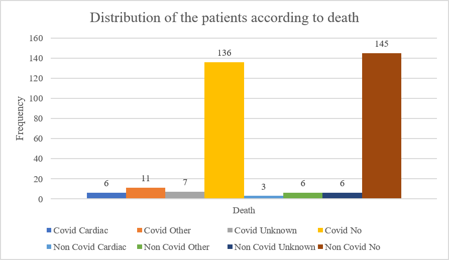
Graph 2. Distribution of the patients according to death.
The Covid group had a greater death rate than the non-Covid group (Graph 2.). Six (3.8%) of 24 (15.0%) Covid-19 deaths were attributable to cardiac causes, 11 (6.9%) to non-cardiac causes, and 7 (4.4%) to unexplained causes. In contrast, the non-Covid cohort reported 15 (9.4%) deaths: 3 (1.9%) cardiac, 6 (3.8%) other, and 6 (3.8%) unknown. In both groups, 136 (85.0%) of Covid patients and 145 (90.6%) of non-Covid patients survived.
Table 4. Associations between the ECG findings and cardiac complication present in covid versus non covid patients.
|
ECG Findings |
Complications |
Total |
p-value |
|
|
Yes |
No |
|||
|
Atrial fibrillation |
5 |
7 |
12 |
.001 |
|
Atrial fibrillation, Prolonged QT |
2 |
12 |
14 |
|
|
Atrial fibrillation, Wide QRS |
4 |
0 |
4 |
|
|
Normal |
24 |
104 |
128 |
|
|
Prolonged QT |
9 |
49 |
58 |
|
|
Sinus tachycardia |
10 |
22 |
32 |
|
|
Sinus tachycardia, Atrial fibrillation |
2 |
4 |
6 |
|
|
Sinus tachycardia, Atrial fibrillation, Prolonged QT |
0 |
1 |
1 |
|
|
Sinus tachycardia, Atrial fibrillation, Wide QRS |
1 |
1 |
2 |
|
|
Sinus tachycardia, Atrial fibrillation, Wide QRS, Prolonged QT |
1 |
0 |
1 |
|
|
Sinus tachycardia, Prolonged QT |
5 |
23 |
28 |
|
|
Sinus tachycardia, Wide QRS |
4 |
3 |
7 |
|
|
Sinus tachycardia, Wide QRS, Prolonged QT |
0 |
2 |
2 |
|
|
Wide QRS |
8 |
11 |
19 |
|
|
Wide QRS, Prolonged QT |
3 |
3 |
6 |
|
|
Total |
78 |
242 |
320 |
|
Associations between ECG abnormalities and complications were statistically significant (p-value 0.001) (Table 4.). Five complications and seven without had atrial fibrillation, whereas nine complications and 49 without had extended QT intervals. In patients without problems, sinus tachycardia with atrial fibrillation or broad QRS complexes were more common. Compared to 24 complications, 104 normal ECG patterns were seen in the group without issues. These results indicate a substantial correlation between the occurrence of complications and specific ECG abnormalities, underscoring the necessity of meticulous ECG monitoring in patients with risk profiles that are elevated.
Table 5. Associations between the ICU admission patients and the Cardiac complications.
|
ICU Admission |
Complications |
Total |
p-value |
|
|
Yes |
No |
|||
|
Yes |
23 |
0 |
23 |
<.001 |
|
No |
55 |
242 |
297 |
|
|
Total |
78 |
242 |
320 |
|
This study found a strong correlation between ICU admission and problems in hospitalized patients (p-value <0.001). No patients without difficulties needed ICU care, whereas all 23 hospitalized patients did. However, most of those without difficulties (n = 242) did not need intensive care admission, and only 55 patients with issues were handled outside the ICU (Table 5.). The substantial predictive value of ICU admission for complications emphasizes the severity of illness in patients requiring intensive care.
Table 6. Association between the Death of patients and the cardiac complications.
|
Death |
Complications |
Total |
p-value |
|
|
Yes |
No |
|||
|
Cardiac |
9 |
0 |
9 |
<.001 |
|
Other |
2 |
15 |
17 |
|
|
Unknown |
2 |
11 |
13 |
|
|
No |
65 |
216 |
281 |
|
|
Total |
78 |
242 |
320 |
|
Statistically significant relationship found between hospitalized patients' cause of death and the presence of complications (p-value <0.001) (Table 6.). No cardiac-related deaths were observed in patients without difficulties, while all nine occurred in the group with issues. Other known (n = 17) and unknown (n = 13) deaths were more common among patients without problems (n = 15 and n = 11, respectively). Most survivors (n = 281) had no complications (n = 216).
Table 7. Comparison of the mean levels of troponin with the Both groups.
|
Troponin Peak (pg/mL) |
Group |
N |
Mean |
Std. Deviation |
Std. Error Mean |
T |
Df |
p-value |
95% Confidence Interval of the Difference |
|
Covid |
160 |
14.9480 |
10.82529 |
.85581 |
11.047 |
191.091 |
<.0001 |
8.15266 to 11.69672 |
|
|
Non Covid |
160 |
5.0233 |
3.45674 |
.27328 |
Peak troponin levels were significantly higher in hospitalized COVID-19 patients than in non-COVID-19 patients (Table 7.). The mean troponin concentration in COVID-19 patients was 14.95±10.83 pg/mL, significantly higher than the non-COVID-19 group (5.02±3.46 pg/mL). The independent samples t-test, adjusted for uneven variances, showed a significant difference between groups (t-value = 11.047, 191.091 degrees of freedom, p <.0001). The mean difference's 95% confidence interval was 8.15 to 11.70 pg/mL, indicating its robustness. These findings indicate that COVID-19 patients have more myocardial injury than those without, stressing the need for cardiac monitoring.
Table 8. Association of cardiac complications affects the mortality.
|
Complications |
Death |
Total |
p-value |
||||
|
Cardiac |
Other |
Unknown |
No |
||||
|
Cerebrovascular |
Stroke |
3 |
0 |
0 |
21 |
24 |
<.001 |
|
TIA |
0 |
1 |
4 |
9 |
14 |
||
|
Inflammatory Heart Disease |
Pericarditis |
0 |
1 |
0 |
5 |
6 |
.505 |
|
Myocarditis |
0 |
1 |
0 |
3 |
4 |
||
|
Arrhythmia |
Tachycardia |
1 |
0 |
0 |
3 |
4 |
.177 |
|
Bradycardia |
0 |
0 |
1 |
3 |
4 |
||
|
Ventricular Arrythmias |
0 |
0 |
0 |
3 |
3 |
||
|
Thrombotic Disorder |
Pulmonary Embolism |
3 |
4 |
1 |
26 |
34 |
.155 |
|
DVT |
0 |
0 |
0 |
7 |
7 |
||
|
Ischemic Heart Disease |
ACD |
2 |
0 |
0 |
2 |
4 |
<.001 |
|
MI |
1 |
1 |
2 |
20 |
24 |
||
|
Ischemic Cardiomyopathy |
1 |
0 |
0 |
9 |
10 |
||
|
Angina |
1 |
0 |
0 |
24 |
25 |
||
|
Other Cardiac Complications |
Heart Failure |
2 |
1 |
4 |
23 |
30 |
.165 |
|
Cardiac Arrest |
1 |
1 |
0 |
20 |
22 |
||
|
Cardiogenic Shock |
0 |
0 |
0 |
6 |
6 |
||
|
Cardiomyopathy |
1 |
0 |
1 |
6 |
8 |
||
The systematic evaluation of the association between specific cardiac complications and mortality outcomes in hospitalized patients was conducted (Table 8.). Stroke is associated with cardiac-related deaths (p <.001), while transient ischemic attacks (TIAs) are more often linked to non-cardiac or unknown causes of death. In contrast, most patients with inflammatory heart disorders including pericarditis and myocarditis survived (p =.505). Although tachycardia caused one cardiac death, the total p-value (.177) reveals no statistically significant connection between arrhythmias and mortality. Despite a high mortality rate from pulmonary embolism (p =.155), other thrombotic events were not substantially linked with death.
Mortality was higher with ischemic heart disease. Acute coronary disease (ACD) significantly linked to cardiac death, with 2/4 cases resulting in death (p <.001). Myocardial infarction (MI), ischemic cardiomyopathy, and angina contributed to death, however their relationships were not statistically significant. Although heart failure and cardiac arrest contributed to death, the relationships were not statistically significant (p =.165). Cardiogenic shock and cardiomyopathy seemed rare and unrelated to mortality.
Table 9. Identify the risk factors for the cardiac complications.
|
Factors |
B |
S.E. |
Wald |
df |
p-value |
Exp(B) |
95% C.I. for EXP(B) |
|
|
Lower |
Upper |
|||||||
|
Covid |
.904 |
.416 |
4.721 |
1 |
.030 |
2.469 |
1.093 |
5.578 |
|
Age |
-.160 |
.117 |
1.868 |
1 |
.172 |
.852 |
.678 |
1.072 |
|
Sex |
.244 |
.363 |
.451 |
1 |
.502 |
1.276 |
.627 |
2.598 |
|
Height(cm) |
.086 |
.091 |
.881 |
1 |
.348 |
1.090 |
.911 |
1.303 |
|
BMI |
.362 |
.328 |
1.223 |
1 |
.269 |
1.436 |
.756 |
2.729 |
|
Hypertension |
-.140 |
.380 |
.135 |
1 |
.713 |
.870 |
.413 |
1.832 |
|
Diabetes Mellitus |
-.046 |
.387 |
.014 |
1 |
.906 |
.955 |
.448 |
2.038 |
|
Hyperlipidaemia |
-.031 |
.403 |
.006 |
1 |
.939 |
.970 |
.440 |
2.135 |
|
Coronary Artery Disease |
35.227 |
3609.414 |
.000 |
1 |
.992 |
1989214414867954.500 |
.000 |
. |
|
Chronic Kidney Disease |
.044 |
.463 |
.009 |
1 |
.925 |
1.044 |
.421 |
2.591 |
|
COPD |
16.872 |
2672.120 |
.000 |
1 |
.995 |
21246233.456 |
.000 |
. |
|
Vitamin D Deficiency |
16.578 |
5626.170 |
.000 |
1 |
.998 |
15844886.162 |
.000 |
. |
|
Heart Failure |
-.179 |
.639 |
.078 |
1 |
.780 |
.836 |
.239 |
2.929 |
|
Constant |
-116.017 |
15810.287 |
.000 |
1 |
.994 |
.000 |
|
|
In order to identify prospective predictors associated with cardiac complications in hospitalized patients, logistic regression analysis was implemented (Table 9.). COVID-19 status, demographics, comorbidities, and clinical features were model covariates. COVID-19 status was the sole independent predictor of cardiac problems. Patients with a history of COVID-19 infection had 2.47 times the risk of cardiac problems compared to those without (OR = 2.469, 95% CI: 1.093–5.578, p = 0.030). The considerable impact of COVID-19 on cardiovascular health is consistent with prior reports of higher cardiac morbidity in this patient population. Other variables like age, sex, height, BMI, hypertension, diabetes mellitus, hyperlipidaemia, coronary artery disease (CAD), chronic kidney disease (CKD), COPD, vitamin D deficiency, and preexisting heart failure did not significantly predict cardiac complications (all p-values > 0.05). CAD, COPD, and vitamin D deficiency had extremely high odds ratios but implausibly large standard errors and non-significant p-values, suggesting sparse data or quasi-complete logistic separation.
DISCUSSION:
This study compared cardiac conditions among hospitalized individuals with and without COVID-19 caused by the SARS-CoV-2 infection. COVID-19, induced by SARS-CoV-2, is understood to be more than a respiratory illness. It may cause significant cardiovascular complications.[18] Acute cardiac damage, heart failure, cardiac arrest, arrhythmias, myocarditis, pericardial effusion, myocardial infarction, cardiomyopathy, and cardiac insufficiency have been reported in COVID-19 patients.[11] Elevated cardiac troponin levels indicate acute cardiac damage, one of the most prevalent cardiac abnormalities which, together with pre-existing cardiovascular disease, increases COVID-19 disease severity and mortality.[14,18] This study compared the cardiovascular risk of hospitalized COVID-19 patients to those without the virus.
Demographic characteristic of a COVID group and without COVID group, each group consist of 160 patients, are described in this study’s descriptive stastistics. The COVID group has a mean age of 56.18±15.955 years, while the Non-COVID group has 57.81±16.547 years. The COVID group averages 167.9±7.251 cm, while the Non-COVID group averages 169.12±6.553 cm. In this study showed a mean BMI of 20.2652±6.79050, while the Non-COVID group had 20.3102±6.06612. Guo et al. (2020) and Xie et al. (2022) place the research group in the middle-aged to older adult range, but other studies focus on younger groups. [6,8,15] A similar mean age and BMI imply both cohorts were similar. Controlling for age, gender, and comorbidities with propensity score matching is recommended for COVID-19 outcomes.[17] Certain meta-analyses employ N=160 per group, whereas others use millions of data points.[2] Some research suggests that the typical BMI (20.3) is lower than overweight or obesity.[6,17] Demographic comparability—similar mean values for age, height, and BMI—is a standard methodological way to improve group comparisons, according to both statistical analysis and research design.[17]
This study demonstrated significant clinical and biochemical differences between COVID and non-COVID groups (Table 1.). COVID patients exhibited higher Troponin Peak (14.9 pg/mL vs 5.0 in Non-COVID) and BNP (62.4 vs 22.3), according to meta-analyses Guo et al. (2020) showed drastically elevated plasma TnT and NT-proBNP in deceased COVID-19 patients, while Toloui et al. linked troponin to cardiac damage. [11,15] The COVID cohort had increased CRP and IL-6, indicating a systemic inflammatory response to SARS-CoV-2.[2] Myoglobin and creatine kinase levels increased, indicating cardiac or muscle damage.[19] Guo et al. (2020) found increased myoglobin and CK-MB in high TnT patients.[15] COVID patients showed increased ferritin levels (mean 172.6 vs 23.6), indicating inflammation.[16] COVID patients had lower heart rates, respiratory rates, and SPO2 (91.7% vs 96.8%), indicating acute respiratory illness and cardiovascular strain.[16] COVID patients had similar mean LVEF, although other investigations show worse heart function.[6] Quantitative biomarker and physiological indicator differences reveal COVID-19's pathogenicity compared to non-infected, supporting systemic and cardiovascular studies.[9]
COVID had 53.8% women and 46.3% men, while non-COVID had 56.3% and 43.8%. Like matching for cohort comparability, demographic balance improves group comparisons.[17] Previous COVID-19 research has linked gender to catastrophic outcomes and heart injury. [15,18] According to Guo et al. (2020), troponin-high patients were mostly men.[15] Huang et al. (2025) suggest feminine gender may worsen post-COVID symptoms. [3] Some meta-analyses, like Jindal et al.'s (2022) Indian cohort, reveal mostly male hospitalized COVID-19 patients, while others find mixed genders. [7] Based on heterogeneity and stratification, Shrestha (2023) and Jindal et al. (2022) recommend identical and reasonably equal gender representation in clinical and laboratory parameter investigations without major gender imbalances. [2,7]
In this study contrasts COVID with non-COVID comorbidities (Graph 1.). COVID patients had 30.6% hypertension and 27.5% diabetes compared. 50.6% and 53.1%. Hyperlipidemia impacted 18.1% of COVID patients and 48.8% of non-COVID patients; Chronic Kidney Disease affected 10.0% and 46.9%. Non-COVID had 1.3% COPD and COVID 5.0%. Table 4 shows similar Coronary artery Disease (44.4% vs 47.5%) and Heart Failure (10.6% vs 10.0%) rates. The meta-analysis by Jindal et al. (2022), the review by Bansal et al. (2020), and the umbrella review by Jafari-Oori et al. (2022) all identify hypertension, diabetes, cardiovascular disease, CKD, and COPD as risk factors for COVID-19 severity and mortality, but this cohort has a lower prevalence of several key.[7,12,14] Disease prevalence differed among trials in Jindal et al.'s (2022) meta-analysis, indicating patient heterogeneity.[7] Similar rates of CAD and Heart Failure and lower rates of other frequent risk factors in the COVID group may reflect selection methods that differ from extensive meta-analyses of COVID-19 outcomes and risk variables.
27.5% of COVID patients developed cardiac complications, compared to 21.3% of non-COVIDs (Table 2.). Extensive study shows SARS-CoV-2 promotes cardiovascular issues. [9,13,14] According to systematic reviews and meta-analyses by Shrestha et al. (2023), Li-wei Huang et al. (2025), and Ting Zhang et al. (2025), COVID-19 patients are more likely to develop cardiac symptoms Sudden cardiac injury, heart failure, arrhythmias, and troponin increase. [2,3,5,13] Jafari-Oori et al. (2022) found 21% acute myocardial damage in COVID-19 patients and more in severe instances. [12] COVID's rising prevalence demonstrates its huge cardiovascular load, which is associated to disease severity and poor outcomes, according to Toloui et al. (2020) and Jafari-Oori et al. (2022). [11,12]
This study found significant differences in numerous cardiac issues between COVID and non-COVID groups (Table 3.). The prevalence of stroke, TIA, and inflammatory heart diseases (Pericarditis, Myocarditis) is low in comparable cohorts, but their effects vary. COVID causes 4.4% higher tachycardia and ventricular arrhythmias than non-COVID. Pulmonary embolism and DVT were more common in COVID (15.6%) than non-COVID (10.1%). As with ACD, MI, Ischemic Cardiomyopathy, and Angina, COVID patients had a higher combined rate of Ischemic Heart Disease (28.1%) than non-COVID patients (21.9%). COVID patients had a higher rate of Heart Failure, Cardiac Arrest, Cardiogenic Shock, and Cardiomyopathy (28.1%) than non-COVID patients (14.4%) (Table 6). These findings indicate COVID-19's cardiac effects. Toloui et al. (2020), E. Brogi et al. (2022), M. Jafari-Oori et al. (2022), A.B. Shrestha et al. (2023) and Ting Zhang et al. (2025) found elevated rates and risks of heart failure, arrhythmias, thromboembolic events, myocardial damage, and cardiac arrest. [2,5,11,12,13] Cardiogenic shock, heart failure, and cardiac arrest are more likely in COVID-19 patients and increase mortality. [13]
COVID patients had 15.1% mortality compared to 9.5% for non-COVID patients. COVID caused 3.8% cardiac fatalities versus 1.9% for non-COVID (Graph 2.). This higher cardiac mortality supports the theory that COVID-19 problems and death are caused by cardiovascular disorders. AMI, cardiac failure, and arrhythmias kill COVID-19 patients, according to M meta-analyses by Jafari-Oori group (2022). [12] According to Shi and Du et al. (2020) many people die from sudden heart damage, cardiac arrest, and acute coronary syndrome. [10] Guo et al. (2020) found that COVID-19-induced cardiac damage killed 69.44% of those with cardiovascular disease and greater troponin levels. [15] A. Toloui et al. (2020), E. Brogi et al. (2022), A.B. Shrestha et al. (2023), and Ting Zhang (2025) found that SARS-CoV-2 infection worsens or causes fatal cardiac events. [2,5,11,13] "Other" and "Unknown" causes also cause mortality, but the COVID group had a higher rate of cardiac fatalities.
The study population analysis found significant ECG problem disparities. Problematic patients had 30.8% fewer Normal ECGs than healthy (43%). Individuals with issues had more specific anomalies. Instead of 24 out of 242, or 9.9%, complications had 19.2% atrial fibrillation (AF), alone or in combination. AF alone was significant (p < .001) (Table 4.). Counting all entries, complications (67.9%) outnumbered non-complications (10.3%). The non-complication group (8.3%) had fewer Wide QRS complexes than the complication group (25.6%), indicating ventricular involvement. This study indicated that COVID-19 patients had greater atrial fibrillation, sinus tachycardia, and ventricular arrhythmias and worse outcomes. Shrestha et al. (2023) observed COVID-19 patients had more atrial, sinus, and ventricular arrhythmias than controls, while Guo et al. (2020) revealed troponin-high patients had more malignant arrhythmias. [2,15] In 2022, Jafari-Oori et al. combined COVID-19 survivor and non-survivor arrhythmia rates. [12] The complication group had 60.3% prolonged QT intervals and the non-complication group 37.2% when counting all entries. Hydroxychloroquine and azithromycin can prolong the QT interval in COVID-19 patients, according to Brogi et al. (2022). [13] The greater occurrence of these ECG anomalies in challenging patients suggests cardiac disease or systemic effects, supporting the idea that severe COVID-19 causes cardiovascular complications.
This study discovered a significant connection between complications and ICU admission (p-value <.001). Although all 23 had problems, no non-complication patients needed ICU stay. This increases awareness of COVID-19 severity and its consequences on organ systems, especially the cardiovascular system, which requires immediate treatment. Table 5. demonstrates that COVID-19 patients with cardiac issues had worse outcomes and needed special care. M. Jafari-Oori et al. (2022) discovered in a meta-analysis that ICU-admitted and critically ill patients had higher pooled AMI and shock rates than non-severe cases. [12] AMI patients were admitted to the ICU more often than non-AMI patients. They also stated ICU and severe/fatal sickness patients had a "several-fold higher likelihood" of elevated troponin levels, a sign of acute heart injury. Bansal et al. (2020) and Guo et al. (2020) also observed that ICU-style mechanical breathing elevated troponin T levels. [14,15]. COVID-19 disease severity is significantly linked to cardiovascular difficulties and cardiac damage, increasing the chance of intensive care, as indicated by 100% of ICU patients having problems.
A significant correlation exists between complications and cardiac mortality (p <.001) (Table 6.), with 9 fatalities in those with issues and none in those without. Significant literature supports this. E. In Brogi et al.'s (2022) systematic review and meta-analysis found greater fatality rates and independent predictors for COVID-19 patients with acute cardiac damage, a common consequence. [13] In a meta-analysis by M. Jafari-Oori et al. (2022) AMI patients had a higher mortality risk (OR = 8.36, 95% CI = 4–12.72, P < 0.001). [12] Dead COVID-19 patients had elevated AMI and arrhythmia rates. Prior vulnerability and cardiac injury kill. T. Guo et al. (2020) identified a 69.44% death rate in cardiovascular disease patients with increased troponin T levels, indicating myocardial injury. [15] Validating these findings, our analysis found only cardiac deaths in the complication group, highlighting cardiac involvement in COVID-19 mortality. We found other causes of death in the group without problems, but the clear association between complications and cardiac-attributed mortality highlights the disease's cardiovascular burden. [5]
COVID group exhibited considerably higher Troponin Peak levels (14.9480 pg/mL) compared to Non-COVID group (5.0233 pg/mL) (p <.0001) (Table 7). The sources' findings that COVID-19 patients had increased cardiac troponin indicate myocardial damage are confirmed. Some studies reveal troponin levels above 3 pg/mL in 71% of recently recovered patients and exceeding 13.9 in 5%. Our COVID group's mean (14.95 pg/mL) exceeds "significantly elevated" study criteria. High troponin predicts poor COVID-19 results and death. According to A. Toloui et al. (2020), COVID-19 patients with abnormal serum troponin readings had a greater death risk. [11] M. Jafari-Oori et al. (2022) observed that severe sickness patients with acute myocardial injury (AMI) had a higher mortality risk and pooled relative risk of AMI. [12] According to M. Sahranavard et al. (2021), expiring patients showed greater uncorrected cardiac Troponin I levels than survivors. [4] In cardiovascular disease patients, elevated troponin T levels increased mortality risk, according to T. Guo et al. (2020). [15] Malignant arrhythmias are associated to high troponin, NT-proBNP, and hsCRP levels. [13,15] Our COVID group's high troponin peaks show cardiac damage is a prevalent COVID-19 consequence linked to disease severity and poor prognosis. [18]
Study analysis links specific difficulties to numerous causes of death, including cardiac death (Table 8.). Complications and cardiovascular mortality are statistically connected. Stroke (p <.001) and Ischemic Heart Disease (Acute Coronary Disease, Myocardial Infarction, Ischemic Cardiomyopathy, and Angina) significantly correlate with cardiac death in this dataset (p <.001). Studies demonstrate that cerebrovascular events and ischemic heart disease are major COVID-19 morbidities that kill. In COVID-19 survivors, T. Zhang et al. (2025) found higher stroke, myocardial infarction, and ischemic cardiomyopathy rates than controls. [5] In addition, M. Jafari-Oori et al. (2022) found that acute myocardial injury (typically ischemia) increased mortality. [12] T. Guo et al. (2020) found that myocardial injury patients with elevated troponin levels had a greater mortality risk, especially with cardiovascular disease. [15] Our data shows that cardiac disorders directly cause death, highlighting their importance. Compared to Inflammatory Heart Disease (Pericarditis, Myocarditis; p = 0.505), Arrhythmia (Tachycardia, Bradycardia, Ventricular Arrhythmias), Thrombotic Disorder (Pulmonary Embolism, DVT; p = 0.155), and Other Cardiac Complications, Myocarditis, heart failure, arrhythmias, and thrombotic events increase COVID-19 morbidity and death, according to Jafari-Oori et al. (2022), A.B. Shrestha et al. (2023). [2,12] This cohort's features, definition, and definition may explain our table's lack of statistical significance for cardiac death. E. According to Brogi et al. (2022) poor reporting prohibited earlier research from calculating heart failure and myocarditis fatality rates. [13] Your data shows 2 heart failure and 1 cardiac arrest deaths, which can be lethal. [17] Some of your data show a clear and statistically significant link to cardiac mortality, showing COVID-19's diverse effects on organ systems and death pathways.
The binary logistic model shows that COVID group membership increases outcome odds (2.469, p = 0.030) (Table 9.). Numerous studies demonstrate COVID-19 infection increases the risk of severe disease, complications, and death (17). COVID-19 patients showed worse cardiac outcomes and mortality than controls, according to T. Zhang et al. (2025). [5] Meta-analyses show COVID-19-comorbidity increases mortality. [7] There was no significant association between result and model demographics and comorbidities. These include hypertension (0.713), Diabetes Mellitus (0.906), Hyperlipidemia (0.939), Chronic Kidney Disease (0.925), and Heart Failure (0.780). Given that age, sex, BMI, hypertension, and diabetes are risk factors for severe COVID-19 and its effects, this model's insignificance is remarkable. Age and male sex raised mortality risk, and heart damage patients had more comorbidities such hypertension and diabetes, according to T. Guo et al. (2020). [15,16] Jindal et al. (2022) meta-analysis found that hypertension, diabetes, and CKD increase mortality risk. [7] Results observed non-significant results may differ from these conclusions due to the cohort analyzed, additional variables in the multivariate model, or insufficient statistical power to identify associations for these factors in this dataset. The model's coefficients for CAD, COPD, and Vitamin D Deficiency have large Exp(B) values, high p-values (p > 0.99) and almost infinite confidence intervals. This trend strongly suggests regression model flaws like quasi-separation or multicollinearity make these coefficients unstable and unreliable for outcome prediction. In other research, CAD, COPD, and Vitamin D Deficiency are risk factors for poor COVID-19 results, but this model identified no independent association. [17] R. Jindal et al. (2022) discovered COPD and CVD increased mortality, while Guo et al. (2020) observed T. Troponin-high patients had more COPD and CHD. [7,15] Weijie Wang et al. (2022) found vitamin D-deficient links in certain data, but its effect on multivariate mortality prediction is unclear. [17] The logistic regression shows a substantial correlation between COVID status and bad outcomes. This model found no significant independent connections for many well-established risk variables and many unanticipated comorbidities. Dataset and model structure must be assessed to interpret such findings.
Our comparative retrospective study demonstrates that acute SARS-CoV-2 hospitalized patients have high cardiovascular load. COVID-19 patients had more cardiac issues than non-COVID-19 patients hospitalized together. Peak troponin levels were greater in COVID-19 patients, indicating myocardial injury. In our cohort, these cardiac issues were strongly linked to higher ICU admission rates and cardiac-related mortality. A binary logistic regression analysis found that COVID-19 status independently predicted cardiac hospitalization. These findings confirm COVID-19's various cardiovascular effects and the need for cardiac monitoring and preventive therapy in acute SARS-CoV-2 patients.
FUTURE DIRECTIONS:
Future research should aim to elucidate the long-term cardiovascular outcomes in COVID-19 survivors through well-designed, longitudinal follow-up studies. These studies are essential to determine the sustained impact of SARS-CoV-2 on cardiac health and to better understand the prognostic significance of acute cardiac complications. In parallel, there is a pressing need to investigate the pathophysiological mechanisms underlying post-COVID cardiovascular sequelae. Exploring potential contributors such as viral persistence, immune dysregulation, endothelial dysfunction, and autoimmunity may provide critical insights. Such mechanistic understanding can facilitate the development of standardized, targeted preventive and therapeutic strategies tailored to COVID-19-related cardiac injury. Moreover, future studies should focus on identifying high-risk subgroups such as elderly individuals, those with severe infection, or those with specific comorbidities who may benefit from intensified surveillance and early intervention.
To enhance the reliability and generalizability of findings, larger, multicenter prospective cohort studies using uniform definitions and robust diagnostic criteria for cardiac complications are warranted. Further investigations should also address less-studied cardiac manifestations, such as cardiomyopathy and cardiac insufficiency, whose true prevalence remains unclear. Additionally, detailed examination of the causes of mortality in COVID-19 patients with cardiovascular complications is necessary to distinguish deaths directly attributable to cardiac events from those resulting from multisystem failure. Differentiating between the incidence and prevalence of cardiac complications can also shed light on both new-onset disease and the exacerbation of pre-existing conditions, thereby refining risk stratification approaches.
LIMITATIONS
This study is subject to several limitations inherent to its retrospective design. The reliance on previously collected data introduces potential biases, including inconsistencies in diagnostic criteria, variation in cardiac evaluation timelines, and differences in institutional practices and healthcare delivery systems. Furthermore, the lack of standardized definitions for cardiac injury across the literature may have influenced data synthesis and outcome interpretation. Our analysis may reflect similar variability, potentially limiting comparability with other cohorts.
Another notable limitation is the absence of extended follow-up data, which constrains the ability to assess the chronic cardiac sequelae of COVID-19. Additionally, the presence of confounding variables such as undiagnosed pre-existing cardiovascular conditions or concurrent risk factors may obscure the attribution of cardiac complications directly to SARS-CoV-2 infection. The inability to use invasive diagnostic modalities, such as endomyocardial biopsy or coronary angiography, restricts precise etiological classification of cardiac injury. Moreover, the study's focus on hospitalized patients may limit the applicability of findings to individuals with milder or asymptomatic disease, and selection bias stemming from the inclusion criteria could further affect the generalizability of the results.
CONCLUSION:
In our comparative retrospective analysis, which aimed to compare the incidence and characteristics of cardiac complications in hospitalised patients with and without COVID-19, our findings reveal a discernible burden of cardiovascular involvement attributable to SARS-CoV-2 infection within the acute care setting. The study demonstrates a statistically higher incidence of overall cardiac complications among hospitalised patients afflicted with COVID-19 compared to a concurrently hospitalised control cohort without the infection. This elevated susceptibility in the COVID-19 group was underscored by significantly higher mean levels of peak troponin, a key indicator of myocardial injury. Furthermore, the presence of these cardiac complications was strongly associated with markers of disease severity and adverse outcomes. Specifically, ICU admission was significantly linked to experiencing complications, with all patients requiring intensive care presenting with complications. Critically, cardiac-attributed mortality was exclusively observed in the group experiencing complications, and specific complications such as stroke and acute coronary disease demonstrated significant associations with cardiac death. The binary logistic regression analysis further substantiated that COVID-19 status itself was an independent and significant predictor of developing cardiac complications during hospitalisation. Collectively, these findings accentuate the significant cardiovascular burden associated with acute SARS-CoV-2 infection in hospitalised patients and underscore the critical need for robust cardiac assessment and monitoring in this population including patients in the study.
REFERENCES





Nikita Gosavi, Kavita Kshirsagar, Rushikesh Kapse, Vishal Patil, Anjali Tajanpure, A Comparative Retrospective Analysis of Cardiac Complications in Hospitalized Patients with Covid-19 v/s without Covid-19, Int. J. of Pharm. Sci., 2026, Vol 4, Issue 7, 3715-3731. https://doi.org/10.5281/zenodo.21433489
 10.5281/zenodo.21433489