

We use cookies to ensure our website works properly and to personalise your experience. Cookies policy
United college of pharmacy, Periyanaickenpalayam, Coimbatore – 641020, Affiliated to the Tamil Nadu Dr. MGR Medical University Chennai.
Novel drug delivery systems have been developed to improve the therapeutical action of drugs by improving bioavailability, reducing side effects, and providing targeted drug delivery. Niosomes have gained considerable attention because of their ability to encapsulate both hydrophilic and lipophilic drugs. It improves drug stability and provides controlled and sustained drug release. Niosomes are also called non-ionic surfactants, and it is biocompatible, biodegradable, cost effective, and suitable for topical, oral, and parenteral route of administration. Incorporating niosomes into a gel base further enhances topical drug delivery by improving skin retention, drug penetration, patient compliance, and local therapeutic efficacy while reduce the systemic adverse effects. All things considered, niosomal gel is a sophisticated and efficient topical medication delivery method that holds great promise for enhancing patient compliance and treatment results in pharmaceutical and dermatological applications. This review discusses the fundamentals of niosomes, including their types, ideal properties, components, advantage, disadvantage, method of preparation, evaluation parameter. It also focuses on gel formulation mechanism, stability studies, and applications of niosomal gel.
A "novel drug delivery system" (NDDS) is any method, formulation, technology, or system that securely distributes a pharmaceutical component throughout the body to achieve the desired therapeutic effect. Drug targeting is the ability to precisely direct a medicinal agent to the intended site of action with little to no contact with non-target tissue. Different drug delivery and targeting systems are currently being developed to reduce drug loss and degradation, avoid negative side effects, and increase drug bioavailability. Since the skin is one of the largest and most accessible organs in the human body, it serves as the primary drug delivery mechanism for topical administration. Skin plays a key impediment for access of many substances keen on the body, and this is largely due to stratum corneum which is outer layer of the skin, it allows only small molecules to enter over a period of time into a systemic circulation. Topical drug delivery system is targeted drug delivery system which is drug medicaments are contact with skin and through directly systemic circulation. Which are avoid first pass metabolism and increases the bioavailability. Oral route of administration drugs is easy to indent but it is serious drawback, especially because of its low bioavailability due to liver metabolism. Topical drugs can be categorized into two primary types: internal and external products. Internal topical formulations are intended for localized effects on mucous membranes and can be administered orally, vaginally, or to anorectal areas. External topical medications are typically applied, sprayed, or otherwise distributed onto the skin to cover the targeted area. The pharmaceutical science was developed many advanced dosages form for treatment of skin infections or diseases. The carriers are designed to encapsulate or immobilize the medication so that it can be delivered to the desired location. Considering biocompatibility, integrity and flexibility. pharmaceuticals are employed in this carrier material with three-dimensional matrices. The gels are identified as the materials of the gels. Because topical gels are applied directly to the skin or site, they are the ideal option for treating local infections and skin issues.
Niosomes are vesicular nanocarriers and have received much attention as potential drug delivery systems in the last 30 years due to their unique advantages. Niosomes can be Unilamellar or multilamellar, are suitable as carriers of both hydrophilic and lipophilic drugs[1] Niosomes, which are created by including cholesterol as an excipient, are also known as non-ionic surfactants. Niosomes are microscopic lamellar structures that contain a non-ionic surfactant of the alkyl diallyl polyglycerol ether class cholesterol charge-inducing molecule. Niosomes can be administered via different routes, such as oral, parenteral, and topical, and using different dosage forms such as powders, suspensions, and semisolids, improving the oral bioavailability of poorly soluble drugs and also enhancing the permeability of drugs through the skin when applied topically[2] the size range of niosomes 10-100nm and it is inexpensive. Non-ionic surfactants are more stable and less toxicity. Niosomes are better than liposomes and its higher chemical stability of surfactants than phospholipids which are easily hydrolysed due to the ester bond and cost effective. Niosomes were formulated by the thin-film hydration method using non-ionic surfactant (Carbopol 40) and cholesterol in optimized ratios, and characterized for vesicle size, zeta potential, entrapment efficiency, and morphology.
NIOSOMES
Niosomes are non-ionic surfactants which are tiny vesicular drug delivery vehicles that are often stabilized with cholesterol. Which have more stable and nontoxic properties in nature. The size range of niosomes is 10 to 100nm. Niosomes are an encapsulated both hydrophilic and lipophilic substance. That is hydrophilic medication encapsulated water core and lipophilic medication encapsulated lipid bilayer. Niosomes are biodegradable, increase the bioavailability of medication, improve their stability, controlled or prolonged drug release. To ensure the targeted site of action and better therapeutical effect. Niosomes can develop the poor soluble of medication and increase the penetration of biological membrane so produce rapid site of action.
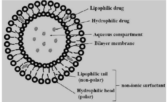
Figure 1: Structure of niosome
TYPES OF NIOSOMES
1] Small Unilamellar vesicles
It is single phospholipid or surfactant bilayer, and particle size range is 10 – 100 nm. It is high tissue penetration in nature and quick medication release.
2] Large Unilamellar vesicles
Which are consist of aqueous core surrounded by single bilayer and particle size range is 100 – 3000 nm. It can hold more hydrophilic medications then the small Unilamellar vesicles.
3] Multilamellar vesicles
It is aqueous core surrounded by several concentric bilayers and particle size range is larger than 500nm. It is made by thin film hydration [ handshaking ] technique.
IDEAL PROPERTIES
1] Niosomes are biocompatible and biodegradable.
2] Nontoxic or irritation.
3] It is bilayer structure so capable for entrap hydrophilic and lipophilic medications.
4] Physically and chemically stable.
5] Enhance bioavailability and penetration. [ Especially for topical and transdermal applications.]
6] Controlled and sustained release for prolonged therapeutical effect.
COMPONENTS OF NIOSOMES [3]
Two main components are used to produce the niosomes are,
1] Cholesterol
2] Nonionic surfactants
Cholesterol
It is giving proper shape and rigidity of the niosome formulations.
Nonionic surfactants:
Nonionic surfactants are surface active agent that are does not carry any charge. The non-ionic surfactants are playing a major role for niosome formulation.
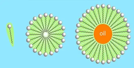
FIGURE 2: Structure of non-ionic surfactant.
ADVANTAGE OF NIOSOME
DISADVANTAGE
FORMULATION OF NIOSOMES
Cholesterol and surfactant (ratio of 1:1) dissolves in the mixture of organic solvent (ether and chloroform). Addition of the aqueous drug solution to this and water in oil emulsion is formed; two phases are sonicated at 4-5ºC. The emulsion is dried in a rotary evaporator at 40°C to form a semisolid gel of large vesicles. Small amounts of phosphate buffered saline (PBS) are added to the clear gel and sonicate again. The organic phase is removed at 40ºC and lower pressure. Viscous niosomal suspension is further diluted with phosphate buffered saline, then heat on a water bath at 60ºC for 10 min to form niosomes.[4]
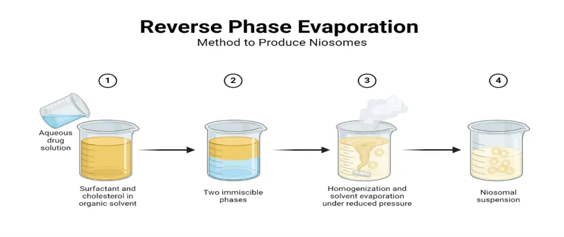
FIGURE3: Method of preparation by reverse phase evaluation.
Cholesterol and a non-ionic surfactant are dissolved in a volatile organic solvent (methanol: chloroform). To create a thin lipid layer, the solvent is evaporated using a rotary evaporator. While shaking, the thin film is hydrated with an aqueous drug solution above the surfactant transition temperature.
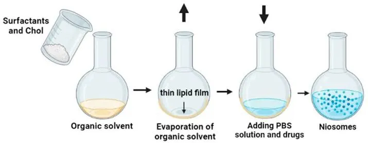
FIGURE 4: Method of preparation by Thin-Film Hydration method.
Diethyl ether is used to dissolve cholesterol and surfactant. A warm aqueous drug solution (approximately 60°C) is injected with the solution gradually. Single-layered niosomes are created when ether instantly evaporates.
Cholesterol and surfactant are mixed in a buffer and heated. As the mixture is homogenized, nitrogen gas is bubbled through it. Organic solvents are not used in the formation of niosomes.
An acidic buffer is used to hydrate a thin lipid film. After adding the drug solution, the pH is adjusted. Drug loading into the vesicles is improved by the pH gradient
The aqueous medication solution, cholesterol, and surfactant are combined.
For a few minutes, the mixture is sonicated at roughly 60°C.
Niosomal vesicles are created that are uniformly small.
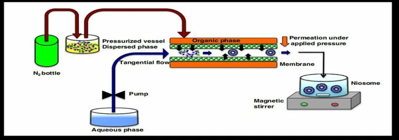
FIGURE4: method of preparation by sonication method.
GEL
Gels are semi solid homogenous preparation used to cure and treat topical diseases. Gels are more hydrophilic in nature, so the rate of released drug or active ingredient was fast. A gel consists of two component, three-dimensional cross-linked material which contain proportionally large amount of liquid medium to form adequate rigid network which immobilized the liquid continuous phase.[5]
IDEAL PROPERTIES
1. The gel need to be uniform and translucent.
2. The container should shatter easily when shear or force is applied during agitation.
3. The gel's composition must be inert.
4. There must be no stickiness in the gel.
5. No other ingredient in the recipe should come into contact with it.
6. The gel must demonstrate reliability.
The most often used polymers are hydroxyethyl cellulose, sodium carboxymethyl cellulose, Poloxamer 407, Carbopol 934, and Carbopol 940.
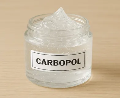
FIGURE 5: pictures of Carbopol and poloxamer
ADVANTAGE
GEL FORMATION MECHANISM
There are three types
A] Chemical crosslinking
B] Physical crosslinking
C] Ionic crosslinking
A. CHEMICAL CROSSLINKING
It involves the assembly of groups where cross-linking agents facilitate the interaction between polymers. This interaction triggers a negative reaction between the additive compounds and free radicals. Once a specific concentration is achieved, this reaction leads to an increase in viscosity, resulting in gel formation. An example of this is polyacrylic acid, which contains polycarboxylic acids. [7]
B. PHYSICAL CROSSLINKING
It can occur due to variations in concentration, temperature fluctuations, or the dissolution of crystalline components. Under these conditions, it is possible to obtain a hydrogen gel solution. Examples of physical crosslinking include cellulose gel and Sephadex. [7]
C. IONIC CROSSLINKING
It happens when polymers or other particles exchange ions during crosslinking, resulting in the creation of gel. Ionic bonds are easier to form when molecules have charges. For example, when calcium ions are present, polysaccharide alginate can produce a gel matrix that can encapsulate different substances, including enzymes.
FORMULATION ELEMENTS
The performance, size, and efficiency of a niosomal drug carrier depend completely on its foundational formulation components. A classic niosomal matrix requires three vital ingredients: non-ionic surfactants, membrane stabilizers, and charge inducers.
A. Non-Ionic Surfactants
The non-ionic surfactant is the primary building block forming the membrane bilayer. Common options include alkyl ethers, alkyl esters, alkyl amides, and fatty acids. Selection is dictated by two mains parameters:
Hydrophilic-Lipophilic Balance (HLB):
This parameter reflects surfactant solubility on a scale from 0 to 20. Surfactants with an HLB value falling strictly between 4 and 8 are typically ideal for spontaneous vesicle formation Hydrophilic surfactants with highly elevated HLB values (such as Tween 20 or Tween 80) have high aqueous solubility and fail to form bilayers alone but can successfully form exceptionally stable niosomes when combined with equimolar amounts of cholesterol.
Critical Packing Parameter (CPP): CPP predicts the dynamic shape of the resulting nanostructure based on molecular geometry via the formula:
CPP = v / (l c × a0)
where v represents the hydrophobic group volume, is the critical hydrophobic chain length, and a0 is the area of the hydrophilic polar headgroup. Stable bilayer vesicles occur precisely when the packing parameter is between
1/2 ≤ CPP ≤ 1
B. Membrane Stabilizers (Cholesterol)
Cholesterol is incorporated to establish hydrogen bonding networks between its 3-OH group and the polar headgroups of the surfactant It serves a dual role: it limits the freedom of liquid-state bilayers and acts as an organizing factor in rigid gel-state bilayers by abolishing the clear phase transition temperature cholesterol enhances structural rigidity, reduces membrane permeability, and prevents rapid payload leakage when exposed to biological fluids like serum or plasma.
C. Charge Inducers
Small quantities (2.5 to 5 mol%) of ionic amphiphiles are added to generate an operational surface charge on the vesicles This induces uniform electrostatic repulsion between adjacent vesicles, providing protection against aggregation, flocculation, or fusion during storage (Ukaegbus & Florence, 1995). Negative charges are induced using dicetyl phosphate (DCP) or Di hexadecyl phosphate, while positive charges are achieved via stearyl amine or methylpyridinium chloride.
DEVELOPMENT OF NIOSOMAL GEL
Carbopol 934 is dispersed in distilled water and allowed to hydrate completely. Propylene glycol containing preservatives is added with continuous stirring. The optimized niosomal suspension is incorporated slowly into the hydrated Carbopol gel. Triethanolamine is added dropwise to adjust the pH to 6.5–7.0 and obtain a smooth, homogeneous gel suitable for topical application.
STABILITY STUDY OF NIOSOMAL GEL
Period the optimized niosomal gel is stored under different conditions such as refrigerated temperature, room temperature, and accelerated conditions for up to three months. Samples are periodically evaluated for changes in appearance, pH, viscosity, drug content, vesicle size, entrapment efficiency, and drug release. A stable formulation shows minimal changes in these parameters throughout the study.
EVALVATION OF NIOSOMES
Wooden block and glass slide apparatus method.26 The mobile top portion of apparatus was kept on the gel (5g), which was placed on the bottom portion of apparatus. The Spreadability was measured by noting the time period for the top portion to move 5cm from the initial point.[8]
In vitro drug release
The niosomal suspension placed over a glass slide and fixed over by drying at room temperature, the dry thin film of niosomal suspension observed in the formation of vesicles. The microphotography of the niosomes also obtained from the microscope by using a digital camera[9]
Morphological characterization
Physical appearance and drug content and pH
The physical appearance of optimized gel was measured visually for colour, uniformity and consistency. The drug content of luliconazole loaded niosomal topical gel was estimated by spectrophotometrically at 299 nm after suitable dilution with solvent. The pH of the gel was determined using a digital pH meter.[8]
Zeta potential
Shows colloidal stability and surface charge. Because of the electrostatic repulsion between vesicles, values greater than ±30 mV indicate good stability.
Viscosity
A 100 g of accurately weighed drug loaded niosomal gel was used to measure a viscosity using a rotational Viscometer with spindle no. 96 (LVDV- III U, Brookfield, WI) at room temperature and estimate by CD-PROGA, DVA-80software.23-25 [8]
Spreadability
The spreadability study of gel was analyzed using It occurs by dialysis bag, Franz diffusion cel, reverse dialysis methods in simulated biological fluids.
Storage Stability
Niosomes are stored at different temperatures (4°C, 25°C, 40°C) and evaluated over time for size, PDI, zeta potential, EE%, and drug content as per ICH guidelines.
EVALUATION OF GEL
1. Measurement of pH
The pH of gel formulations is determined by digital pH meter. One gram of gel is dissolved in 100 ml distilled water and stored for two hours. The measurement of pH of each formulation is done in triplicate and average values are calculated. [10]
2. Drug content
1 g of the prepared gel is mixed with 100ml of suitable solvent. Aliquots of different concentration are prepared by suitable dilutions after filtering the stock solution and absorbance is measured. Drug content is calculated using the equation, which is obtained by linear regression analysis of calibration curve.[10]
3.Viscosity
The Brookfield Viscometer is used to measure the prepared gel's viscosity. The gel is rotated at 0.3, 0.6, and 1.5 revolutions per minute, and the appropriate dial reading is recorded at each speed. The dial reading is multiplied using the factor listed in the Brooke field Viscometer catalogues to determine the gel's viscosity.
4.Extrudability
Once the gels have set in the container, the formulas are put into the collapsible tubes. The weight in grams needed to extrude a 0.5 cm is used to calculate the formulation's extrudability. gel ribbon in ten seconds.
5.Consistency
Freeze-thaw cycling is used to conduct stability studies for every gel formulation. In order to see this synthesis, the product is heated to 4°C for one month, followed by 25°C and 40°C. Liquid exudates are observed to separate after this gel is exposed to room temperature.
EVALUATION PARAMETERS
|
EVALUATION PARAMETER |
STANDARD ANALYTICAL METHODOLOGY |
CRITICAL PHARMACEUTICAL SIGNIFICANCE |
|
Vesicle charge |
Dynamic light scattering [DLS], scanning/ transmission electron microscopy [SEM/TEM] optical microscopy. |
Governs in vivo clearance rates, biological distribution, and cellular uptake mechanisms. |
|
Surface charge |
Zetasizer instrument tracking electrokinetic mobility [Zeta potential]. |
High absolute value [ >> ±30 mV) confirm long-term electrostatic stability against vesicle fusion. |
|
Entrapment efficiency [EE%] |
Exhaustive dialysis, centrifugation, or gel chromatography to isolate free drug; vesicle lysis via Triton X-100/propan-1-ol |
Measures the exact percentage of initial drug successfully locked within the core or bilayer matrix. |
|
Bilayer properties |
Small-Angle X ray scattering [SAXS], atomic force microscopy [AFM], Fluorescence polarization via DPH probe. |
Determines overall membrane thickness, lamellarity counts, and micro viscosity/ rigidity indices. |
|
In vitro Drug Release |
Membrane diffusion cell or dialysis bag techniques in physiological butter media maintained at 37°C |
Profiles release kinetics over time to establish sustained efficacy. |
APPICATIONS OF NIOSOMAL GEL
Treatment for fungal infections
It is used to deliver antifungal drugs such as clotrimazole, improving penetration under the skin and increasing therapeutical effect.
Management of bacterial skin
Delivers antibiotics locally, providing sustained drug release and reducing systemic side effect.
Pain management
Anti inflammatory drugs for localized relief of muscle and join pain. And delivery analgesic effect.
Wound healing
Enhance delivery of antimicrobial and healing agents, promoting faster tissue repair.
Cosmetic and dermatological products
Used to deliver antioxidants, vitamins, and anti-aging compounds, improving skin hydration and reducing signs of aging.
Transdermal drug delivery
Enables controlled release of drugs through the skin, reducing dosing frequency and improving patient compliance.
Targeted topical therapy
Increases drug concentration at the site of action while minimizing systemic exposure and adverse effects.
Improved bioavailability
Enhances the absorption of poorly water-soluble drugs by increasing their penetration through the skin.
CONCLUSION
Niosomal gels are promising topical drug delivery system that are combines the advantage of niosome with the ease of application of gel. Niosomes increase medication penetration via the skin while improving stability, bioavailability, and controlled release. Improved patient compliance extended pharmacological action, and fewer systemic side effects are all benefits of incorporating niosomes into a gel foundation. The creation of a stable and efficient formulation is ensured by a variety of preparation techniques and evaluation criteria. Niosomal gels are widely used in the treatment of bacterial skin illnesses, fungal infections, wound healing, pain management, and cosmetic formulations because of these benefits. All things considered, niosomal gel is a sophisticated and successful topical medication delivery method with a great deal of promise for use in pharmaceutical and dermatological applications in the future.
REFERENCES

Jeevan Nithish S, Christopher Vimalson D, Alagarraja M, Naveen Yadav J. K, Naveen V, Farisha. P, Sinthan. S Chandru. M, Madhan. G, Swarnalatha. J, Taufiq Ahmed. S, Jayachandran. S, A Comprehensive Review on Niosomal Gel, Int. J. of Pharm. Sci., 2026, Vol 4, Issue 7, 3014-3024, https://doi.org/10.5281/zenodo.21374814
 10.5281/zenodo.21374814